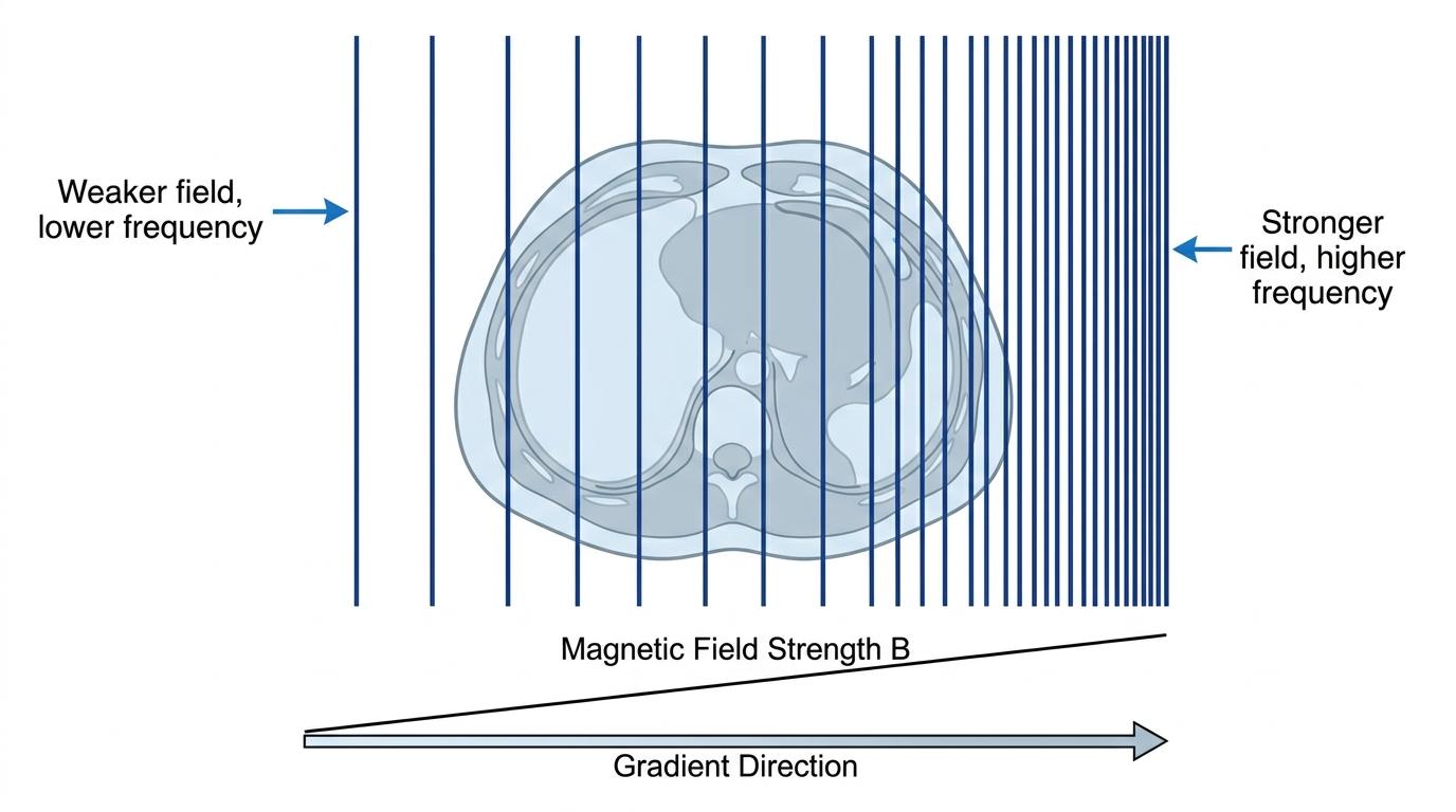
By sending an RF pulse of a very specific, narrow frequency, the scanner will only excite a single, thin "slice" (or successive small regions) of the patient's cross-section where the protons happen to be precessing at that exact frequency.
By varying the frequency of the RF pulses and using multiple gradient coils in different directions, the scanner can scan the entire cross-section of the patient step-by-step. Finally, the detected RF signals are processed by a computer to construct a detailed 3D visual image.
Worked Example: Explaining the MR process
Question: An MR scanner is used to image a patient's brain. Explain how the protons in the patient's brain produce the detected signal, and how gradient coils allow a cross-sectional image to be formed. (6 marks)
Answer:
- Protons (hydrogen nuclei) in the brain are initially aligned with their spins parallel to the strong magnetic field of the superconducting magnet.
- These spinning protons precess about the magnetic field lines.
- The scanner transmits short radio frequency (RF) pulses.
- The protons absorb this RF energy (excitation) and undergo a change of spin state.
- When the RF pulse stops, the excited protons de-excite (relax) and emit RF signals, which are detected by the receiver coils.
- Gradient field coils are used to vary the magnetic field strength across the cross-section, which changes the precession frequency of protons in different regions. This ensures the RF pulse only causes excitation in successive small regions, allowing the computer to map the origin of the signals and produce a visual image.
In the exam
When asked to describe the principles of MR scanners in an AQA exam, examiners are looking for very specific keywords. Make sure you hit these checklist items:
- Always state that it is hydrogen nuclei or protons that are involved.
- Mention the superconducting magnet and state that protons initially align with their spins parallel to the field.
- Use the word precess (not just spin or rotate) to describe their motion about the magnetic field lines.
- Remember the sequence: short RF pulses cause excitation and a change of spin state.
- Crucially, state that the protons emit RF signals as they de-excite.
- Remember the role of gradient field coils: they allow scanning of successive small regions (localising the signal) to process a visual image.
Check yourself
- What fundamental particle in the human body is targeted by an MR scanner?
- What happens to the spin state of these particles when subjected to the correct RF pulse?
- What is the purpose of the gradient field coils?
- Does the patient emit or reflect the RF signals that the scanner detects?