Physics of vision (A-level only)
What you'll learn
- How the eye refracts light to form a real, inverted image on the retina.
- The difference in sensitivity and spectral response between rod cells and cone cells.
- How the neural connections in the eye dictate our spatial resolution and our ability to see fine detail.
The eye as an optical refracting system
When you look at an object, your eye must gather the diverging light rays and bend them so that they converge perfectly on the back of your eye. The eye acts as a complex positive (convex) lens system.
There are two main refractive components in the eye:
- The cornea: The transparent, curved front surface of the eye.
- The lens: A flexible, crystalline structure located just behind the pupil.
You might assume that the lens does most of the work. In fact, the cornea is responsible for about 80%80\%80% of the eye's total refractive power. Refraction occurs when light changes speed as it passes from one medium to another. Because the difference in refractive index between air (n≈1.0n \approx 1.0n≈1.0) and the cornea (n≈1.38n \approx 1.38n≈1.38) is large, the light bends significantly here.
The lens (n≈1.4n \approx 1.4n≈1.4) is surrounded by the aqueous and vitreous humours (which have similar refractive indices to the cornea). Because the change in refractive index is small here, the lens bends the light much less. However, the lens is crucial because its shape can be adjusted by the ciliary muscles (a process called accommodation) to fine-tune the focal length and keep objects at different distances in sharp focus.
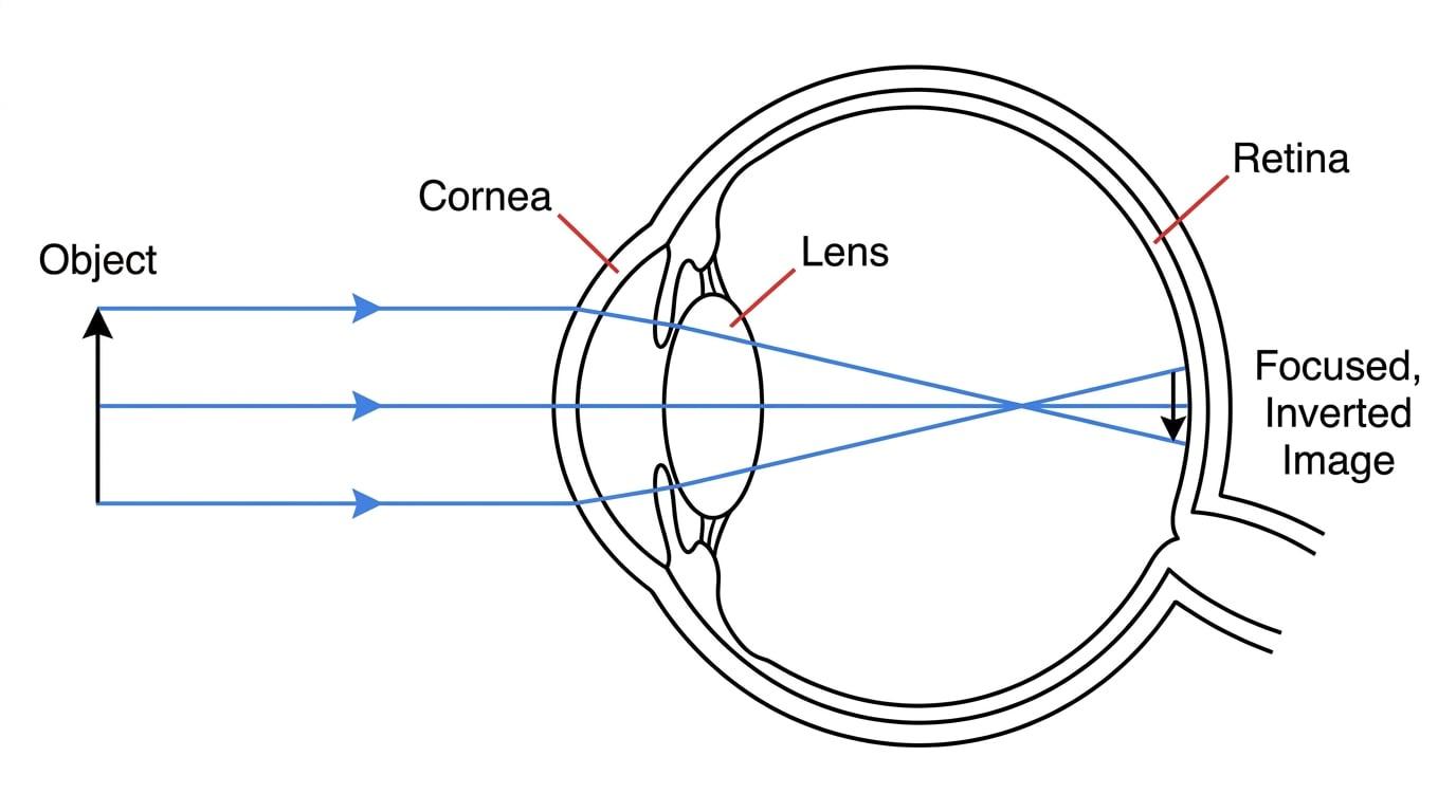
As shown in the diagram above, the light rays cross over before they hit the light-sensitive back wall of the eye (the retina). This means the image formed on the retina is real, inverted, and diminished (smaller than the object). Your brain automatically flips the image so that you perceive the world the right way up!
Refraction in the eye
The cornea provides most of the eye's refractive power due to the large change in refractive index between the air and the cornea. The lens provides the fine-tuning necessary for focusing on objects at different distances.
Sensitivity and spectral response
The retina is covered in millions of specialised light-detecting cells called photoreceptors. The eye acts as a biological photodetector, converting incident light energy (photons) into electrical impulses that travel down the optic nerve to the brain. There are two types of photoreceptor, named after their shapes: rods and cones.
Sensitivity
Sensitivity refers to how much light is required to trigger a response in the cell.
- Rods have very high sensitivity. They can be triggered by just a few photons, making them perfect for seeing in very dim light (nighttime vision). However, rods cannot distinguish between different colours.
- Cones have lower sensitivity. They require a much higher intensity of light to trigger an impulse, which is why they operate primarily in daylight. Cones are responsible for colour vision; humans have three types of cones, each sensitive to a different range of wavelengths (broadly corresponding to red, green, and blue).
Spectral Response
Spectral response
The spectral response of a photodetector (like the eye) is a measure of its sensitivity to different wavelengths of light across the electromagnetic spectrum.
The eye is not equally sensitive to all colours of visible light. The response depends heavily on whether you are using your rods or your cones.
- Photopic vision (daylight vision using cones) has a peak spectral response at around 555 nm555\text{ nm}555 nm, which is in the yellow-green region.
- Scotopic vision (nighttime vision using rods) has a peak spectral response at around 505 nm505\text{ nm}505 nm, which is in the blue-green region.
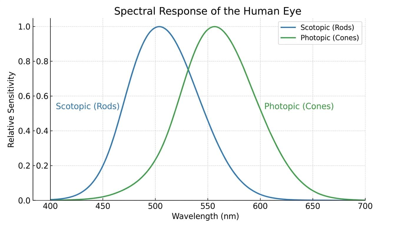
This shift in peak sensitivity as light levels drop is why a blue object and a red object that appear equally bright in daylight will look very different at twilight. As your eyes switch from using cones to using rods, the blue object will suddenly appear significantly brighter than the red object, because rods are much more sensitive to shorter (blue) wavelengths and almost entirely insensitive to red.
Worked Example: Spectral Response
A student observes a red flower and a blue flower that appear equally bright at midday. Explain, with reference to the spectral response of the eye, why the blue flower appears brighter than the red flower at twilight. (3 marks)
- At midday, light intensity is high, so the student's eyes use cones for photopic vision. The cones are sensitive to both red and blue wavelengths, allowing the flowers to appear equally bright.
- At twilight, light intensity is low, so the student's eyes switch to using rods for scotopic vision.
- The peak spectral response of rods is at approximately 505 nm505\text{ nm}505 nm (blue-green). Rods are highly sensitive to blue light but have virtually zero sensitivity to the longer wavelengths of red light, causing the blue flower to appear much brighter.
Spatial resolution
While rods are great for seeing in the dark, you wouldn't want to use them to read a book. This comes down to spatial resolution.
Spatial resolution
Spatial resolution is the ability to distinguish two separate points or details as distinct from one another. In the eye, it depends on how densely packed the photoreceptors are and how they connect to the optic nerve.
For two tiny spots of light to be resolved (seen as two separate spots rather than one blurry blob), their images must fall on the retina such that there is at least one unstimulated photoreceptor between the two stimulated ones.
Cones and high resolution
Cones are densely packed in the centre of the retina, in a small pit called the fovea. Crucially, each individual cone cell in the fovea has its own dedicated nerve fibre connecting it to the brain. Because there is a one-to-one connection, the brain knows exactly which specific cone was stimulated. This provides extremely high spatial resolution, allowing us to see fine detail when we look directly at an object.
Rods and retinal convergence
Rods are distributed mostly around the periphery (the outer edges) of the retina. Unlike cones, rods share nerve fibres. Several rods will connect to a single bipolar cell, which then connects to a single ganglion cell in the optic nerve. This sharing is called retinal convergence.
If light from two separate points hits two different rods that share the same nerve fibre, the brain only receives a single combined impulse. It cannot tell which specific rod was hit, only that one of the rods in that cluster was stimulated. Therefore, the brain perceives it as a single point of light. This means rods provide very poor spatial resolution.
Why do rods do this? It is an evolutionary trade-off to boost sensitivity. By pooling the signals from many rods, a single nerve fibre can reach its threshold potential and fire an impulse even if each individual rod only absorbed a tiny amount of light.
Mixing up sensitivity and resolution
Students frequently mix these two up!
- Rods: High sensitivity (due to retinal convergence pooling signals), but low spatial resolution (brain can't pinpoint the exact source).
- Cones: Low sensitivity (one-to-one connections mean they need a lot of light to fire), but high spatial resolution (brain can pinpoint exact source).
Worked Example: Explaining spatial resolution
Explain why reading small text in a dimly lit room is very difficult, referring to the distribution and neural connections of photoreceptors. (4 marks)
- In dim light, the eye relies on rods because cones require a higher light intensity to be stimulated.
- Rods are primarily located at the periphery of the retina, meaning they are not concentrated at the fovea where we usually focus images for reading.
- Rods exhibit retinal convergence, where multiple rod cells connect to a single nerve fibre.
- Because multiple rods share a neural pathway, the brain cannot distinguish between closely spaced details (such as small letters), resulting in poor spatial resolution.
The peripheral star trick
If you ever want to see a very faint star at night, don't look directly at it. Look slightly to the side! Looking directly puts the faint light on your fovea (which only has cones, and thus poor sensitivity). Looking to the side puts the light on your peripheral retina, which is packed with highly sensitive rods.
In the exam
- Be precise with vocabulary: Always use the terms "retinal convergence", "photopic", "scotopic", "spatial resolution", and "sensitivity" exactly as defined.
- Numbers matter: If an AQA question asks about spectral response, explicitly state the peak wavelengths (approx 555 nm555\text{ nm}555 nm for cones, 505 nm505\text{ nm}505 nm for rods).
- Link structure to function: Whenever discussing the one-to-one connection of cones or the many-to-one connection of rods, immediately follow up by stating the consequence (high resolution or high sensitivity, respectively).
- Identify the refracting surface: Remember that the largest change in refractive index happens at the air-cornea boundary, not at the lens.
Check yourself
- What type of image is formed on the retina by the eye's refracting system?
- Which part of the eye is responsible for the majority of its refracting power, and why?
- What is the peak wavelength for scotopic vision, and which photoreceptors are responsible for it?
- Explain the term 'retinal convergence' and state its effect on both sensitivity and spatial resolution.