Imaging techniques (A-level only)
Welcome to medical imaging! While you might already be familiar with X-rays from GCSE, X-rays only show us anatomy (the physical structure of bones and tissues). In this topic, we are going to look at how we can image function—how organs are actually working in real time.
What you'll learn:
- What a radioactive tracer is and the ideal properties of medical isotopes.
- The specific roles of Technetium-99m, Iodine-131, and Indium-111.
- How hospitals generate short-lived isotopes on-site using a Mo-Tc generator.
- The physics behind PET scans and matter-antimatter annihilation.
Radioactive Tracers
To see inside a working body, we need a signal to come out of it. We do this by putting a radioactive source into the patient and detecting the radiation that escapes.
Radioactive tracer
A radioactive tracer is a chemical compound in which one or more atoms have been replaced by a radioactive isotope. It is designed to be absorbed by a specific target organ or tissue in the body.
When the tracer is injected or swallowed, it travels through the bloodstream and concentrates in the target organ. As the isotope decays, it emits radiation. A detector outside the body (usually a gamma camera) picks up this radiation to build up an image of where the tracer has gathered.
The ideal tracer
Not just any radioactive isotope makes a good tracer. To get a clear image without harming the patient, an ideal medical tracer must meet four strict criteria:
- Radiation emitted: It should ideally emit only gamma radiation. Alpha and beta particles are highly ionizing and have very short ranges. They would be absorbed entirely by the patient's tissue, giving them a large, dangerous dose of radiation without any signal escaping the body to be detected. Gamma rays are highly penetrating and easily escape the body.
- Gamma energy: The gamma photons need a specific "goldilocks" energy—usually between 100 and 200 keV. If the energy is too low, the photons are absorbed by the patient. If the energy is too high, the photons pass straight through the detector outside without being registered.
- Half-life: It needs a half-life of hours or days. If the half-life is too short (seconds), it decays before you can even run the scan. If it is too long (years), the patient remains radioactive for an unsafe amount of time.
- Labelling: It must be chemically capable of being "labelled" (attached) to a compound that has a biological affinity for the specific organ you want to study.
Three Key Medical Isotopes
AQA requires you to know the specific properties of three isotopes.
1. Technetium-99m (Tc-99m)
Technetium-99m is the absolute superstar of medical imaging, used in over 80% of all scans. The "m" stands for metastable, meaning the nucleus is in an excited energy state and remains there for a relatively long time before dropping to its ground state.
- Radiation: It emits only gamma photons (no alpha or beta), meaning it delivers a very low radiation dose to the patient.
- Half-life: 6 hours. This is perfect: long enough to prepare the tracer and scan the patient, but short enough that the patient's radioactivity drops to safe levels within a couple of days.
- Gamma energy: Exactly 140 keV. This easily escapes the body but interacts strongly with the crystal inside a gamma camera.
- Labelling: It is chemically versatile and can be attached to dozens of different compounds to target the brain, bones, lungs, or kidneys.
2. Iodine-131 (I-131)
Iodine naturally accumulates in the thyroid gland (in your neck). Because of this biological fact, we don't even need to chemically label it to complex molecules!
- Radiation: Emits beta and gamma radiation.
- Half-life: 8 days.
- Use: The gamma radiation allows us to image the thyroid. However, because it emits beta particles (which are absorbed locally and destroy tissue), I-131 is often used for radiotherapy to treat hyperthyroidism or thyroid cancer.
3. Indium-111 (In-111)
- Radiation: Emits gamma radiation.
- Half-life: 2.8 days.
- Labelling: Indium-111 can be labelled to white blood cells or antibodies. When re-injected into the patient, these tagged white blood cells naturally flock to areas of infection or inflammation, allowing doctors to locate hidden abscesses or tumours.
Summary of the Big Three
- Tc-99m: The all-rounder. 6-hour half-life, gamma-only (140 keV), highly versatile labelling.
- I-131: The thyroid specialist. 8-day half-life, beta and gamma emitter.
- In-111: The infection hunter. 2.8-day half-life, gamma emitter, attaches to white blood cells.
The Molybdenum-Technetium Generator
We have a logistical problem. Technetium-99m has a half-life of just 6 hours. If a central nuclear reactor produces Tc-99m and puts it in a delivery van to a hospital, nearly all of it will have decayed into useless ground-state Technetium-99 by the time it arrives!
The solution is the Mo-Tc generator. Hospitals don't buy Tc-99m directly; they buy Molybdenum-99 (Mo-99).
Mo-99 has a half-life of 66 hours (nearly 3 days). This is long enough to easily transport from the reactor to the hospital. As the Mo-99 sits in the hospital, it decays via beta emission into the much-desired Tc-99m.
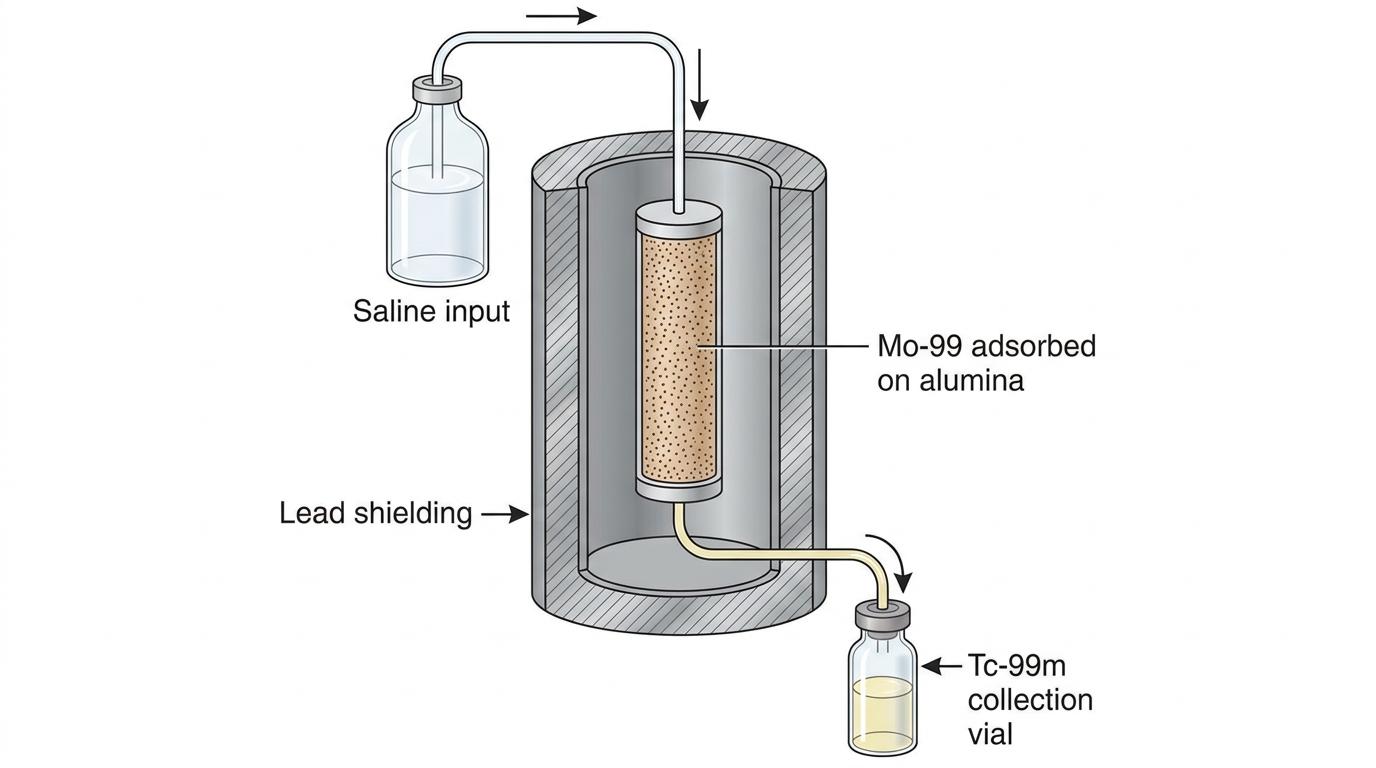
How it is used: The generator (sometimes affectionately called a "moly cow") consists of an alumina column containing the Mo-99.
- When the hospital needs Tc-99m for a scan, they wash a saline (saltwater) solution through the column.
- The Tc-99m dissolves in the saline, while the Mo-99 remains stuck to the alumina.
- The resulting radioactive saline drops into a collection vial, ready to be labelled and injected.
- This process is called elution (or "milking the cow").
AQA-style question: Mo-Tc generator
Question: Explain why hospitals use a Molybdenum-Technetium generator rather than having Technetium-99m delivered directly from a nuclear facility. (3 marks)
Model Answer:
- Technetium-99m has a very short half-life of 6 hours.
- If it were transported directly, too much of its activity would be lost during transit.
- Molybdenum-99 has a much longer half-life (66 hours), so it can be transported to the hospital with high activity, acting as an on-site source as it continuously decays into Tc-99m.
PET Scans (Positron Emission Tomography)
So far, we've talked about tracers that emit single gamma rays. PET scans use a much cleverer physical trick involving antimatter.
In a PET scan, the patient is injected with a tracer that is a positron emitter (a β+\beta^+β+ emitter). The most common tracer is FDG (a type of radioactive glucose). Because cancer cells and active brain cells use a lot of glucose, the FDG gathers in these highly active areas.
The Physics of the Scan
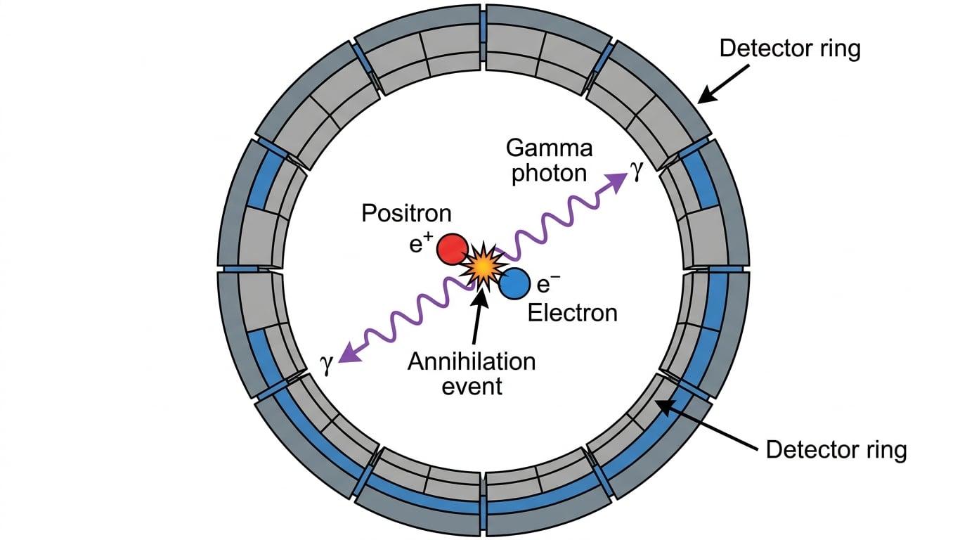
- The tracer decays inside the organ, emitting a positron (β+\beta^+β+).
- The positron travels roughly 1 mm1\text{ mm}1 mm through the tissue until it hits an electron (e−e^-e−).
- Annihilation occurs. The positron and electron destroy each other, converting their entire mass into pure energy.
- To conserve momentum, this energy is released as two identical gamma photons travelling in exactly opposite directions (180∘180^\circ180∘ to each other).
Detecting the signal
The patient lies inside a ring of gamma detectors. When the two gamma photons burst out of the patient in opposite directions, they hit the detector ring at exactly the same time.
The computer uses coincidence detection. It only registers a signal if two detectors on opposite sides of the ring trigger simultaneously. The computer then draws a straight line between those two detectors (a "line of response"). It knows the annihilation event must have happened somewhere along that exact line.
By analyzing millions of these lines intersecting, the computer can build an incredibly high-resolution 3D image of the organ's activity.
PET vs Gamma Camera
Don't confuse a PET scanner with a standard gamma camera!
- A standard tracer (like Tc-99m) emits one gamma photon at a time in a random direction.
- A PET tracer emits a positron, which causes an annihilation, resulting in two gamma photons travelling in exactly opposite directions.
Calculating PET Photon Energy
Because PET relies on matter-antimatter annihilation, it is a perfect opportunity for examiners to test your knowledge of rest mass energy from the Particles topic (E=mc2E = mc^2E=mc2).
Worked Example: Energy of PET photons
Question: In a PET scan, a positron annihilates with an electron. Calculate the frequency of one of the emitted gamma photons. (Rest mass of an electron/positron = 9.11×10⁻³¹ kg9.11 \times 10^{-31}\text{ kg}9.11×10⁻³¹ kg, c=3.00×10⁸ m s−1c = 3.00 \times 10^8\text{ m s}^{-1}c=3.00×10⁸ m s−1, h=6.63×10⁻³⁴ J sh = 6.63 \times 10^{-34}\text{ J s}h=6.63×10⁻³⁴ J s)
Step-by-step solution:
- Recognise that two particles are annihilating (an electron and a positron). They have the same mass. The total mass destroyed is:
- Calculate the total energy released using E=mc2E = mc^2E=mc2:
- Because two identical gamma photons are produced to conserve momentum, the energy is split equally between them. The energy of one photon is half the total (which is simply the rest energy of one electron!):
- Finally, use E=hfE = hfE=hf to find the frequency of the gamma photon:
Shortcut for Annihilation Math
Because the total mass (2me2m_e2me) turns into two photons (2γ2\gamma2γ), the energy of one gamma photon is exactly equal to the rest mass energy of one electron (mec2m_e c^2mec2). You don't actually need to multiply by two and then divide by two! Just evaluate E=mec2E = m_e c^2E=mec2.
In the exam
- Be specific about gamma. If asked why a tracer is used, explicitly state that gamma is highly penetrating (escapes the body) and minimally ionizing (reduces patient dose). Do not just say "it is safe".
- Know your half-life times. You must be able to state that Tc-99m is roughly 6 hours, Mo-99 is roughly 66 hours, In-111 is days, and I-131 is 8 days.
- Mo-Tc generator questions are common. The key mark is always for stating that Mo-99's longer half-life allows it to be transported without dropping to low activity levels.
- PET conservation. If asked why two photons are produced in a PET scan, the required A-level physics phrase is "to conserve momentum" (the original electron-positron pair had roughly zero net momentum).
Check yourself
- Can you list three reasons why Technetium-99m is considered the ideal medical tracer?
- Why is Iodine-131 suitable for imaging the thyroid without needing complex chemical labelling?
- What is the specific role of the saline wash in a Mo-Tc generator?
- In a PET scan, what particle is emitted by the tracer, and what is the angle between the two resulting gamma rays?