Time is of the essence
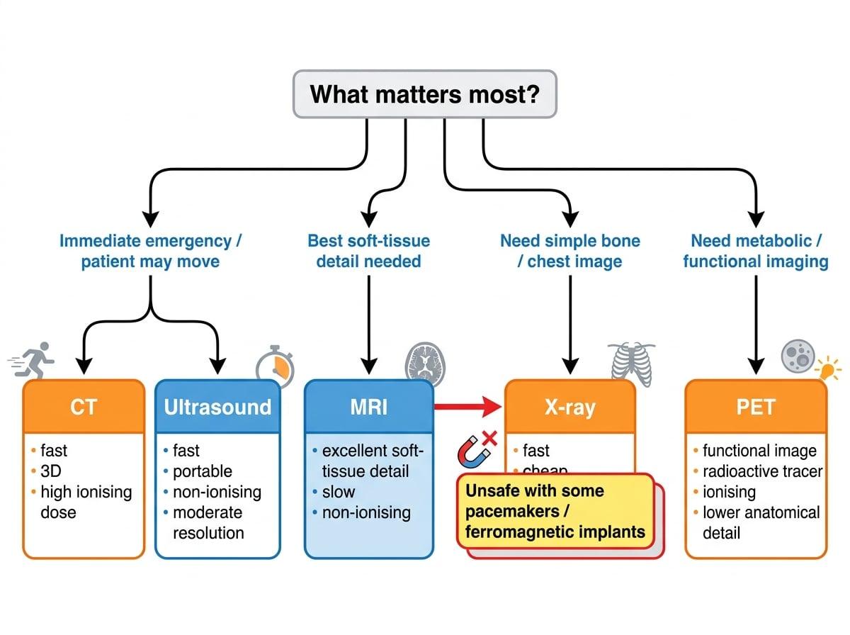
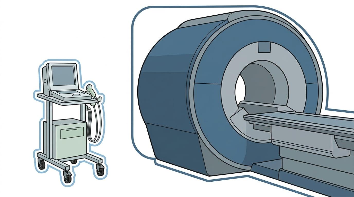
If an exam question mentions an "emergency" or a "restless" patient (like a young child or someone in pain), lean heavily towards the speed of CT or Ultrasound over the slow, movement-sensitive MRI.
Example 2: Justifying Convenience
Why might a hospital choose to perform a CT scan rather than an MRI for a patient who has just arrived in A&E with severe head trauma following a car crash?
- Identify the patient's state: They have severe trauma, meaning they are likely in pain, possibly uncooperative, or require immediate life-saving intervention.
- Compare the scan times: An MRI scan takes 30+ minutes and requires the patient to remain perfectly still, whereas a CT scan takes only a few minutes.
- Compare the environment: MRI machines restrict access to the patient and limit the use of metallic life-support equipment. CT allows faster, easier access.
- Conclude: CT is far more convenient and appropriate for rapid emergency triage, despite MRI potentially offering better soft-tissue detail of the brain.
3. Comparing Safety Issues
Medical imaging introduces energy into the human body. Assessing safety is about weighing the diagnostic benefit against the potential harm caused by that energy.
The biggest dividing line in imaging safety is ionising vs non-ionising radiation.
Ionising Radiation Risks
Ionising radiation (X-rays, gamma rays) carries enough energy to remove electrons from atoms, which can damage cellular DNA and increase the long-term risk of cancer. Its use must always be medically justified.
Non-Ionising Techniques (The Safest)
- Ultrasound: Uses high-frequency sound waves. It is non-ionising and has no known harmful side effects at diagnostic intensities. This is why it is universally the standard for monitoring fetal development during pregnancy.
- MRI: Uses strong magnetic fields and radio-frequency waves. It is entirely non-ionising. However, the immense magnetic field (often 1.51.51.5 to 3.0 T3.0 \text{ T}3.0 T) is a severe safety hazard for patients with magnetic implants (like older pacemakers, cochlear implants, or surgical pins). The field can forcefully rip magnetic metals through tissue or induce currents that destroy delicate electronics.
Ionising Techniques (Require Careful Dose Management)
- Conventional X-ray: Uses a low dose of ionising radiation. The risk is very small, but still present.
- CT Scan: Because a CT scan takes hundreds of X-ray slices to build a 3D image, the radiation dose is significantly higher than a standard X-ray (sometimes equivalent to hundreds of chest X-rays). It carries a measurable increased lifetime cancer risk, especially in children.
- Radionuclide Imaging (PET / Gamma): Involves injecting the patient with a radioactive isotope that emits gamma radiation from inside the body. The patient becomes a temporary radiation source and continues to receive a dose (and emit radiation) until the tracer decays or is excreted.
Confusing the source of radiation
Students often write that "MRI uses radiation". It uses radio waves (which are part of the electromagnetic spectrum), but in physics exams, the word "radiation" without qualification usually implies ionising radiation. Always specify that MRI is non-ionising.
Example 3: Weighing Safety Factors
A patient requires imaging to investigate a suspected brain tumour. The patient has a metallic cardiac pacemaker fitted. Compare the safety of using MRI versus a CT scan for this patient, and state which must be used.
- State the safety profile of MRI: MRI is non-ionising but uses exceptionally strong magnetic fields.
- Apply this to the patient: The magnetic field would induce currents in the pacemaker, destroying it, or physically move it, posing a direct threat to the patient's life.
- State the safety profile of CT: CT uses ionising radiation (X-rays), which carries a small long-term risk of inducing cancer.
- Compare and conclude: The immediate, fatal risk posed by the MRI's magnetic field to the pacemaker far outweighs the long-term statistical risk of the CT's ionising radiation. Therefore, the CT scan must be used.
Summary Matrix
When preparing for long-answer comparative questions, keep this mental checklist handy:
| Technique | Resolution | Convenience | Safety |
|---|---|---|---|
| Ultrasound | Moderate (depends on freq) | Excellent (cheap, portable, fast) | Excellent (non-ionising) |
| X-ray | Good (2D structural) | High (fast, accessible) | Good (low dose ionising) |
| CT | Excellent (3D structural) | Good (fast scan, large machine) | Poor (high dose ionising) |
| MRI | Excellent (soft tissue) | Poor (slow, expensive, noisy) | Moderate (non-ionising, but magnetic hazard) |
| PET | Poor (but functional) | Poor (cyclotron needed, slow) | Poor (internal ionising dose) |
In the exam
- Read the clinical context: The exam board doesn't just ask "Compare MRI and CT". They ask "Evaluate MRI vs CT for a restless 5-year-old child". The context dictates the marks.
- Hit all three criteria: If asked for a full comparison, explicitly use the headings "Resolution", "Convenience", and "Safety" to ensure you don't miss any specification points.
- Be specific with hazards: Don't just say "radiation is bad". Say "CT uses high-dose ionising radiation which can damage DNA and increase cancer risk."
- Define the trade-off: Always conclude by clearly stating which factor overrides the others (e.g., "Therefore, despite the poorer resolution, Ultrasound is chosen for its safety.")
Check yourself
- Which imaging technique provides the best soft-tissue resolution without using ionising radiation?
- Why is an on-site cyclotron often necessary for a hospital offering PET scans?
- If a patient has a piece of metallic shrapnel embedded in their leg, which high-resolution imaging technique becomes completely unsafe?
- Why might a doctor choose a simple X-ray over a CT scan for a suspected broken arm, despite CT offering 3D imagery?