

What you'll learn
- How radioactive decay changes atomic number and mass number, including positron emission and electron capture.
- How α, β and γ radiation behave, how half-life works, and why radioisotopes are useful but risky.
- How ionisation energies reveal electronic structure.
- How orbitals, electron configurations and atomic spectra link to quantised electron energy levels.
1. Atoms, nuclei and why models matter
An atom has a tiny, positively charged nucleus containing protons and neutrons, with electrons arranged around it in energy levels. Different models of the atom explain different evidence: the nuclear model explains radioactivity, while shells and orbitals help explain ionisation energies and spectra.
Scientific models are not “guesses”; they are evidence-based explanations that improve over time as new observations are made.
Atomic number and mass number
- Atomic number, ZZZ, is the number of protons in the nucleus.
- Mass number, AAA, is the total number of protons and neutrons.
- Isotopes are atoms of the same element with the same ZZZ but different AAA.
- A radioisotope is an isotope with an unstable nucleus that undergoes radioactive decay.
2. Radioactive decay
Radioactive decay
Radioactive decay is the spontaneous, random change of an unstable nucleus into a more stable nucleus, with emission of radiation.
“Random” means you cannot predict exactly when one nucleus will decay. However, with a very large number of nuclei, you can predict the overall rate statistically.
Types of nuclear decay
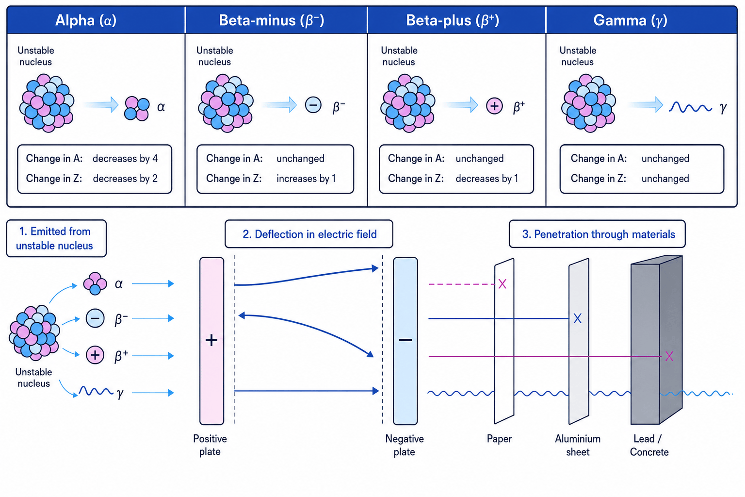
In nuclear equations, the total mass number and total atomic number must balance.
Alpha emission:
ZAX→Z−2A−4Y+24He{}^{A}_{Z}\text{X} \to {}^{A-4}_{Z-2}\text{Y} + {}^{4}_{2}\text{He}ZAX→Z−2A−4Y+24HeBeta-minus emission:
ZAX→Z+1AY+−10e{}^{A}_{Z}\text{X} \to {}^{A}_{Z+1}\text{Y} + {}^{0}_{-1}\text{e}ZAX→Z+1AY+−10ePositron emission:
ZAX→Z−1AY++10e{}^{A}_{Z}\text{X} \to {}^{A}_{Z-1}\text{Y} + {}^{0}_{+1}\text{e}ZAX→Z−1AY++10eElectron capture:
ZAX+−10e→Z−1AY{}^{A}_{Z}\text{X} + {}^{0}_{-1}\text{e} \to {}^{A}_{Z-1}\text{Y}ZAX+−10e→Z−1AYGamma emission releases energy only, so AAA and ZZZ are unchanged.
Balancing an electron-capture equation
Complete the equation for electron capture by potassium-40.
- In electron capture, an inner electron is captured by the nucleus, so the electron appears on the left-hand side:
- Balance mass numbers:
so A=40A = 40A=40.
- Balance atomic numbers:
so Z=18Z = 18Z=18.
- Atomic number 18 is argon, so:
Beta decay and mass number
In beta-minus, beta-plus and electron capture processes, the mass number does not change. The particle involved has mass number 0, so only the atomic number changes.
3. Alpha, beta and gamma radiation
Alpha, beta and gamma radiation differ in charge, mass, ionising power and penetrating power.
- Alpha radiation is made of helium nuclei, 24He{}^{4}_{2}\text{He}24He. It has charge +2, is strongly ionising, but is stopped by paper or skin.
- Beta-minus radiation is made of fast electrons. It has charge −1, is moderately penetrating, and is stopped by a few millimetres of aluminium.
- Beta-plus radiation is made of positrons. A positron has the same mass as an electron but charge +1.
- Gamma radiation is high-energy electromagnetic radiation. It has no mass or charge, is weakly ionising per collision, but is very penetrating and needs thick lead or concrete to reduce it.
In an electric field, charged radiation is deflected: positive particles bend towards the negative plate, negative particles bend towards the positive plate, and gamma radiation is not deflected. In a magnetic field, alpha and beta radiation are also deflected in opposite directions; gamma is unaffected.
4. Half-life
Half-life
The half-life of a radioisotope is the time taken for half the unstable nuclei in a sample to decay, or for the corrected count rate to fall to half its initial value.
Half-life is not affected by temperature, pressure or chemical state. It is a nuclear property, not a chemical one.
Background radiation
A detector records background radiation as well as radiation from the sample. In half-life calculations using count rate, subtract the background count rate before using ratios.
Using half-life with count rate
A radioactive sample has a measured count rate of 820 counts min⁻¹. The background count rate is 20 counts min⁻¹. Later, the measured count rate is 70 counts min⁻¹. The half-life is 6.0 h. Find the time elapsed.
-
Correct both count rates by subtracting background:
initial corrected count rate = 820 − 20 = 800 counts min⁻¹
later corrected count rate = 70 − 20 = 50 counts min⁻¹ -
Find the ratio:
Since 16=2416 = 2^416=24, four half-lives have passed.
- Calculate the time:
5. Radiation: risks and uses
Ionising radiation can remove electrons from atoms and molecules in living cells. This can damage DNA and proteins, causing mutations, cancer, burns or cell death. Risk depends on dose, exposure time, radiation type and whether the source is outside or inside the body.
Benefit versus risk
Radioisotopes are useful because their radiation is detectable and their decay is predictable, but their use must be justified by benefit, minimised dose, shielding, distance, exposure-time control and safe disposal.
Important uses include:
- Medicine: technetium-99m for diagnostic imaging; iodine-131 for thyroid treatment; cobalt-60 for radiotherapy or sterilising equipment.
- Health and biological analysis: tracers can follow movement of substances through organs or biochemical pathways.
- Radio-dating: carbon-14 dating estimates the age of once-living material using known half-life.
- Industry: thickness gauges, leak detection in pipes and checking material integrity.
- Analysis: emitted radiation can help identify trace elements, for example in neutron activation analysis.
Ethically, you should weigh diagnostic or industrial benefit against radiation dose, consent, worker safety, environmental contamination and radioactive waste.
6. Ionisation energy and electronic structure
First ionisation energy
The first standard molar ionisation energy is the enthalpy change when one mole of gaseous atoms each loses one electron to form one mole of gaseous 1+ ions.
X(g)→X+(g)+e−\text{X(g)} \to \text{X}^{+}\text{(g)} + \text{e}^{-}X(g)→X+(g)+e−Ionisation energies are positive because energy is needed to remove an electron attracted to the nucleus.
Across a period, first ionisation energy generally increases because nuclear charge increases while shielding changes only slightly. Down a group, it generally decreases because the outer electron is farther from the nucleus and more shielded.
Successive ionisation energies always increase, but a large jump shows that the next electron is being removed from an inner shell. This reveals the number of electrons in the outer shell.
Using successive ionisation energies
An element has successive ionisation energies of 738, 1451, 7733 and 10543 kJ mol⁻¹. Deduce the group.
-
Compare the increases between values. The biggest jump is between the second and third ionisation energies: from 1451 to 7733 kJ mol⁻¹.
-
The first two electrons are removed relatively easily, but the third is much harder to remove because it is from an inner shell.
-
Therefore the atom has two outer-shell electrons, so it is in Group 2.
7. Orbitals and electron configurations
Atomic orbital
An atomic orbital is a region around the nucleus that can hold up to two electrons with opposite spins.
Electrons occupy shells, labelled by principal quantum number nnn, and sub-shells, labelled s, p and d.
- An s sub-shell contains one orbital, so it holds 2 electrons.
- A p sub-shell contains three orbitals, so it holds 6 electrons.
- A d sub-shell contains five orbitals, so it holds 10 electrons.
An s orbital is spherical. A p orbital is dumbbell-shaped, with three orientations: pxp_xpx, pyp_ypy and pzp_zpz.
For elements 1–36, the usual filling order is:
1s, 2s, 2p, 3s, 3p, 4s, 3d, 4p1s,\ 2s,\ 2p,\ 3s,\ 3p,\ 4s,\ 3d,\ 4p1s, 2s, 2p, 3s, 3p, 4s, 3d, 4p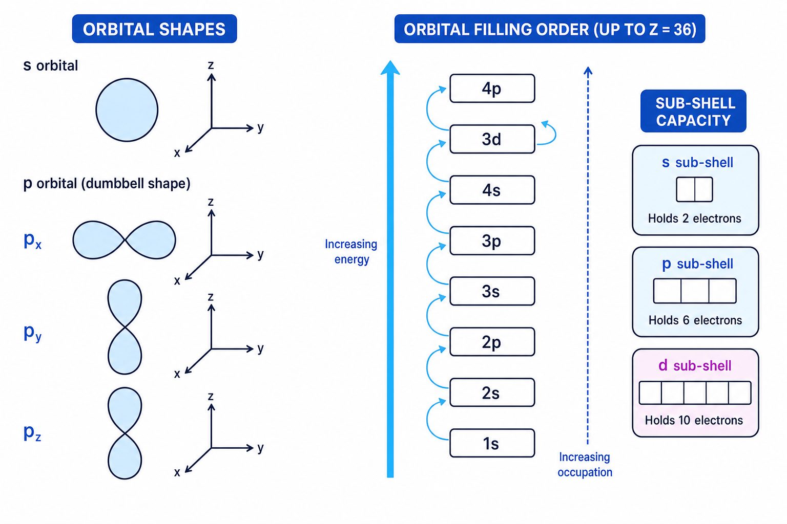
Writing an electron configuration
Write the electron configuration of bromine, atomic number 35.
-
A neutral bromine atom has 35 electrons, because it has 35 protons.
-
Fill sub-shells in order up to argon, which accounts for 18 electrons:
-
Continue using the filling order: add 4s24s^24s2 to reach 20 electrons, then 3d103d^{10}3d10 to reach 30 electrons.
-
Put the remaining 5 electrons in 4p4p4p:
Chromium and copper
For neutral atoms, chromium is [Ar] 3d5 4s1\text{[Ar] }3d^5\,4s^1[Ar] 3d54s1 and copper is [Ar] 3d10 4s1\text{[Ar] }3d^{10}\,4s^1[Ar] 3d104s1, rather than the simple predicted arrangements. Half-filled and full d sub-shells are especially stable.
8. Spectra, photons and electron transitions
Atoms only allow electrons to have certain energies. These are quantised energy levels.
Line spectrum
A line spectrum is a pattern of separate wavelengths of light emitted or absorbed by atoms, caused by electron transitions between quantised energy levels.
In an emission spectrum, an electron falls from a higher energy level to a lower one and emits a photon. In an absorption spectrum, an electron absorbs a photon and moves to a higher energy level.
The photon energy equals the energy gap:
E=hfE = hfE=hfwhere EEE is energy, hhh is the Planck constant and fff is frequency. Frequency and wavelength are related by:
f=cλf = \frac{c}{\lambda}f=λcwhere ccc is the speed of light and λ\lambdaλ is wavelength.
Increasing energy order is:
Einfrared<Evisible<EultravioletE_\text{infrared} < E_\text{visible} < E_\text{ultraviolet}Einfrared<Evisible<EultravioletSo ultraviolet has higher frequency and shorter wavelength than visible light; infrared has lower frequency and longer wavelength.
Hydrogen emission spectrum and the Lyman limit
Hydrogen has one electron, so its spectrum is especially important evidence for quantised energy levels. Transitions down to n=1n = 1n=1 form the Lyman series, which is in the ultraviolet. Transitions down to n=2n = 2n=2 form the Balmer series, much of which is visible.
The lines in a series get closer together and converge. The Lyman convergence limit corresponds to an electron falling from n=∞n = \inftyn=∞ to n=1n = 1n=1. Its frequency is linked to the ionisation energy of hydrogen from the ground state:
IEH=hflimitNAIE_\text{H} = h f_\text{limit} N_AIEH=hflimitNA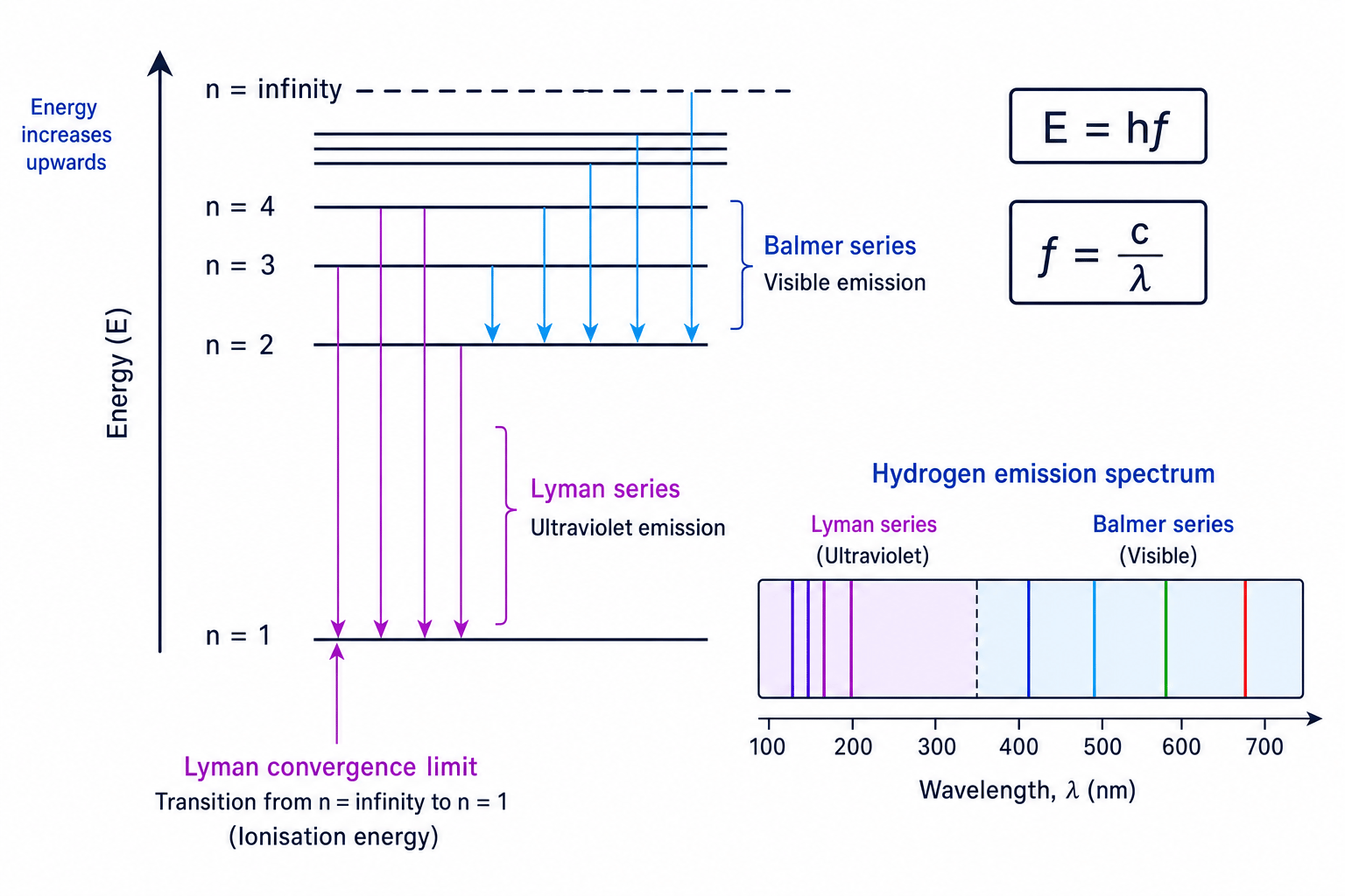
Calculating hydrogen ionisation energy from the Lyman limit
The Lyman convergence limit has wavelength 91.2 nm. Calculate the molar ionisation energy of hydrogen. Use h=6.626×10⁻³⁴ J sh = 6.626 \times 10^{-34}\ \text{J s}h=6.626×10⁻³⁴ J s, c=3.00×10⁸ m s−1c = 3.00 \times 10^8\ \text{m s}^{-1}c=3.00×10⁸ m s−1 and NA=6.022×10²³ mol−1N_A = 6.022 \times 10^{23}\ \text{mol}^{-1}NA=6.022×10²³ mol−1.
- Convert wavelength into metres:
- Calculate frequency:
- Calculate energy per atom:
- Convert to molar ionisation energy:
In the exam
- For nuclear equations, balance mass number and atomic number separately; remember that electron capture puts an electron on the reactant side.
- For half-life questions, subtract background count rate first, then use powers of two or repeated halving.
- For ionisation energy and spectra calculations, watch units carefully: nm to m, J mol⁻¹ to kJ mol⁻¹, and identify big jumps or convergence limits.
Check yourself
- A nuclide emits a positron. What happens to its mass number and atomic number?
- A set of successive ionisation energies has a large jump after the fifth electron is removed. What does that tell you about the outer shell?
- How would you use the Lyman convergence limit to calculate the ionisation energy of hydrogen?
