The diagram shows three types of neurones involved in a spinal reflex arc: Neurone P, Neurone Q, and Neurone R.
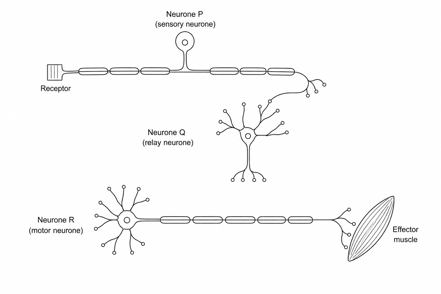
Which of the following statements correctly describes a structural or functional feature of these neurones?
In Neurone P, impulses travel from the receptor to the cell body along a long, myelinated axon, and then from the cell body to the synaptic terminals along a short dendron.
The cell body of Neurone P is located within the grey matter of the spinal cord, whereas the cell body of Neurone R is located within the dorsal root ganglion.
Action potentials travel via saltatory conduction in both Neurone P and Neurone R, whereas conduction along Neurone Q is slower due to the absence of a myelin sheath.
During a reflex action, impulses are transmitted from Neurone R to Neurone Q, which then coordinates the response and excites Neurone P.

