Cellular control
What you'll learn
- How gene mutations can change protein production and protein function.
- How cells control gene expression at the transcriptional, post-transcriptional and post-translational levels.
- How Homeobox and Hox genes help control body plan development.
- Why mitosis and apoptosis are both essential for forming the correct body shape.
Starting point: why cellular control matters
A cell’s behaviour depends on which genes are being used, when they are used, and how strongly they are used. This matters because many metabolic reactions are controlled by enzymes, and enzymes are proteins coded for by genes.
Most specialised cells in one organism contain the same DNA, but they do not all make the same proteins. A neurone, a muscle cell and an epithelial cell behave differently because they express different sets of genes.
Gene expression
Gene expression is the process by which information in a gene is used to make a functional product, usually a protein. It includes transcription of DNA into mRNA and, for protein-coding genes, translation of mRNA into a polypeptide.
Gene mutations
A nucleotide is a DNA or RNA monomer made from a sugar, a phosphate group and a nitrogen-containing base. A gene mutation is a change in the nucleotide sequence of a gene.
Gene mutation
A gene mutation is a change in the sequence of one or more nucleotides in DNA. It may affect the sequence of codons, the amino acid sequence of a protein, and therefore the protein’s structure and function.
Substitution mutations
A substitution is when one nucleotide is replaced by another.
Because the genetic code is degenerate, meaning that more than one codon can code for the same amino acid, a substitution does not always change the protein. Possible outcomes include:
- Silent effect: the codon changes but still codes for the same amino acid.
- Amino acid change: the codon now codes for a different amino acid.
- Premature stop codon: the codon becomes a stop codon, so translation stops early.
If the changed amino acid is in an enzyme’s active site or affects folding, the protein may lose function. If the change is in a less important region, the effect may be neutral.
Insertion and deletion mutations
An insertion is when one or more nucleotides are added. A deletion is when one or more nucleotides are removed.
Codons are read in groups of three nucleotides. If an insertion or deletion is not in a multiple of three, it causes a frameshift. This changes the reading frame, so every codon after the mutation may be different.
Insertions or deletions of three nucleotides, or multiples of three, may add or remove amino acids without shifting the whole reading frame.
Mutations are not automatically harmful
A mutation can be harmful, neutral or beneficial. Its effect depends on how it changes the protein and on the environment. For example, a mutation that improves survival in one environment may be useless or harmful in another.
Predicting the effect of a nucleotide insertion
Original mRNA sequence: 5′-AUG GAA UUU CCG UAA-3′
Mutated mRNA sequence after inserting C after the first codon: 5′-AUG CGA AUU UCC GUA A...-3′
-
The original mRNA is read in triplets: AUG, GAA, UUU, CCG, UAA. This gives a defined amino acid sequence followed by a stop codon.
-
After the insertion, the first codon remains AUG, but all codons after it are regrouped: CGA, AUU, UCC, GUA. The reading frame has shifted.
-
Because the downstream codons have changed, the amino acid sequence after the mutation will be different, and the original stop codon may no longer be in the correct frame. This is likely to produce a non-functional protein, especially if the altered region affects folding or an active site.
Control of gene expression
Cells can control gene expression at several stages:
- Transcriptional level: controlling whether mRNA is made from DNA.
- Post-transcriptional level: controlling what happens to the primary mRNA after transcription.
- Post-translational level: controlling the activity of the protein after translation.
This allows cells to respond to signals and control metabolism without changing the DNA sequence itself.
Transcriptional control: the lac operon
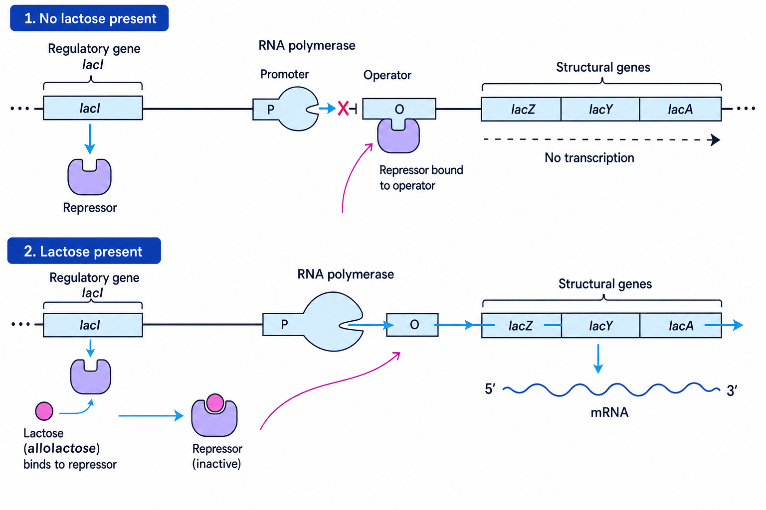
The lac operon is a classic example of gene control in bacteria. It controls genes needed for lactose metabolism.
An operon is a group of genes controlled together by the same regulatory region. The lac operon includes:
- a promoter, where RNA polymerase binds;
- an operator, where a repressor protein can bind;
- structural genes, including lacZ, lacY and lacA, which code for proteins involved in lactose use.
A nearby regulatory gene, lacI, codes for a repressor protein.
When lactose is absent
The repressor binds to the operator. This blocks RNA polymerase from transcribing the structural genes, so the enzymes for lactose metabolism are not produced.
When lactose is present
A form of lactose called allolactose binds to the repressor. This changes the repressor’s shape, so it can no longer bind to the operator. RNA polymerase can now transcribe the structural genes, and the bacterium produces enzymes needed to use lactose.
Predicting lac operon activity
A bacterium is moved into a medium containing lactose.
-
Lactose is converted into allolactose, which binds to the lac repressor and changes its shape.
-
The repressor can no longer bind to the operator, so the operator is unblocked and RNA polymerase can move along the structural genes.
-
The structural genes are transcribed, producing mRNA that is translated into proteins needed for lactose uptake and breakdown.
Regulatory gene versus operator
Do not confuse the regulatory gene with the operator. The regulatory gene codes for the repressor protein; the operator is a DNA sequence where the repressor binds.
Transcription factors in eukaryotes
A transcription factor is a protein that binds to specific DNA sequences and affects transcription.
In eukaryotes, transcription factors can:
- help RNA polymerase bind to DNA, increasing transcription;
- block transcription, reducing gene expression;
- allow different cell types to express different genes;
- respond to signals from inside or outside the cell.
You are not expected to recall named transcription factors for this specification point. Focus on their general role: they regulate whether genes are transcribed and how much mRNA is produced.
Good wording for transcription factors
If asked for the role of transcription factors, say that they bind to specific DNA sequences and increase or decrease the rate of transcription of target genes.
Post-transcriptional control: editing primary mRNA
In eukaryotic cells, transcription produces a primary mRNA transcript. This is not always ready for translation.
The primary mRNA contains:
- exons, which are sequences usually kept in the mature mRNA;
- introns, which are non-coding sequences removed before translation.
During post-transcriptional control, the primary mRNA is edited. Introns are removed and exons are joined together to produce mature mRNA. This mature mRNA can leave the nucleus and be translated by ribosomes.
If introns are not removed correctly, the codon sequence may change, a premature stop codon may appear, or the final protein may be non-functional.
Post-translational control: activation by cyclic AMP
Post-translational control happens after a protein has been made. The cell can activate or inhibit proteins that already exist, which is often faster than making new proteins from scratch.
Cyclic AMP, often written as cAMP, is a small signalling molecule called a second messenger. It can activate proteins inside the cell. For example, cAMP may activate a protein kinase, which is an enzyme that adds phosphate groups to other proteins. This phosphorylation can change a target protein’s shape and activity.
Different levels, different speeds
Transcriptional and post-transcriptional control alter how much protein is produced. Post-translational control alters the activity of proteins that have already been produced, so it can create a rapid response.
Genetic control of body plans
A body plan is the overall arrangement of an organism’s body structures, such as the positions of the head, limbs, segments or organs.
Development depends on cells dividing, differentiating and organising themselves in the right places. This is controlled by patterns of gene expression.
Homeobox genes
Homeobox genes contain similar, highly conserved DNA sequences. Highly conserved means that the sequence has changed very little during evolution.
Homeobox genes are found in plants, animals and fungi. They are involved in regulating gene expression, often by coding for proteins that act as transcription factors.
The fact that similar Homeobox sequences occur in very different groups of organisms supports the idea that these organisms share ancient evolutionary relationships.
Hox genes
Hox genes are a subset of Homeobox genes. In animals, they help control the development of the body plan, especially the identity of body regions along the anterior–posterior axis.
The anterior end is the head end; the posterior end is the tail end.
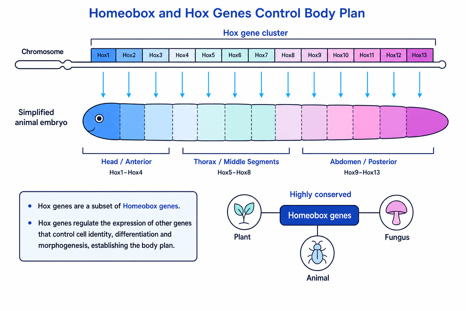
Hox genes do not directly build body parts. Instead, they regulate other genes, switching them on or off in particular regions of the embryo. This affects cell identity and development.
Interpreting a Hox gene mutation
A mutation prevents a Hox gene from functioning in a region where it is normally active.
-
The Hox gene normally helps regulate target genes that give cells positional identity in that region of the embryo.
-
If the Hox protein is not functional, those target genes may not be switched on or off correctly.
-
The affected cells may develop as the wrong type of body region, so the organism’s body plan may be altered.
Mitosis and apoptosis in body form
Mitosis is nuclear division that produces two genetically identical daughter nuclei. It is important in development because it increases cell number for growth.
The cell cycle is the ordered sequence of events a cell goes through as it grows and divides, including DNA replication and mitosis.
Apoptosis is programmed cell death. It is controlled and organised, unlike accidental cell death caused by injury. During apoptosis, a cell is dismantled and removed without damaging surrounding tissue.
Both mitosis and apoptosis are needed to form the correct body shape:
- mitosis adds cells where growth is needed;
- apoptosis removes unwanted cells;
- together, they sculpt tissues and organs during development.
For example, apoptosis helps remove cells between developing digits, separating fingers or toes.
Genes that regulate the cell cycle and apoptosis can respond to internal stimuli, such as DNA damage or chemical signals inside the cell, and external stimuli, such as growth factors, hormones or signals from neighbouring cells.
Mitosis alone does not create shape
Mitosis increases cell number, but body form also depends on where cells divide, how they differentiate, and where apoptosis removes cells.
Explaining webbed digits
During limb development, early digits may begin as part of a broader tissue paddle.
-
Mitosis increases the number of cells in the developing limb, producing enough tissue for the digits to form.
-
Signals activate apoptosis in the cells between the future digits, so these cells are removed in a controlled way.
-
If apoptosis is reduced or fails to occur between the digits, extra tissue remains, producing webbing.
In the exam
-
For mutation questions, link the mutation type to its effect on codons, amino acid sequence, protein structure and protein function.
-
For gene regulation, name the level of control: lac operon and transcription factors are transcriptional; mRNA editing is post-transcriptional; cAMP activation is post-translational.
-
For development questions, use the chain: genes regulate gene expression, gene expression affects cell behaviour, and cell behaviour shapes the body plan.
Check yourself
- Why can a one-nucleotide deletion be more disruptive than a one-nucleotide substitution?
- What happens to the lac operon when lactose is absent, and what changes when lactose is present?
- How do Hox genes, mitosis and apoptosis each contribute to body plan development?