Synaptic transmission (A-level only)
What you'll learn
- The detailed structure of a cholinergic synapse and a neuromuscular junction.
- The step-by-step sequence of events when acetylcholine crosses a synapse.
- Why synapses transmit in one direction, and how summation and inhibition affect whether an impulse continues.
- How to predict the effects of drugs on synaptic transmission using the information given.
Before you start: the electrical basics
A neurone communicates electrically along its own membrane using an action potential: a brief change in membrane potential that travels along the axon. At a synapse, that electrical signal must be converted into a chemical signal, then back into an electrical signal in the next cell.
Key electrical words
- Membrane potential is the voltage difference across a cell-surface membrane.
- Depolarisation means the inside of the membrane becomes less negative.
- Threshold is the membrane potential that must be reached to trigger a new action potential.
This detailed synapse work is A-level-only detail, so it goes beyond the simpler AS idea of “an impulse passes from one neurone to another”.
What is a synapse?
Synapse
A synapse is a junction where one neurone communicates with another cell across a tiny gap, usually using a chemical messenger called a neurotransmitter.
A cholinergic synapse is a synapse that uses acetylcholine, often shortened to ACh, as its neurotransmitter. The neurone before the synapse is the presynaptic neurone. The cell after the synapse is the postsynaptic cell.
Structure of a cholinergic synapse
A typical cholinergic synapse has several key parts:
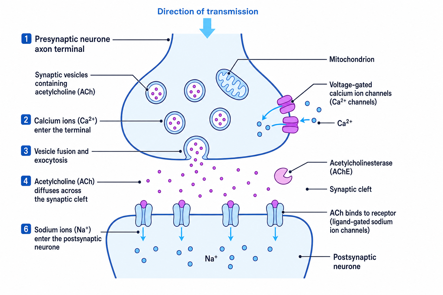
- The presynaptic axon terminal contains synaptic vesicles, which are small membrane-bound sacs filled with ACh.
- The presynaptic membrane contains voltage-gated calcium ion channels, which open when the membrane is depolarised.
- The synaptic cleft is the narrow gap between the two cells, often around 20–30 nm wide.
- The postsynaptic membrane contains receptor proteins. In this case, they are usually ligand-gated sodium ion channels: channels that open when ACh binds.
- The cleft contains acetylcholinesterase, an enzyme that breaks down ACh.
- Mitochondria in the presynaptic terminal provide ATP for processes such as vesicle movement and neurotransmitter recycling.
The diagram shows how these structures are arranged across the synaptic cleft.
Exocytosis
Exocytosis is the release of substances from a cell when vesicles fuse with the cell-surface membrane.
Transmission across a cholinergic synapse
Here is the sequence you need to know carefully:
- An action potential arrives at the presynaptic axon terminal.
- Depolarisation of the presynaptic membrane opens voltage-gated calcium ion channels.
- Calcium ions, Ca²⁺, diffuse into the presynaptic terminal.
- Ca²⁺ causes synaptic vesicles to move towards and fuse with the presynaptic membrane.
- ACh is released into the synaptic cleft by exocytosis.
- ACh diffuses across the synaptic cleft.
- ACh binds to receptor proteins on the postsynaptic membrane.
- Ligand-gated sodium ion channels open.
- Sodium ions, Na⁺, diffuse into the postsynaptic cell, causing depolarisation.
- If threshold is reached, a new action potential is generated in the postsynaptic neurone.
- Acetylcholinesterase hydrolyses ACh into choline and ethanoic acid.
- Choline is taken back into the presynaptic neurone and reused to make more ACh.
Why the enzyme matters
Acetylcholinesterase stops the postsynaptic membrane being stimulated continuously. Without removal of ACh, sodium channels may stay open for too long.
The impulse does not jump the gap
The action potential travels along neurone membranes. Across the synaptic cleft, the signal is carried by a neurotransmitter such as ACh.
Why synaptic transmission is unidirectional
Transmission across a cholinergic synapse is normally unidirectional, meaning it goes in one direction only: from presynaptic neurone to postsynaptic cell.
This happens because the two sides are structurally different. Synaptic vesicles containing ACh are found in the presynaptic terminal, while the receptor proteins for ACh are on the postsynaptic membrane. So ACh is released from one side and detected on the other.
One-way communication
Synapses are one-way because neurotransmitter release machinery is on the presynaptic side, while the matching receptor proteins are on the postsynaptic side.
Summation: combining synaptic inputs
A single synapse may not release enough neurotransmitter to bring the postsynaptic membrane to threshold. Instead, the postsynaptic neurone may need to combine several small depolarisations.
Postsynaptic potentials
- An excitatory postsynaptic potential, or EPSP, is a small depolarisation that makes an action potential more likely.
- An inhibitory postsynaptic potential, or IPSP, makes an action potential less likely, usually by hyperpolarising the membrane.
Temporal summation
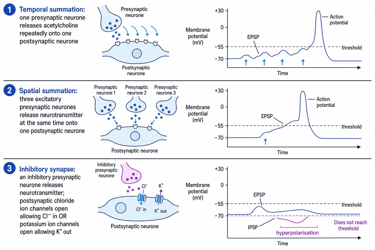
Temporal summation happens when one presynaptic neurone releases neurotransmitter repeatedly in a short time. The EPSPs overlap in time and add together.
Spatial summation
Spatial summation happens when several presynaptic neurones release neurotransmitter onto the same postsynaptic neurone at about the same time. Their EPSPs add together.
The diagrams below show temporal summation, spatial summation, and how inhibitory synapses can prevent threshold being reached.
Deciding whether summation reaches threshold
A postsynaptic neurone is at -70 mV. Threshold is -55 mV. It receives EPSPs of +6 mV, +5 mV and +4 mV, plus an IPSP of -5 mV.
- Combine the changes in membrane potential:
(+6 + 5 + 4 - 5) mV = +10 mV. - Apply the net change to the resting membrane potential:
-70 mV + 10 mV = -60 mV. - Compare with threshold: -60 mV is still more negative than -55 mV, so threshold is not reached and no action potential is produced.
Inhibitory synapses
An inhibitory synapse reduces the chance that the postsynaptic neurone will fire.
Inhibitory neurotransmitters may cause:
- chloride ion channels to open, so Cl⁻ enters the postsynaptic neurone
- potassium ion channels to open, so K⁺ leaves the postsynaptic neurone
Both effects can make the inside of the postsynaptic membrane more negative. This is called hyperpolarisation. The membrane potential moves further away from threshold, so a new action potential is less likely.
Inhibition is not just ‘no stimulation’
An inhibitory synapse actively makes firing less likely. It does not simply fail to excite the postsynaptic neurone.
Neuromuscular junctions
Neuromuscular junction
A neuromuscular junction, or NMJ, is a synapse between a motor neurone and a skeletal muscle fibre.
The postsynaptic membrane at an NMJ is the sarcolemma, which is the cell-surface membrane of a muscle fibre. The specialised folded region of sarcolemma at the NMJ is called the motor end plate. These folds increase the surface area for ACh receptor proteins.
A motor neurone action potential usually triggers ACh release, which depolarises the muscle fibre. This produces a muscle action potential that spreads along the sarcolemma and leads towards contraction.
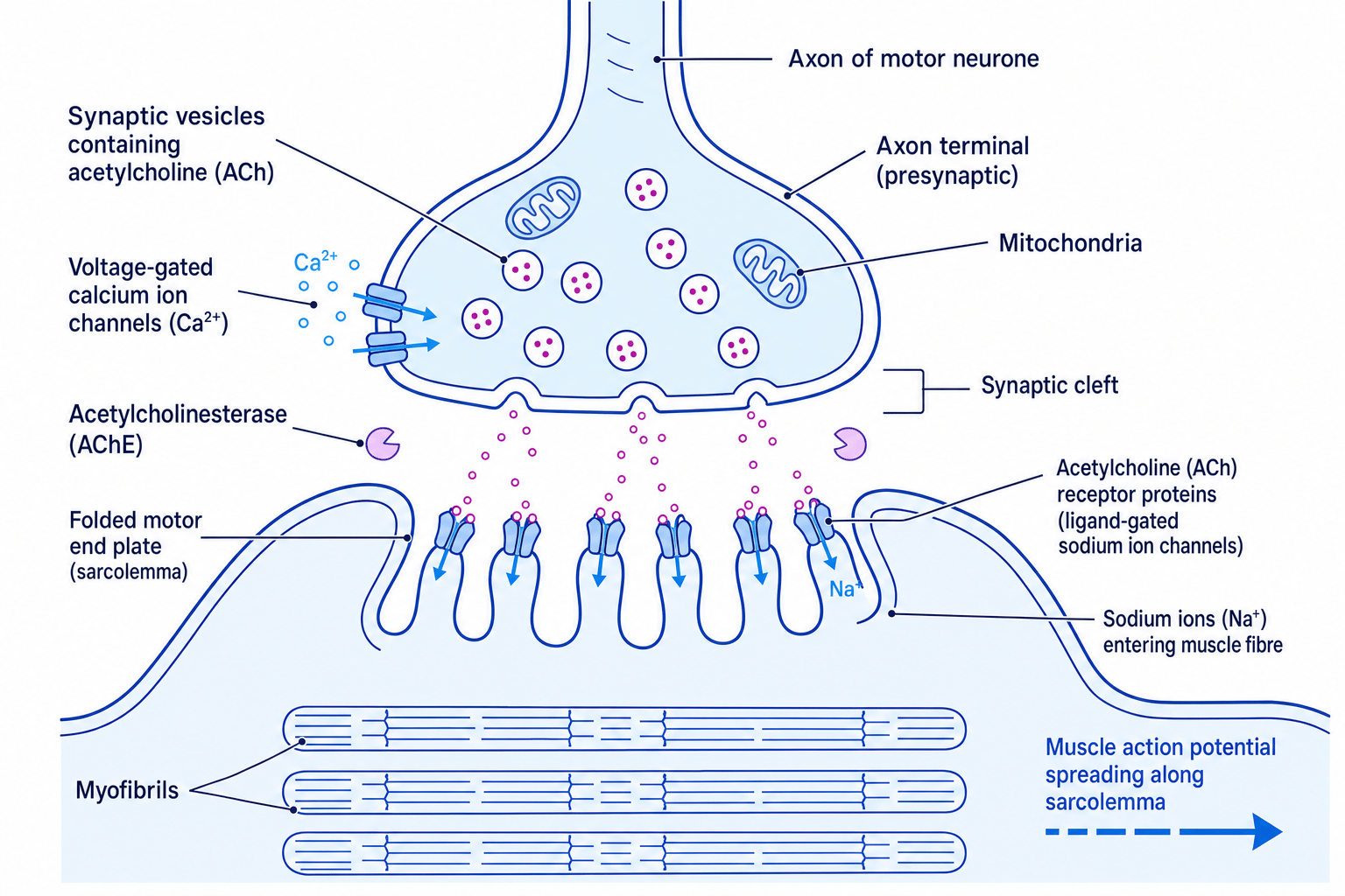
Cholinergic synapse compared with a neuromuscular junction
| Feature | Cholinergic synapse between neurones | Neuromuscular junction |
|---|---|---|
| Presynaptic cell | Neurone | Motor neurone |
| Postsynaptic cell | Usually another neurone | Skeletal muscle fibre |
| Neurotransmitter | Acetylcholine | Acetylcholine |
| Postsynaptic membrane | Neurone membrane, often dendrite or cell body | Folded motor end plate of the sarcolemma |
| Main ion movement | Na⁺ enters after ACh binds | Na⁺ enters after ACh binds |
| Outcome | May or may not trigger a new action potential | Usually triggers a muscle action potential |
| Summation | Often important | Usually not needed for one motor neurone impulse to activate the muscle fibre |
| Enzyme | Acetylcholinesterase breaks down ACh | Acetylcholinesterase breaks down ACh |
Comparing synapses
For comparison questions, separate structure from sequence. The sequence is very similar, but the postsynaptic cell and final response are different.
Predicting effects of drugs on synapses
You do not need to memorise the names of individual synaptic drugs for this section. Instead, you need to use the information given and reason from the synapse sequence.
Agonists and antagonists
- An agonist binds to a receptor and activates it, mimicking the normal neurotransmitter.
- An antagonist binds to a receptor but does not activate it, blocking the normal neurotransmitter.
Common drug targets include:
- Voltage-gated calcium ion channels: blocking these reduces Ca²⁺ entry, so less ACh is released.
- Vesicle fusion or exocytosis: blocking this reduces neurotransmitter release.
- ACh receptors: an agonist may cause depolarisation; an antagonist prevents ACh from binding.
- Acetylcholinesterase: inhibiting this means ACh remains in the cleft for longer.
- Reuptake or recycling of neurotransmitter components: disrupting this may reduce future neurotransmitter release.
Predicting an acetylcholinesterase inhibitor
A drug inhibits acetylcholinesterase at a neuromuscular junction. Predict its likely effect.
- Identify the normal role of the enzyme: acetylcholinesterase breaks down ACh in the synaptic cleft.
- If the enzyme is inhibited, ACh remains in the synaptic cleft for longer and can keep binding to receptors on the motor end plate.
- More sodium ion channels stay open for longer, so the muscle fibre is depolarised for longer.
- The likely effect is prolonged stimulation of the muscle fibre, which could cause repeated contraction or sustained contraction, depending on the data in the question.
Use the information given
A drug that increases stimulation at first can sometimes prevent normal signalling later if the membrane cannot reset properly. If the question gives data, use it rather than assuming “more neurotransmitter always means more action potentials”.
In the exam
- For a sequence question, write the events in order: action potential, Ca²⁺ entry, vesicle fusion, ACh diffusion, receptor binding, Na⁺ entry, depolarisation, breakdown by acetylcholinesterase.
- For summation questions, state whether inputs are repeated from one neurone or simultaneous from several neurones, then compare the net effect with threshold.
- For drug questions, locate the drug’s target first, then explain how that changes neurotransmitter release, receptor activation or neurotransmitter breakdown.
Check yourself
- Why does Ca²⁺ entry into the presynaptic terminal lead to neurotransmitter release?
- How are temporal summation and spatial summation different?
- What structural features make a neuromuscular junction different from a synapse between two neurones?