Brain structure and function
- Alongside dopamine, the biological explanation covers how the structure and functions of the brain relate to cognitions and behaviour.
- Scans show brain dysfunction in two ways: differences in brain volume, meaning how much tissue there is, and differences in brain activity, meaning how hard an area is working.
- MRI scans measure volume, while fMRI and PET scans measure activity by tracking blood flow and oxygen use.
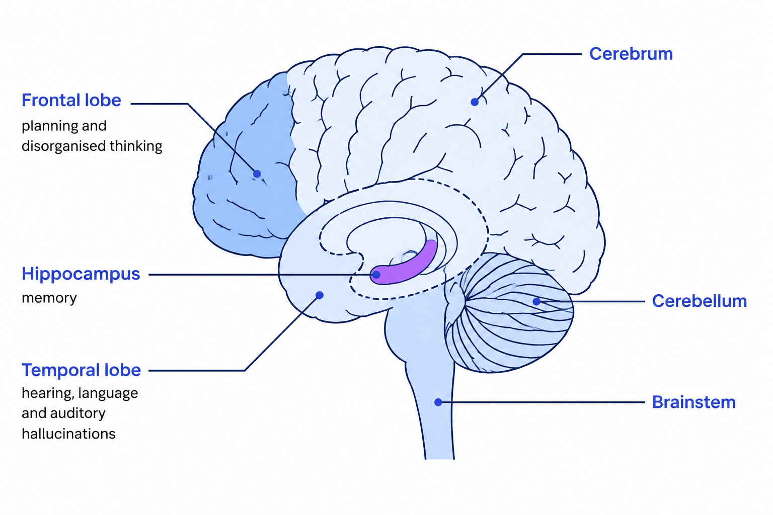
- The three areas involved are the frontal lobes, the hippocampus and the temporal lobes.
- The argument is that a difference or damage in these areas disrupts thinking, memory and perception, and those disruptions are the symptoms.
Key Idea
- Scans find two kinds of difference in schizophrenia: less tissue in some areas, and unusual levels of activity in others.
- Low activity in the frontal lobes matches the disorganised thinking and the low motivation.
- Overactive temporal lobes match hearing a voice, because those lobes handle sound and speech.
- All of these are group averages, so a great many people diagnosed with schizophrenia have scans that look entirely ordinary.
Brain volume
- Many people diagnosed with schizophrenia have enlarged ventricles, the fluid-filled spaces inside the brain.
- Larger ventricles mean less tissue around them, so the finding points to a loss of grey matter.
- Overall brain volume tends to be slightly smaller, and the reduction is clearest in the temporal lobes and the hippocampus.
- These are averages across groups, so many individuals diagnosed with schizophrenia have scans that look entirely ordinary.
Note
- Volume and activity are measured by different scans, so an MRI result and an fMRI result are two separate pieces of evidence.
- Differences this small cannot be used to diagnose anyone, and a diagnosis still rests on the symptoms.
The frontal lobes, hippocampus and temporal lobes
- Frontal lobes
- They handle logic, reasoning, planning, problem solving and judgement.
- Activity here is often low, and people with schizophrenia perform poorly on the Wisconsin Card Sort Test, which requires a person to switch sorting rule when the rule changes without warning.
- Low frontal activity fits the disorganised thinking and the negative symptoms such as avolition.
- Hippocampus
- It stores and retrieves memories and helps set an experience in its context.
- It is often reduced in volume, which fits the difficulty many people report in using and accessing memories.
- Heckers (2001) reported abnormal hippocampal activity while people were experiencing auditory hallucinations.
- Temporal lobes
- They process hearing and language, including telling a person's own inner speech apart from a voice in the room.
- They have been found to be overactive, reported by Mandal (2002), which fits hallucinations, delusions and disordered thinking.
Example
- One scan can show all three at once: wider ventricles, a smaller hippocampus and little activity across the frontal lobes.
- In Daniel et al. (1991) amphetamine raised prefrontal blood flow in ten in-patients, and their card sorting scores improved at the same time.
Neurological damage
- Neurological damage means physical harm to brain tissue, from a head injury, an infection, a stroke or a tumour.
- Damage to the frontal lobes leaves planning, judgement and self-control impaired, which shows up as disorganised behaviour.
- Damage to the hippocampus leaves memory impaired, so new information is not laid down or retrieved reliably.
- Damage to the temporal lobes makes speech and sound harder to interpret, and can produce hallucination-like experiences.
- Because damage to these areas produces symptoms that resemble schizophrenia, the theory treats the same areas as involved in the condition itself.
Common Mistake
- A scan difference found alongside symptoms is not proof that the difference caused them.
- A finding about volume and a finding about activity are two different findings, so which of the two a result concerns changes what it shows.
- The debate that goes with this theory is nature and nurture, not reductionism or free will.
The three main divisions of the brain
- Cerebrum
- The largest division, split into two hemispheres and four lobes, responsible for thinking, memory, language and perception.
- Cerebellum
- Sits at the back and beneath the cerebrum, and controls balance, co-ordination and fine movement.
- Brainstem
- Joins the brain to the spinal cord and controls automatic functions such as breathing, heart rate and the sleep and waking cycle.
- The frontal and temporal lobes are parts of the cerebrum, and the hippocampus lies deep inside the temporal lobe.
Criticisms of the biological theory
Definition
Nature-nurture debate
The discussion of how far behaviour is shaped by inherited biology (nature) compared with experiences and environment (nurture).
- The theory sits on the nature side, explaining schizophrenia through dopamine and physical brain differences.
- It gives little weight to nurture, such as a stressful upbringing, bereavement, poverty, city living or discrimination.
- Scan findings are correlational, so a difference measured at the same time as symptoms cannot show which came first.
- The differences may be a result of living with schizophrenia, or of years of anti-psychotic medication, rather than its cause.
- Not everyone diagnosed shows these differences, and some people who have never been diagnosed do, so the findings cannot explain every case.
- Nature and nurture both contribute, so biology on its own is an incomplete explanation.
Note
- The debate that belongs with this theory is nature and nurture, and social drift is the psychological explanation that carries the social side.
- Anti-psychotic medication is itself a possible source of some brain differences, which is why scans taken before treatment starts matter.
Turning this into marks
- When two brain findings are asked for, make them genuinely separate rather than one finding rephrased.
- Name the area and the symptom it is linked to, such as overactive temporal lobes and auditory hallucinations, because a general reference to brain differences does not secure the knowledge mark.
- The mark most often lost is writing that a brain difference causes schizophrenia when the evidence shows only that the two occur together.
Self review
- What is the difference between a finding about brain volume and one about brain activity?
- What do the frontal lobes normally do, and how does their activity differ in schizophrenia?
- Which symptom is linked to overactive temporal lobes?
- Name the three main divisions of the brain and one function of each.
- Give two reasons brain differences might be an effect of schizophrenia rather than its cause.