The nervous system
Nervous system
The nervous system is the network of neurones, made up of the central nervous system and the nerves connected to it, that detects stimuli and coordinates the body's responses by carrying electrical impulses.
Central nervous system
The central nervous system is the brain and spinal cord, which receive impulses from sensory neurones, coordinate a response and send impulses out along motor neurones.
Spinal cord
The spinal cord is the column of nervous tissue running inside the backbone that carries impulses between the body and the brain and coordinates many reflexes.
- The nervous system allows you to detect changes in your surroundings and inside your body, and to respond to them within a fraction of a second.
- It is built from neurones, which are nerve cells specialised to carry electrical impulses.
- It has two parts: the central nervous system (CNS), which is the brain and the spinal cord, and the peripheral nervous system, which is all the nerves running between the CNS and the rest of the body.
- A nerve is a bundle of hundreds of neurone fibres wrapped in a protective sheath, which is why one nerve can carry impulses in both directions at the same time.
- Impulses travelling into the CNS are carried by sensory neurones, and impulses travelling out of the CNS are carried by motor neurones.
- The CNS is the only place where information is compared and a decision is made, so every response is coordinated from there.
- Because the signal is electrical rather than chemical, it travels at up to about 100 m s−1100\ \text{m s}^{-1}100 m s−1, which is why nervous responses are so much faster than messages carried in the blood.
The nervous system detects stimuli using receptors, carries the information as electrical impulses along neurones, and coordinates a response through the brain and spinal cord.
How a neurone is built
Neurone
A neurone is a nerve cell adapted to carry electrical impulses rapidly from one part of the body to another.
Myelin sheath
The myelin sheath is a fatty layer surrounding the axon of a neurone that insulates it electrically and increases the speed at which an impulse travels.
- A neurone has a cell body containing the nucleus, the cytoplasm and the mitochondria that supply the energy needed to send impulses.
- Short branched fibres called dendrites spread out from the cell body and receive impulses from other neurones.
- One long fibre called the axon carries the impulse away from the cell body, and in a motor neurone running to your foot that axon can be over 1 m1\ \text{m}1 m long.
- Most axons are wrapped in a myelin sheath, a fatty layer made by cells that coil themselves around the fibre.
- The myelin sheath acts as an electrical insulator, so the impulse jumps from gap to gap along the axon instead of travelling along every part of the membrane.
- Jumping between the gaps raises the speed of conduction from roughly 1 m s−11\ \text{m s}^{-1}1 m s−1 in an unmyelinated fibre to roughly 100 m s−1100\ \text{m s}^{-1}100 m s−1 in a myelinated one.
- The axon ends in many fine branches with swollen tips called axon terminals, so one neurone can pass its impulse on to many others.
A neurone is long and thin so that a single cell can stretch all the way from a receptor to the spinal cord without the impulse having to be handed on part way.
Sensory, relay and motor neurones
Sensory neurone
A sensory neurone is a neurone that carries electrical impulses from a receptor to the central nervous system.
Relay neurone
A relay neurone is a neurone found inside the central nervous system that carries impulses from a sensory neurone to a motor neurone.
Motor neurone
A motor neurone is a neurone that carries electrical impulses from the central nervous system to an effector.
- A sensory neurone carries impulses from a receptor into the CNS.
- Its cell body sits in a small bulge part way along the fibre, off to one side of the main pathway, rather than at either end.
- On the receptor side of the cell body it has one very long fibre called the dendron, and on the CNS side it has a shorter axon.
- A relay neurone lies entirely inside the CNS and links a sensory neurone to a motor neurone.
- It is short, has no myelin sheath, and carries many dendrites and branches, so it can connect to large numbers of other neurones.
- A motor neurone carries impulses out of the CNS to an effector.
- Its cell body is large and lies inside the CNS, with many dendrites reaching out from it to collect impulses from relay neurones.
- From that cell body a single very long myelinated axon runs all the way to the muscle or gland, ending in branches that spread across the effector.
- Do not draw or describe a sensory neurone with its cell body at the end of the fibre, because the mark is for placing it in a bulge part way along, to one side.
- Do not write that a relay neurone runs from the CNS to a muscle, because that is the job of the motor neurone.
Sensory receptors
Receptor
A receptor is a cell or group of cells that detects a stimulus and converts it into an electrical impulse.
Stimulus
A stimulus is a change in the environment, inside or outside the body, that is detected by a receptor.
- A receptor detects a stimulus, which is any change inside or outside the body.
- Its job is to convert the energy of the stimulus into an electrical impulse, so light, sound, heat, pressure and chemicals all end up as the same kind of signal.
- Each type of receptor responds to only one type of stimulus, so a light receptor in the retina cannot detect sound and a sound receptor in the ear cannot detect light.
- A stronger stimulus makes the receptor send impulses more frequently, which is how the CNS can tell a light touch from a hard press.
- Some receptors are single specialised cells in the skin, and others are grouped together inside a sense organ such as the eye, the ear, the nose or the tongue.
- The receptor is always the start of the pathway, so an answer that begins with the sensory neurone has already dropped a mark.
A touch receptor in your fingertip is squashed by the edge of a desk, and that change in shape starts a burst of impulses in the sensory neurone attached to it.
Synapses
Synapse
A synapse is the junction between two neurones, where the impulse is carried across a small gap by a chemical neurotransmitter.
Neurotransmitter
A neurotransmitter is a chemical released into a synapse that diffuses across the gap and starts an impulse in the next neurone.
- Neurones do not touch each other, so where two of them meet there is a tiny gap of about 20 nm20\ \text{nm}20 nm called the synapse.
- The electrical impulse arrives at the axon terminal of the first neurone and cannot cross the gap on its own.
- The impulse makes small sacs in the axon terminal, called vesicles, move to the membrane and release a neurotransmitter.
- The neurotransmitter molecules diffuse across the gap because they are at a high concentration on one side and a low concentration on the other.
- They bind to receptor molecules on the membrane of the next neurone, and this starts a new electrical impulse in that neurone.
- The neurotransmitter is then broken down or reabsorbed, so the second neurone is ready for the next impulse rather than firing without stopping.
- Vesicles are found on only one side of the gap, so a synapse allows impulses to travel in one direction only.
- Crossing a synapse by diffusion is slower than travelling along an axon, so every extra synapse in a pathway adds a short delay.
Describing a synapse
- Name the gap as the synapse before you describe anything crossing it, because the label carries a mark of its own.
- Use the word diffuses for the neurotransmitter, since travels or moves is too vague to be credited.
- Finish the chain by saying that a new impulse starts in the next neurone, which is the step most answers leave out.
- If you are asked why transmission is one way, say that the neurotransmitter is released from only one side of the synapse.
Effectors and the response
Effector
An effector is a muscle or a gland that carries out a response when it receives an impulse from a motor neurone.
- An effector is the part of the body that carries out the response, and it is always either a muscle or a gland.
- A muscle responds by contracting, which pulls on a bone and moves part of the body.
- A gland responds by secreting a substance, such as a salivary gland releasing saliva or a sweat gland releasing sweat.
- The motor neurone meets the muscle at a junction that works like a synapse, releasing a neurotransmitter that makes the muscle fibres contract.
- One muscle receives branches from many motor neurones, so the CNS controls how strongly the muscle contracts by controlling how many of them fire.
Putting the parts together
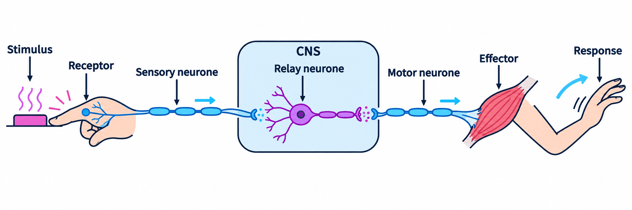
- The components always act in the same order, which you can write as one chain.
- stimulus→receptor→sensory neurone→CNS→motor neurone→effector→response\text{stimulus} \rightarrow \text{receptor} \rightarrow \text{sensory neurone} \rightarrow \text{CNS} \rightarrow \text{motor neurone} \rightarrow \text{effector} \rightarrow \text{response}stimulus→receptor→sensory neurone→CNS→motor neurone→effector→response
- Every join between two neurones in that chain is a synapse, so the pathway is a series of electrical steps linked by chemical ones.
- Naming all seven stages in order is what turns a two-mark answer into a full-mark one.
Tracing an impulse when your name is called
- The stimulus is the sound of your name, which is a change in the air pressure around you.
- Sound receptors in your ear convert that pressure change into electrical impulses.
- A sensory neurone in the auditory nerve carries the impulses to your brain.
- The CNS compares the sound with what it has stored, recognises it and decides to turn your head.
- A motor neurone carries impulses from the brain to the muscles in your neck.
- Those neck muscles are the effectors, and they contract.
- The response is that your head turns towards the sound.
Why the structure suits the job
- Long axons mean one cell carries the signal from your toe to your spinal cord, which avoids the delay of many handovers.
- Myelin sheaths mean the signal arrives fast enough for you to react before a hazard causes damage.
- Branched dendrites and axon terminals mean each neurone connects to thousands of others, so the CNS can link any receptor to any effector.
- One-way synapses mean impulses cannot run backwards into the receptor they came from.
- Name the two organs that make up the central nervous system.
- Describe where the cell body is found in a sensory neurone and in a motor neurone.
- Explain how the myelin sheath increases the speed of an impulse.
- Describe how an impulse is passed from one neurone to the next across a synapse.
- State the two types of effector and how each one responds.
- Write out the pathway from stimulus to response, naming all seven stages.