The kidney contains an outer cortex and inner medulla
Definition
Nephron
The tiny tubule that is the functional (filtering) unit of the kidney, where urine is made by ultrafiltration and selective reabsorption. Each kidney has about a million of them.
- Each kidney is supplied with blood by a renal artery and drained by a renal vein.
- The cortex is the outer region and contains the Bowman's capsules and convoluted parts of many nephrons.
- The medulla is the inner region and contains loops of Henlé and collecting ducts.
- The central renal pelvis collects urine before it passes into the ureter.
- The ureter carries urine from the kidney to the bladder, and the urethra carries urine from the bladder out of the body.
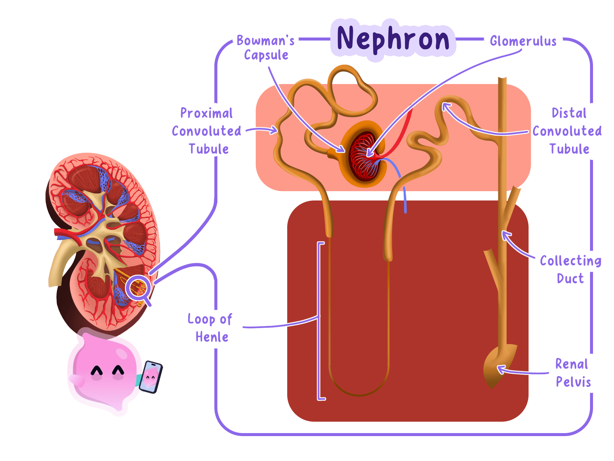
A nephron is the kidney tubule
Definition
Nephron
The tiny tubule that is the functional (filtering) unit of the kidney, where urine is made by ultrafiltration and selective reabsorption. Each kidney has about a million of them.
- A nephron is the microscopic filtering unit of a kidney.
- Each nephron begins with a cup-shaped Bowman's capsule surrounding a knot of capillaries called the glomerulus.
- The tubule then forms the proximal convoluted tubule, which is a tightly coiled section in the cortex.
- It continues as the loop of Henlé, which descends into the medulla and returns towards the cortex.
- The tubule joins a collecting duct, which passes through the medulla towards the renal pelvis.
- Many nephrons empty into the same collecting duct.
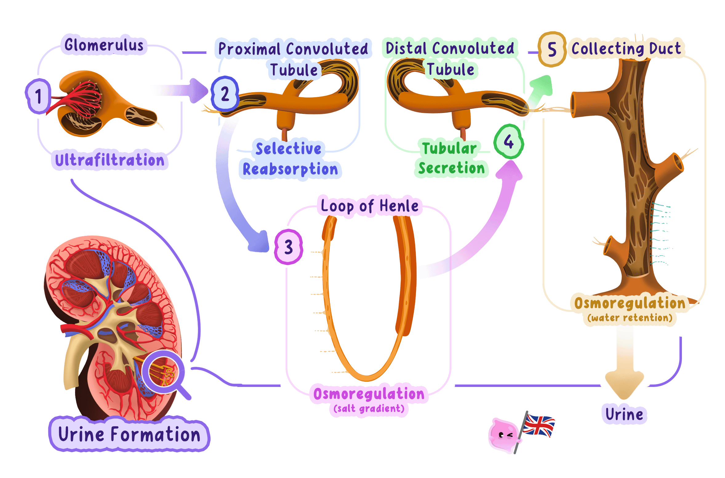
Bowman’s capsule receives the filtrate
Definition
Glomerulus
A tight knot of capillaries at the start of a nephron, inside the Bowman's capsule, where the blood is filtered under high pressure during ultrafiltration.
- The glomerulus receives blood through an afferent arteriole and drains through an efferent arteriole.
- Its capillary walls and the wall of Bowman's capsule form a filtration barrier.
- High pressure forces water and small dissolved molecules into Bowman's capsule.
- Blood cells and large plasma proteins remain inside the capillaries.
- The fluid entering the nephron is called the filtrate.
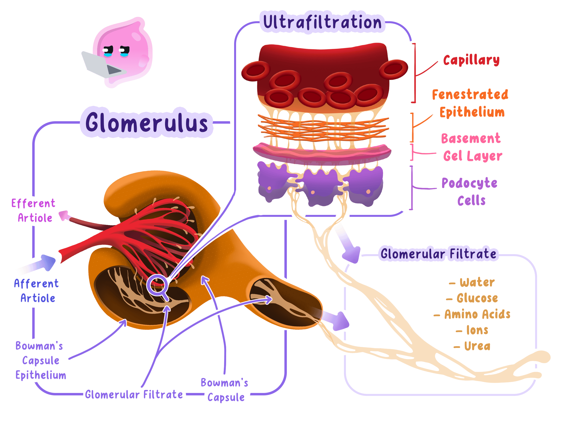
Common Mistake
- Do not label the glomerulus as part of the tubule.
- The glomerulus is a capillary network enclosed by Bowman’s capsule.
Tubule regions have distinct positions
- The proximal convoluted tubule lies mainly in the cortex and is closely surrounded by capillaries.
- The loop of Henlé extends into the medulla before turning back towards the cortex.
- The collecting duct runs through the medulla and carries tubule fluid towards the renal pelvis.
- Blood capillaries run alongside the tubule, allowing substances and water to move between the filtrate and blood.
- Knowing the order of the regions helps trace the route taken by filtrate through the nephron.
- Bowman’s capsule→proximal convoluted tubule→loop of Henleˊ→collecting duct\text{Bowman's capsule} \rightarrow \text{proximal convoluted tubule} \rightarrow \text{loop of Henlé} \rightarrow \text{collecting duct}Bowman’s capsule→proximal convoluted tubule→loop of Henleˊ→collecting duct
Exam technique
On a nephron diagram, use shape and position: the cup is Bowman’s capsule, the first coil is the proximal convoluted tubule, the long hairpin is the loop of Henlé and the straighter tube is the collecting duct.
Self review
- Which kidney region contains Bowman’s capsules?
- What structures form the start of a nephron?
- What is the order of the named tubule regions?
- Which nephron region extends into the medulla?
- Where does the collecting duct carry the tubule fluid?