The eye as a sense organ
Sense organ
A sense organ is an organ containing groups of receptor cells that detect one particular type of stimulus.
Retina
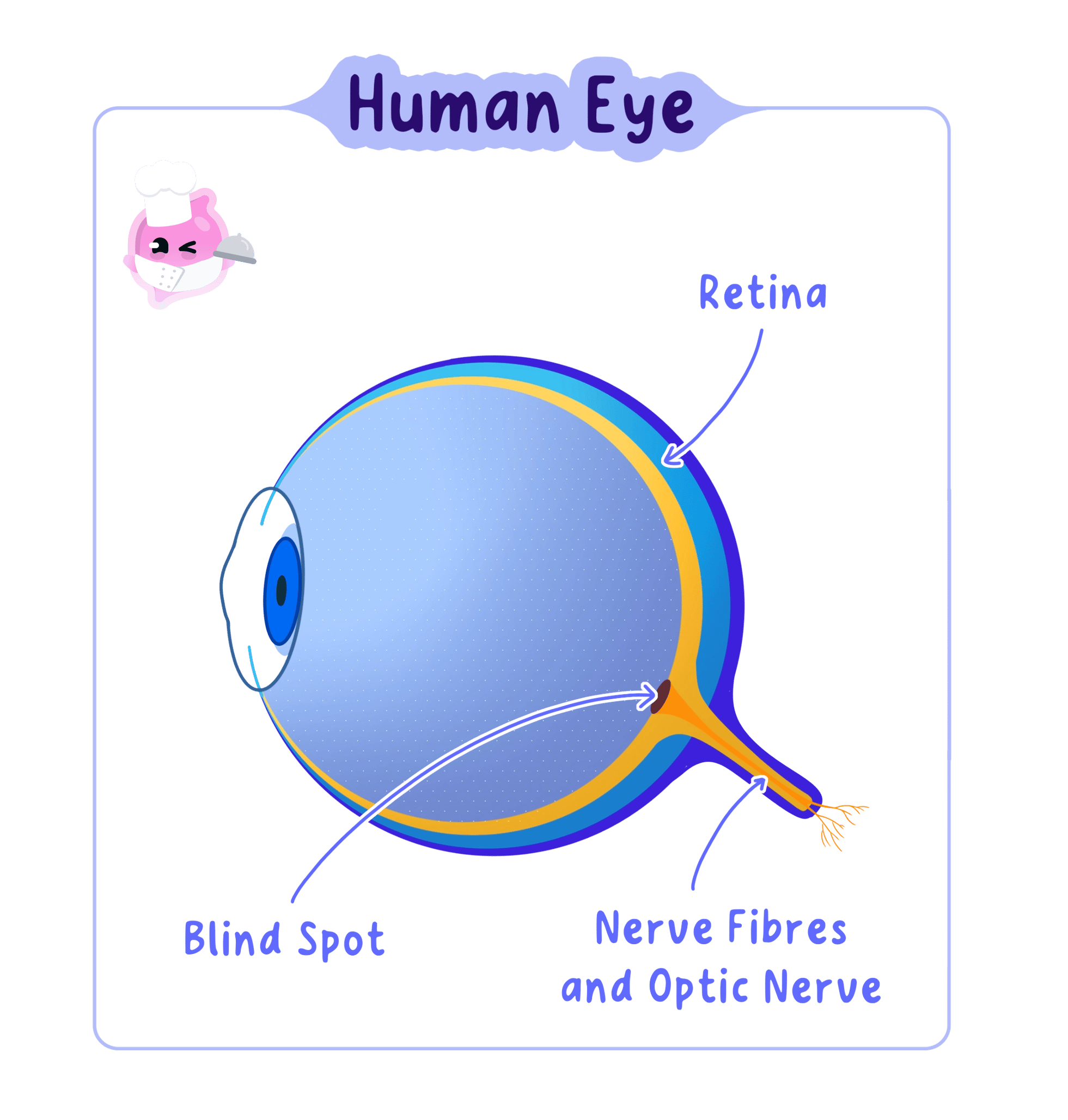
The retina is the light-sensitive layer lining the back of the eye that contains the rod and cone receptor cells.
- The eye is the sense organ that detects light, and its receptor cells are held in the retina at the back.
- Everything in front of the retina exists to get light onto those receptors in the right amount and sharply focused.
- Light is bent, or refracted, whenever it passes from one transparent material into another of different density.
- The eye uses that effect twice, at the cornea and at the lens, to bring rays from one point on an object back to a single point on the retina.
Every structure in the eye is shaped either to control how much light enters or to bend that light so it focuses exactly on the retina.
The structures of the eye
- The cornea is the transparent curved layer at the front of the eye, and because it is strongly curved and light enters it from air, it does most of the focusing.
- The sclera is the tough white outer coat around the rest of the eyeball, and it protects the eye and holds its shape.
- The iris is the coloured ring of muscle behind the cornea, and it changes the size of the pupil to control how much light gets in.
- The pupil is the hole in the centre of the iris, so it is a gap rather than a structure, and it looks black because almost no light comes back out.
- The lens is a clear, flexible, elastic disc that fine-tunes the focusing by changing its own shape.
- The ciliary body is a ring of muscle around the lens, and contracting or relaxing it changes the diameter of that ring.
- The suspensory ligaments are inelastic fibres that join the ciliary body to the edge of the lens and hold it in place.
- The retina lines the inside of the back of the eye and contains the light receptor cells that convert light into electrical impulses.
- The optic nerve leaves the back of the eye carrying those impulses to the brain.
- The aqueous humour is the watery fluid between the cornea and the lens, and the vitreous humour is the jelly filling the space behind the lens.
- Both fluids press outwards, which keeps the eyeball firm so the distance from the lens to the retina stays the same.
The cornea does the bulk of the bending and cannot change, while the lens does the smaller adjustable part, which is why the lens is the structure that alters shape.
How an image is formed
Optic nerve
The optic nerve is the bundle of sensory neurones that carries electrical impulses from the retina to the brain.
- Light from an object reaches the eye and passes first through the cornea, where it is refracted strongly inwards.
- It then passes through the pupil, which sets how much of that light is allowed through.
- The lens refracts the light again by the extra amount needed for that particular distance.
- The rays meet on the retina, forming an image that is upside down and reversed from left to right.
- The receptor cells absorb the light and convert it into electrical impulses.
- The optic nerve carries the impulses to the brain, which interprets them and turns the upside-down image the right way up.
- Where the optic nerve leaves the retina there are no receptor cells, so light landing on that small patch produces no impulses at all.
- Do not write that the eye sees the object, because the retina only detects light and it is the brain that produces the image you experience.
- Do not say the pupil widens or narrows by itself, because the pupil is a hole and it is the iris muscles that change its size.
Accommodation
Accommodation
Accommodation is the change in the shape of the lens that allows the eye to focus light from objects at different distances onto the retina.
- Light from a near object spreads out as it reaches the eye, so it needs to be bent a lot to meet on the retina.
- Light from a distant object arrives almost parallel, so it needs to be bent much less.
- Since the distance from the lens to the retina is fixed, the eye adjusts by changing how strongly the lens refracts light.
- To focus on a near object the ciliary muscles contract, which makes the ring of muscle smaller.
- A smaller ring means the suspensory ligaments slacken, so they stop pulling on the edge of the lens.
- The elastic lens then springs into a fatter, more curved shape, which refracts the light more strongly and brings the spreading rays to a focus on the retina.
- To focus on a distant object the ciliary muscles relax, so the ring of muscle widens.
- A wider ring pulls the suspensory ligaments tight, and they stretch the lens outwards.
- The lens becomes thinner and less curved, so it refracts the light less and the nearly parallel rays still meet on the retina.
- The lens becomes stiffer with age, so it cannot become as fat as it once did, which is why many people need reading glasses in later life.
Looking from the board down to your book
- While you read the board, the ciliary muscles are relaxed, the suspensory ligaments are taut and the lens is thin.
- You look down at your book, and light from the page now reaches your eye spreading out rather than parallel.
- For a moment the light focuses behind the retina, so the words look blurred.
- The ciliary muscles contract, the ring narrows and the suspensory ligaments slacken.
- The lens becomes fatter and refracts the light more strongly.
- The rays now meet exactly on the retina and the words become sharp, all within about a second.
The iris and the pupil reflex
Pupil reflex
The pupil reflex is the automatic change in the size of the pupil, caused by the muscles of the iris, that controls how much light enters the eye.
- The iris contains two sets of muscle: circular muscles arranged in rings and radial muscles arranged like the spokes of a wheel.
- In bright light the circular muscles contract and the radial muscles relax, so the iris closes in and the pupil becomes smaller.
- A smaller pupil lets in less light, which protects the receptor cells of the retina from being damaged.
- In dim light the radial muscles contract and the circular muscles relax, so the iris is pulled outwards and the pupil becomes larger.
- A larger pupil lets in more light, so enough of it reaches the receptors for you to see.
- The change is a reflex, so light receptors in the retina are the receptor and the iris muscles are the effector, and it happens without you deciding anything.
- Do not say only that the circular muscles contract in bright light, because the mark scheme usually wants the radial muscles relaxing as well.
- Do not mix up the two sets: circular muscles close the pupil, and radial muscles open it.
Rod and cone cells
Rod cells
Rod cells are light receptor cells in the retina that respond in dim light but do not detect colour.
Cone cells
Cone cells are light receptor cells in the retina that detect colour and need bright light to respond.
Fovea
The fovea is the small central area of the retina, packed with cone cells, where an image is seen in the sharpest detail.
- The retina holds two kinds of light receptor cell, named after their shapes.
- Rod cells are very sensitive, so they respond in dim light, but they cannot tell colours apart and give only shades of grey.
- Cone cells need bright light to respond, and there are three types, sensitive to red, green and blue light.
- The brain compares the impulses arriving from the three types of cone, and the mixture tells it which colour you are looking at.
- Cones are packed most densely at the fovea, the small central patch of retina that lies straight in line with the lens.
- When you look directly at something its image falls on the fovea, which is why that part of the scene is the sharpest and most colourful.
- Rods are spread across the rest of the retina and become more common towards its edges, so faint objects are easier to see slightly off to one side.
Walking home on a dark evening you can make out the shapes of parked cars but not their colours, because only the rod cells are responding at that light level.
Linking structure to function
- Questions on the eye almost always ask you to explain how a structure is related to its function, so pair the feature with what it makes possible.
- The cornea is transparent and curved, so light can enter and be strongly refracted.
- The lens is elastic, so it can change shape and alter how much it refracts light.
- The iris is muscular, so it can change the size of the pupil in either direction.
- The retina is packed with receptor cells, so a detailed pattern of light can be converted into a detailed pattern of impulses.
Describing accommodation
- Give all three steps in order: what the ciliary muscles do, what happens to the suspensory ligaments, then what shape the lens takes.
- Finish with the effect on the light, because the final mark is usually for saying the light is refracted more or less so it focuses on the retina.
- Use slacken and tighten for the ligaments rather than contract and relax, since ligaments are not muscles.
- The pairing candidates most often reverse is near with relaxed muscles, so learn it as near means contract and fat.
Protecting the eye
- The eyeball sits in a bony socket in the skull, which shields it from knocks.
- Tears wash the front of the cornea, keeping it moist and clear and carrying away dust.
- Eyelashes and the blinking reflex stop most particles reaching the cornea in the first place.
- State which structure of the eye does most of the focusing and explain why.
- Describe the path taken by light from entering the eye to reaching the brain.
- Explain what happens to the ciliary muscles, suspensory ligaments and lens when you focus on a near object.
- Explain how the iris makes the pupil smaller in bright light.
- Give two differences between rod cells and cone cells.
- Explain why an image falling on the fovea is the sharpest part of what you see.