3.1.1 Structure of the nervous system
The nervous system
Nervous system
The nervous system is the network of neurones, made up of the central nervous system and the nerves connected to it, that detects stimuli and coordinates the body's responses by carrying electrical impulses.
Central nervous system
The central nervous system is the brain and spinal cord, which receive impulses from sensory neurones, coordinate a response and send impulses out along motor neurones.
Spinal cord
The spinal cord is the column of nervous tissue running inside the backbone that carries impulses between the body and the brain and coordinates many reflexes.
- The nervous system allows you to detect changes in your surroundings and inside your body, and to respond to them within a fraction of a second.
- It is built from neurones, which are nerve cells specialised to carry electrical impulses.
- It has two parts: the central nervous system (CNS), which is the brain and the spinal cord, and the peripheral nervous system, which is all the nerves running between the CNS and the rest of the body.
- A nerve is a bundle of hundreds of neurone fibres wrapped in a protective sheath, which is why one nerve can carry impulses in both directions at the same time.
- Impulses travelling into the CNS are carried by sensory neurones, and impulses travelling out of the CNS are carried by motor neurones.
- The CNS is the only place where information is compared and a decision is made, so every response is coordinated from there.
- Because the signal is electrical rather than chemical, it travels at up to about 100 m s−1100\ \text{m s}^{-1}100 m s−1, which is why nervous responses are so much faster than messages carried in the blood.
The nervous system detects stimuli using receptors, carries the information as electrical impulses along neurones, and coordinates a response through the brain and spinal cord.
How a neurone is built
Neurone
A neurone is a nerve cell adapted to carry electrical impulses rapidly from one part of the body to another.
Myelin sheath
The myelin sheath is a fatty layer surrounding the axon of a neurone that insulates it electrically and increases the speed at which an impulse travels.
- A neurone has a cell body containing the nucleus, the cytoplasm and the mitochondria that supply the energy needed to send impulses.
- Short branched fibres called dendrites spread out from the cell body and receive impulses from other neurones.
- One long fibre called the axon carries the impulse away from the cell body, and in a motor neurone running to your foot that axon can be over 1 m1\ \text{m}1 m long.
- Most axons are wrapped in a myelin sheath, a fatty layer made by cells that coil themselves around the fibre.
- The myelin sheath acts as an electrical insulator, so the impulse jumps from gap to gap along the axon instead of travelling along every part of the membrane.
- Jumping between the gaps raises the speed of conduction from roughly 1 m s−11\ \text{m s}^{-1}1 m s−1 in an unmyelinated fibre to roughly 100 m s−1100\ \text{m s}^{-1}100 m s−1 in a myelinated one.
- The axon ends in many fine branches with swollen tips called axon terminals, so one neurone can pass its impulse on to many others.
A neurone is long and thin so that a single cell can stretch all the way from a receptor to the spinal cord without the impulse having to be handed on part way.
Sensory, relay and motor neurones
Sensory neurone
A sensory neurone is a neurone that carries electrical impulses from a receptor to the central nervous system.
Relay neurone
A relay neurone is a neurone found inside the central nervous system that carries impulses from a sensory neurone to a motor neurone.
Motor neurone
A motor neurone is a neurone that carries electrical impulses from the central nervous system to an effector.
- A sensory neurone carries impulses from a receptor into the CNS.
- Its cell body sits in a small bulge part way along the fibre, off to one side of the main pathway, rather than at either end.
- On the receptor side of the cell body it has one very long fibre called the dendron, and on the CNS side it has a shorter axon.
- A relay neurone lies entirely inside the CNS and links a sensory neurone to a motor neurone.
- It is short, has no myelin sheath, and carries many dendrites and branches, so it can connect to large numbers of other neurones.
- A motor neurone carries impulses out of the CNS to an effector.
- Its cell body is large and lies inside the CNS, with many dendrites reaching out from it to collect impulses from relay neurones.
- From that cell body a single very long myelinated axon runs all the way to the muscle or gland, ending in branches that spread across the effector.
- Do not draw or describe a sensory neurone with its cell body at the end of the fibre, because the mark is for placing it in a bulge part way along, to one side.
- Do not write that a relay neurone runs from the CNS to a muscle, because that is the job of the motor neurone.
Sensory receptors
Receptor
A receptor is a cell or group of cells that detects a stimulus and converts it into an electrical impulse.
Stimulus
A stimulus is a change in the environment, inside or outside the body, that is detected by a receptor.
- A receptor detects a stimulus, which is any change inside or outside the body.
- Its job is to convert the energy of the stimulus into an electrical impulse, so light, sound, heat, pressure and chemicals all end up as the same kind of signal.
- Each type of receptor responds to only one type of stimulus, so a light receptor in the retina cannot detect sound and a sound receptor in the ear cannot detect light.
- A stronger stimulus makes the receptor send impulses more frequently, which is how the CNS can tell a light touch from a hard press.
- Some receptors are single specialised cells in the skin, and others are grouped together inside a sense organ such as the eye, the ear, the nose or the tongue.
- The receptor is always the start of the pathway, so an answer that begins with the sensory neurone has already dropped a mark.
A touch receptor in your fingertip is squashed by the edge of a desk, and that change in shape starts a burst of impulses in the sensory neurone attached to it.
Synapses
Synapse
A synapse is the junction between two neurones, where the impulse is carried across a small gap by a chemical neurotransmitter.
Neurotransmitter
A neurotransmitter is a chemical released into a synapse that diffuses across the gap and starts an impulse in the next neurone.
- Neurones do not touch each other, so where two of them meet there is a tiny gap of about 20 nm20\ \text{nm}20 nm called the synapse.
- The electrical impulse arrives at the axon terminal of the first neurone and cannot cross the gap on its own.
- The impulse makes small sacs in the axon terminal, called vesicles, move to the membrane and release a neurotransmitter.
- The neurotransmitter molecules diffuse across the gap because they are at a high concentration on one side and a low concentration on the other.
- They bind to receptor molecules on the membrane of the next neurone, and this starts a new electrical impulse in that neurone.
- The neurotransmitter is then broken down or reabsorbed, so the second neurone is ready for the next impulse rather than firing without stopping.
- Vesicles are found on only one side of the gap, so a synapse allows impulses to travel in one direction only.
- Crossing a synapse by diffusion is slower than travelling along an axon, so every extra synapse in a pathway adds a short delay.
Describing a synapse
- Name the gap as the synapse before you describe anything crossing it, because the label carries a mark of its own.
- Use the word diffuses for the neurotransmitter, since travels or moves is too vague to be credited.
- Finish the chain by saying that a new impulse starts in the next neurone, which is the step most answers leave out.
- If you are asked why transmission is one way, say that the neurotransmitter is released from only one side of the synapse.
Effectors and the response
Effector
An effector is a muscle or a gland that carries out a response when it receives an impulse from a motor neurone.
- An effector is the part of the body that carries out the response, and it is always either a muscle or a gland.
- A muscle responds by contracting, which pulls on a bone and moves part of the body.
- A gland responds by secreting a substance, such as a salivary gland releasing saliva or a sweat gland releasing sweat.
- The motor neurone meets the muscle at a junction that works like a synapse, releasing a neurotransmitter that makes the muscle fibres contract.
- One muscle receives branches from many motor neurones, so the CNS controls how strongly the muscle contracts by controlling how many of them fire.
Putting the parts together
- The components always act in the same order, which you can write as one chain.
- stimulus→receptor→sensory neurone→CNS→motor neurone→effector→response\text{stimulus} \rightarrow \text{receptor} \rightarrow \text{sensory neurone} \rightarrow \text{CNS} \rightarrow \text{motor neurone} \rightarrow \text{effector} \rightarrow \text{response}stimulus→receptor→sensory neurone→CNS→motor neurone→effector→response
- Every join between two neurones in that chain is a synapse, so the pathway is a series of electrical steps linked by chemical ones.
- Naming all seven stages in order is what turns a two-mark answer into a full-mark one.
Tracing an impulse when your name is called
- The stimulus is the sound of your name, which is a change in the air pressure around you.
- Sound receptors in your ear convert that pressure change into electrical impulses.
- A sensory neurone in the auditory nerve carries the impulses to your brain.
- The CNS compares the sound with what it has stored, recognises it and decides to turn your head.
- A motor neurone carries impulses from the brain to the muscles in your neck.
- Those neck muscles are the effectors, and they contract.
- The response is that your head turns towards the sound.
Why the structure suits the job
- Long axons mean one cell carries the signal from your toe to your spinal cord, which avoids the delay of many handovers.
- Myelin sheaths mean the signal arrives fast enough for you to react before a hazard causes damage.
- Branched dendrites and axon terminals mean each neurone connects to thousands of others, so the CNS can link any receptor to any effector.
- One-way synapses mean impulses cannot run backwards into the receptor they came from.
- Name the two organs that make up the central nervous system.
- Describe where the cell body is found in a sensory neurone and in a motor neurone.
- Explain how the myelin sheath increases the speed of an impulse.
- Describe how an impulse is passed from one neurone to the next across a synapse.
- State the two types of effector and how each one responds.
- Write out the pathway from stimulus to response, naming all seven stages.
3.1.2 Coordinated response
What a coordinated response is
Stimulus
A stimulus is a change in the environment, inside or outside the body, that is detected by a receptor.
Coordination
Coordination is the process by which the nervous system links information from receptors to the correct effectors so that different parts of the body work together as one response.
- A coordinated response is one in which the right effectors act, in the right order and by the right amount, because the CNS has processed information from receptors first.
- Without coordination each organ would act on its own, so muscles could pull against each other and a response could work against what the body needs.
- Coordination needs three things: information arriving from receptors, a central point where that information is compared, and a route out to the effectors.
- The nervous system supplies all three, which is why it can turn a single stimulus into a whole-body response.
The nervous system coordinates a response because every receptor sends its information to one place, and that same place controls every effector.
The pathway from stimulus to response
- Every coordinated response follows the same sequence of components.
- stimulus→receptor→sensory neurone→CNS→motor neurone→effector→response\text{stimulus} \rightarrow \text{receptor} \rightarrow \text{sensory neurone} \rightarrow \text{CNS} \rightarrow \text{motor neurone} \rightarrow \text{effector} \rightarrow \text{response}stimulus→receptor→sensory neurone→CNS→motor neurone→effector→response
- The receptor detects the change and turns it into an electrical impulse.
- The sensory neurone carries that impulse into the brain or spinal cord.
- Inside the CNS the impulse is passed across synapses to relay neurones, where it is compared with information from other receptors and with what is stored in the brain.
- The CNS then selects which motor neurones to fire, and each one carries impulses out to a particular effector.
- The effectors contract or secrete, and together their actions make up the response.
- Do not name the effector as the stimulus, because the stimulus is the change that was detected, not the part that acts.
- Do not miss out the CNS, because a pathway that runs straight from a sensory neurone to a motor neurone is not coordinated by anything.
Receptors in different parts of the body
Receptor
A receptor is a cell or group of cells that detects a stimulus and converts it into an electrical impulse.
Sense organ
A sense organ is an organ containing groups of receptor cells that detect one particular type of stimulus.
- The nervous system can respond to many different kinds of change because it has several different types of receptor, each tuned to one stimulus.
- The eyes contain light receptors in the retina.
- The ears contain sound receptors, and also receptors that detect the position of your head so you can balance.
- The nose contains receptors that detect chemicals carried in the air, and the tongue contains receptors that detect chemicals dissolved in saliva.
- The skin contains separate receptors for touch, pressure, temperature and pain, which is why you can feel a light brush and a hot surface as different things.
- Receptors inside the body monitor internal conditions such as blood temperature, blood glucose concentration and the stretch of a muscle.
- All of them send the same kind of electrical impulse, so the CNS identifies the stimulus by which neurone the impulse arrives on, not by what the impulse looks like.
Pressing gently on a closed eyelid makes you see a faint patch of light, because the brain treats any impulse arriving down the optic nerve as light however it was produced.
Why the nervous system can coordinate
- It reaches every part of the body, because nerves branch out from the spinal cord to every organ, muscle and area of skin.
- It has an enormous number of links between neurones, since one neurone in the CNS forms synapses with thousands of others.
- Those links let the impulse from one receptor be routed to any effector or to several effectors at once.
- It receives information from many types of receptor at the same time, so the CNS can weigh up what it sees, hears and feels before choosing a response.
- It can also block a response, because a relay neurone that is not stimulated enough does not pass the impulse on, so unimportant signals are filtered out.
- One stimulus can therefore produce a whole set of matched actions, such as several muscles contracting in sequence while a gland releases a substance.
Catching a ball thrown towards you
- Light receptors in both retinas detect the ball and send impulses along the optic nerves.
- The brain compares the two images to judge how far away the ball is and how fast it is moving.
- Stretch receptors in your leg and back muscles report where your body already is.
- The brain sends impulses along motor neurones to leg muscles, arm muscles and hand muscles together.
- Those effectors move you into position, raise your arms and close your fingers at the moment the ball arrives.
- The catch works only because one control centre timed all of those separate muscles against each other.
Voluntary responses
Voluntary response
A voluntary response is a response that is consciously decided by the cerebrum before impulses are sent to the effector.
- In a voluntary response the impulse travels all the way up to the cerebrum, where a conscious decision is made before any motor neurone fires.
- That extra journey means the impulse crosses many more synapses, and each one adds a delay while the neurotransmitter diffuses.
- A voluntary response is therefore slower than an automatic one, but it can be changed to suit the situation and it can be learnt and improved with practice.
Reaction time
Reaction time
Reaction time is the time taken between a stimulus being detected by a receptor and the response being carried out by an effector.
- Reaction time measures how long the whole pathway takes, from the receptor detecting the stimulus to the effector acting.
- A typical voluntary reaction time is about 0.20 s0.20\ \text{s}0.20 s to 0.25 s0.25\ \text{s}0.25 s, and most of that time is spent crossing synapses rather than travelling along axons.
- Reaction time gets longer with tiredness, with alcohol, with some drugs, with distraction such as a phone, and with increasing age.
- Reaction time gets shorter with practice at the same task, and caffeine also shortens it for a while.
- This matters in real situations such as driving, where a longer reaction time means a longer thinking distance before the brakes are even touched.
Measuring reaction time with a falling ruler
- Aim: to measure reaction time and to find out how one chosen factor, such as which hand is used or whether the person is distracted, changes it.
- Apparatus: a 30 cm30\ \text{cm}30 cm ruler marked in millimetres, a chair with a flat-topped table, a results table and a conversion table or calculator.
- Variables: the chosen factor is the independent variable, the distance the ruler falls is the dependent variable, and the person being tested, the hand used, the starting position of the ruler, the height of the table and the surroundings are all controlled.
- Method, setting up:
- The person being tested sits upright with one forearm resting flat along the table so that only the hand hangs over the edge.
- They hold the thumb and first finger about 2 cm2\ \text{cm}2 cm apart, ready to close them but not moving.
- A partner holds the ruler vertically so that the 0 cm0\ \text{cm}0 cm mark is level with the top of the thumb, and checks that it hangs straight down between the fingers without touching them.
- The partner gives no countdown and no warning word, because a warning lets the brain prepare and shortens the measured time.
- Method, taking readings:
- The partner releases the ruler at a moment the tested person cannot predict, without pushing it downwards.
- The tested person catches the ruler as soon as they see it fall, keeping their eyes on the ruler rather than on the partner's hand.
- Read the mark level with the top of the thumb, in millimetres, with your eye at the same height as the thumb to avoid a parallax error.
- Record the distance, then repeat the drop at least ten times and calculate a mean, discarding any drop the person clearly anticipated or fumbled.
- Change only the chosen factor and repeat the whole set of drops with everything else kept the same.
- Results: the ruler falls further when reaction time is longer, so a distracted person or a tired person catches the ruler lower down.
- Maths: convert the mean distance ddd in metres into a time using t=2dgt=\sqrt{\dfrac{2d}{g}}t=g2d with g=9.8 m s−2g=9.8\ \text{m s}^{-2}g=9.8 m s−2, then compare the mean times for the two conditions.
- Watch out: anticipating the drop, resting the hand on the ruler, changing the starting gap between the fingers and letting the person watch the partner's arm instead of the ruler all make the results unreliable.
- Improving it: a computer test that records the time between a shape appearing on screen and a key being pressed removes the human reading of the scale and measures the time directly.
- Safety: use a plastic ruler rather than a metal one, keep the falling path clear of faces and never test anyone who does not want to take part.
Working out a reaction time
- The ruler measures a distance, so the distance has to be converted before you can compare reaction times.
- Always calculate the mean distance first, because converting each drop separately and then averaging the times gives a slightly different answer.
- Convert centimetres to metres before substituting, since 18 cm=0.18 m18\ \text{cm}=0.18\ \text{m}18 cm=0.18 m.
Converting a ruler drop into a time
- Five drops give catch distances of 19 cm19\ \text{cm}19 cm, 22 cm22\ \text{cm}22 cm, 20 cm20\ \text{cm}20 cm, 21 cm21\ \text{cm}21 cm and 18 cm18\ \text{cm}18 cm.
- The mean distance is 19+22+20+21+185=20 cm\dfrac{19+22+20+21+18}{5}=20\ \text{cm}519+22+20+21+18=20 cm.
- In metres this is d=0.20 md=0.20\ \text{m}d=0.20 m.
- Substituting into t=2dgt=\sqrt{\dfrac{2d}{g}}t=g2d gives t=2×0.209.8t=\sqrt{\dfrac{2\times0.20}{9.8}}t=9.82×0.20.
- This works out as t=0.0408=0.20 st=\sqrt{0.0408}=0.20\ \text{s}t=0.0408=0.20 s.
- The reaction time is 0.20 s0.20\ \text{s}0.20 s, which is close to the value expected for an alert person.
Explaining a coordinated response
- Start with the stimulus in the words of the question, then name the sense organ or receptor that detects it.
- Say impulse rather than message or signal, because the mark scheme wants the correct term.
- Name the specific muscle or gland as the effector, since a vague ending loses the last mark in an explain question.
- When a question asks why one factor changed reaction time, quote the two mean values from the data before you explain them.
- If asked to improve the ruler method, name the change and its effect together, such as using an electronic timer so the reading no longer depends on judging a scale.
Limits of nervous coordination
- Nervous coordination is fast but short-lived, because a muscle relaxes as soon as the impulses stop arriving.
- It is also targeted, because a motor neurone ends on one particular effector rather than reaching the whole body.
- Changes that need to be held steady for hours or years are controlled by chemical messengers carried in the blood instead.
- Write out the seven stages of a coordinated response in order.
- Name the stimulus detected by receptors in the ear, the nose and the tongue.
- Give two features of the nervous system that allow one stimulus to produce a whole-body response.
- Explain why a voluntary response takes longer than an automatic one.
- Describe how you would use a falling ruler to compare two people's reaction times fairly.
- State three factors that make reaction time longer.
3.1.3 The reflex arc
Reflex actions
Reflex action
A reflex action is a rapid, automatic response to a stimulus that happens without conscious thought.
- A reflex action has three features that always appear in mark schemes: it is rapid, it is automatic, and it is protective.
- It is automatic because the response happens without you choosing it, so the same stimulus produces the same response every time.
- It is rapid because the impulse does not travel to the conscious part of the brain before the effector acts.
- It is protective because the responses it produces reduce damage, such as moving skin away from a hot surface or shutting the eyelid before dust reaches the eye.
- You still become aware of the stimulus, but only after the response, because a separate set of impulses travels up the spinal cord to the brain while the reflex is happening.
- That is why you pull your hand back from a hot pan first and feel the pain a moment later.
A reflex is fast because the decision is taken in the spinal cord instead of the conscious brain, which removes most of the synapses from the pathway.
The reflex arc
Reflex arc
A reflex arc is the pathway of neurones running from receptor, through sensory neurone, relay neurone and motor neurone, to an effector that brings about a reflex action.
Relay neurone
A relay neurone is a neurone found inside the central nervous system that carries impulses from a sensory neurone to a motor neurone.
- The reflex arc has five components joined in a fixed order.
- receptor→sensory neurone→relay neurone→motor neurone→effector\text{receptor} \rightarrow \text{sensory neurone} \rightarrow \text{relay neurone} \rightarrow \text{motor neurone} \rightarrow \text{effector}receptor→sensory neurone→relay neurone→motor neurone→effector
- The receptor detects the stimulus, such as a pain receptor in the skin detecting a sharp point, and starts an electrical impulse.
- The sensory neurone carries the impulse into the spinal cord.
- At the first synapse a neurotransmitter diffuses across the gap and starts a new impulse in the relay neurone.
- The relay neurone (interneuron) is short and stays inside the spinal cord, and it passes the impulse across a second synapse to the motor neurone.
- The motor neurone carries the impulse back out to the effector along one long myelinated axon.
- The effector carries out the response, so a muscle contracts or a gland secretes.
- The relay neurone also passes impulses on to other neurones running up the spinal cord, which is how the brain is informed of what has just happened.
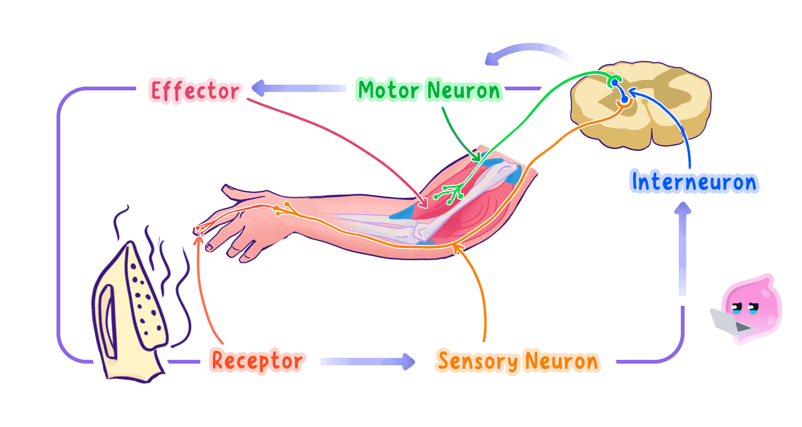
The word arc describes the shape of the route, because the impulse travels in to the spinal cord and straight back out again to a part of the body near where it started.
Inside the spinal cord
Spinal cord
The spinal cord is the column of nervous tissue running inside the backbone that carries impulses between the body and the brain and coordinates many reflexes.
- The spinal cord runs down inside the vertebrae of the backbone, which shield it from being crushed or cut.
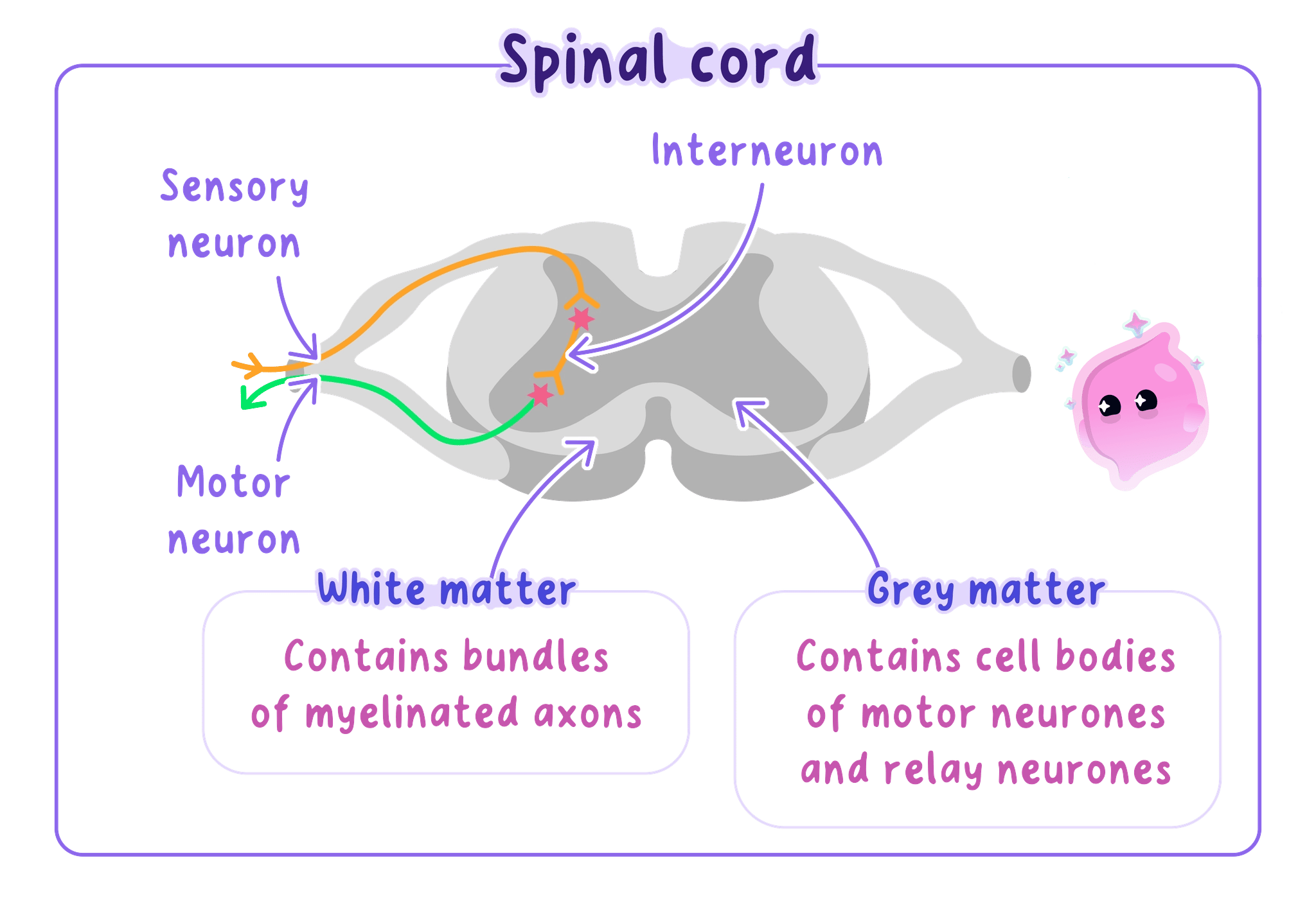
- A cross-section shows a butterfly-shaped inner region of grey matter, which contains the cell bodies of relay and motor neurones and the synapses between them.
- Around it lies white matter, which contains myelinated axons carrying impulses up to the brain and back down again.
- The white matter looks pale because the myelin sheaths around those axons are made of fat.
- Sensory neurones enter through the back of the spinal cord, and their cell bodies sit in a swelling just outside the cord itself.
- Motor neurones leave through the front of the spinal cord, with their cell bodies in the grey matter, so impulses in and impulses out use separate routes.
- Do not write that a reflex misses out the central nervous system, because the spinal cord is part of the CNS and is where the relay neurone sits.
- What the impulse misses out is the conscious part of the brain, so write that instead.
Why the structure makes the response fast
- The pathway contains only two synapses, and since crossing a synapse by diffusion is the slowest step, keeping the number low keeps the whole response short.
- The route is short, because the impulse only reaches the nearest part of the spinal cord instead of travelling up to the head and back.
- The sensory and motor neurones are myelinated, so the impulse travels along them at roughly 100 m s−1100\ \text{m s}^{-1}100 m s−1.
- No decision is made, so no time is spent comparing the stimulus with memory or weighing up choices.
- A withdrawal reflex therefore takes roughly 0.02 s0.02\ \text{s}0.02 s, while a voluntary response to the same stimulus takes roughly 0.25 s0.25\ \text{s}0.25 s.
- Saving that fraction of a second is enough to limit a burn to reddened skin rather than a blister.
Tracing the withdrawal reflex
- You take hold of a saucepan handle that is far hotter than you expected.
- Temperature and pain receptors in the skin of your fingers detect the heat and start impulses.
- A sensory neurone carries those impulses along your arm and into the back of your spinal cord.
- A relay neurone in the grey matter receives them across a synapse and passes them straight to a motor neurone.
- The motor neurone carries impulses out to the biceps muscle in your upper arm.
- The biceps contracts, bending your elbow and pulling your hand off the handle.
- Only after your hand has moved do impulses reaching your brain make you aware of the pain.
Reflex and voluntary responses compared
Voluntary response
A voluntary response is a response that is consciously decided by the cerebrum before impulses are sent to the effector.
- Route: a reflex passes through the spinal cord or the base of the brain, while a voluntary response passes through the cerebrum.
- Synapses: a reflex crosses two, while a voluntary response crosses very many more.
- Speed: a reflex takes a small fraction of a second, while a voluntary response takes about ten times as long.
- Control: a reflex is the same every time and cannot normally be stopped, while a voluntary response can be changed to suit the situation.
- Learning: a reflex is present from birth and needs no practice, while a voluntary skill improves the more you repeat it.
- Awareness: in a reflex you notice the stimulus after the response, while in a voluntary response you notice it before.
Reflexes in the body
- The withdrawal reflex pulls a limb away from something sharp or hot.
- The blinking reflex closes the eyelid when something touches the eyelashes or moves quickly towards the face, which keeps grit off the cornea.
- The pupil reflex changes the size of the pupil when the light level changes, which protects the retina.
- The knee-jerk reflex straightens the leg when the tendon below the kneecap is tapped, and doctors test it because a missing response points to damage somewhere in the arc.
- The swallowing and coughing reflexes keep food and dust out of the airway.
Writing about a reflex arc
- Name all five components in order, because the marks are usually one for each stage rather than one for the overall idea.
- When the question says explain how the structure is related to its function, pair each structural point with the speed it produces, such as few synapses and therefore little delay.
- Say the impulse does not pass through the conscious brain, not that the brain is not involved at all.
- If you are given a diagram, use the arrows on it to identify which neurone is which before you start writing.
- The most common lost mark is naming the muscle only as a muscle, so say which muscle contracts in the example given.
When a reflex arc is damaged
- If any one component stops working, the whole response fails, because each part depends on the impulse arriving from the part before it.
- Damage to the receptor means the stimulus is never detected, so no impulse starts.
- Damage to a neurone or to the spinal cord breaks the route, so impulses cannot reach the effector.
- Damage to the effector means the impulses arrive but the muscle cannot contract.
- Testing a reflex therefore tells a doctor that every part of that particular arc is intact.
- Give the three features of a reflex action.
- Name the five components of a reflex arc in order.
- Explain two structural reasons why a reflex is faster than a voluntary response.
- Describe what the grey matter and the white matter of the spinal cord each contain.
- Explain why you feel the pain of a burn only after your hand has moved.
- Give two protective reflexes and state what each one prevents.
3.1.4 Structure of the eye
The eye as a sense organ
Sense organ
A sense organ is an organ containing groups of receptor cells that detect one particular type of stimulus.
Retina
The retina is the light-sensitive layer lining the back of the eye that contains the rod and cone receptor cells.
- The eye is the sense organ that detects light, and its receptor cells are held in the retina at the back.
- Everything in front of the retina exists to get light onto those receptors in the right amount and sharply focused.
- Light is bent, or refracted, whenever it passes from one transparent material into another of different density.
- The eye uses that effect twice, at the cornea and at the lens, to bring rays from one point on an object back to a single point on the retina.
Every structure in the eye is shaped either to control how much light enters or to bend that light so it focuses exactly on the retina.
The structures of the eye
- The cornea is the transparent curved layer at the front of the eye, and because it is strongly curved and light enters it from air, it does most of the focusing.
- The sclera is the tough white outer coat around the rest of the eyeball, and it protects the eye and holds its shape.
- The iris is the coloured ring of muscle behind the cornea, and it changes the size of the pupil to control how much light gets in.
- The pupil is the hole in the centre of the iris, so it is a gap rather than a structure, and it looks black because almost no light comes back out.
- The lens is a clear, flexible, elastic disc that fine-tunes the focusing by changing its own shape.
- The ciliary body is a ring of muscle around the lens, and contracting or relaxing it changes the diameter of that ring.
- The suspensory ligaments are inelastic fibres that join the ciliary body to the edge of the lens and hold it in place.
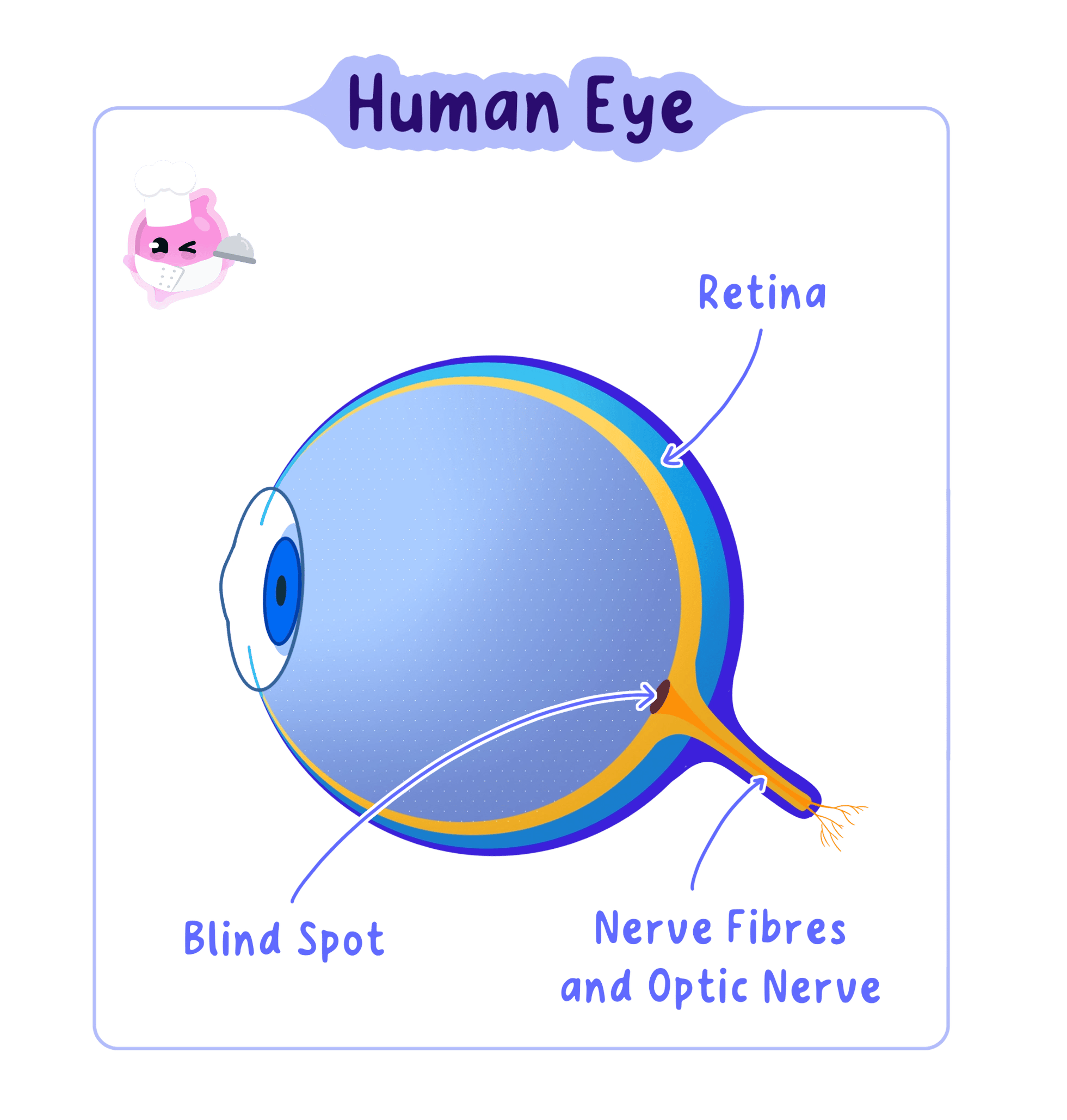
- The retina lines the inside of the back of the eye and contains the light receptor cells that convert light into electrical impulses.
- The optic nerve leaves the back of the eye carrying those impulses to the brain.
- The aqueous humour is the watery fluid between the cornea and the lens, and the vitreous humour is the jelly filling the space behind the lens.
- Both fluids press outwards, which keeps the eyeball firm so the distance from the lens to the retina stays the same.
The cornea does the bulk of the bending and cannot change, while the lens does the smaller adjustable part, which is why the lens is the structure that alters shape.
How an image is formed
Optic nerve
The optic nerve is the bundle of sensory neurones that carries electrical impulses from the retina to the brain.
- Light from an object reaches the eye and passes first through the cornea, where it is refracted strongly inwards.
- It then passes through the pupil, which sets how much of that light is allowed through.
- The lens refracts the light again by the extra amount needed for that particular distance.
- The rays meet on the retina, forming an image that is upside down and reversed from left to right.
- The receptor cells absorb the light and convert it into electrical impulses.
- The optic nerve carries the impulses to the brain, which interprets them and turns the upside-down image the right way up.
- Where the optic nerve leaves the retina there are no receptor cells, so light landing on that small patch produces no impulses at all.
- Do not write that the eye sees the object, because the retina only detects light and it is the brain that produces the image you experience.
- Do not say the pupil widens or narrows by itself, because the pupil is a hole and it is the iris muscles that change its size.
Accommodation
Accommodation
Accommodation is the change in the shape of the lens that allows the eye to focus light from objects at different distances onto the retina.
- Light from a near object spreads out as it reaches the eye, so it needs to be bent a lot to meet on the retina.
- Light from a distant object arrives almost parallel, so it needs to be bent much less.
- Since the distance from the lens to the retina is fixed, the eye adjusts by changing how strongly the lens refracts light.
- To focus on a near object the ciliary muscles contract, which makes the ring of muscle smaller.
- A smaller ring means the suspensory ligaments slacken, so they stop pulling on the edge of the lens.
- The elastic lens then springs into a fatter, more curved shape, which refracts the light more strongly and brings the spreading rays to a focus on the retina.
- To focus on a distant object the ciliary muscles relax, so the ring of muscle widens.
- A wider ring pulls the suspensory ligaments tight, and they stretch the lens outwards.
- The lens becomes thinner and less curved, so it refracts the light less and the nearly parallel rays still meet on the retina.
- The lens becomes stiffer with age, so it cannot become as fat as it once did, which is why many people need reading glasses in later life.
Looking from the board down to your book
- While you read the board, the ciliary muscles are relaxed, the suspensory ligaments are taut and the lens is thin.
- You look down at your book, and light from the page now reaches your eye spreading out rather than parallel.
- For a moment the light focuses behind the retina, so the words look blurred.
- The ciliary muscles contract, the ring narrows and the suspensory ligaments slacken.
- The lens becomes fatter and refracts the light more strongly.
- The rays now meet exactly on the retina and the words become sharp, all within about a second.
The iris and the pupil reflex
Pupil reflex
The pupil reflex is the automatic change in the size of the pupil, caused by the muscles of the iris, that controls how much light enters the eye.
- The iris contains two sets of muscle: circular muscles arranged in rings and radial muscles arranged like the spokes of a wheel.
- In bright light the circular muscles contract and the radial muscles relax, so the iris closes in and the pupil becomes smaller.
- A smaller pupil lets in less light, which protects the receptor cells of the retina from being damaged.
- In dim light the radial muscles contract and the circular muscles relax, so the iris is pulled outwards and the pupil becomes larger.
- A larger pupil lets in more light, so enough of it reaches the receptors for you to see.
- The change is a reflex, so light receptors in the retina are the receptor and the iris muscles are the effector, and it happens without you deciding anything.
- Do not say only that the circular muscles contract in bright light, because the mark scheme usually wants the radial muscles relaxing as well.
- Do not mix up the two sets: circular muscles close the pupil, and radial muscles open it.
Rod and cone cells
Rod cells
Rod cells are light receptor cells in the retina that respond in dim light but do not detect colour.
Cone cells
Cone cells are light receptor cells in the retina that detect colour and need bright light to respond.
Fovea
The fovea is the small central area of the retina, packed with cone cells, where an image is seen in the sharpest detail.
- The retina holds two kinds of light receptor cell, named after their shapes.
- Rod cells are very sensitive, so they respond in dim light, but they cannot tell colours apart and give only shades of grey.
- Cone cells need bright light to respond, and there are three types, sensitive to red, green and blue light.
- The brain compares the impulses arriving from the three types of cone, and the mixture tells it which colour you are looking at.
- Cones are packed most densely at the fovea, the small central patch of retina that lies straight in line with the lens.
- When you look directly at something its image falls on the fovea, which is why that part of the scene is the sharpest and most colourful.
- Rods are spread across the rest of the retina and become more common towards its edges, so faint objects are easier to see slightly off to one side.
Walking home on a dark evening you can make out the shapes of parked cars but not their colours, because only the rod cells are responding at that light level.
Linking structure to function
- Questions on the eye almost always ask you to explain how a structure is related to its function, so pair the feature with what it makes possible.
- The cornea is transparent and curved, so light can enter and be strongly refracted.
- The lens is elastic, so it can change shape and alter how much it refracts light.
- The iris is muscular, so it can change the size of the pupil in either direction.
- The retina is packed with receptor cells, so a detailed pattern of light can be converted into a detailed pattern of impulses.
Describing accommodation
- Give all three steps in order: what the ciliary muscles do, what happens to the suspensory ligaments, then what shape the lens takes.
- Finish with the effect on the light, because the final mark is usually for saying the light is refracted more or less so it focuses on the retina.
- Use slacken and tighten for the ligaments rather than contract and relax, since ligaments are not muscles.
- The pairing candidates most often reverse is near with relaxed muscles, so learn it as near means contract and fat.
Protecting the eye
- The eyeball sits in a bony socket in the skull, which shields it from knocks.
- Tears wash the front of the cornea, keeping it moist and clear and carrying away dust.
- Eyelashes and the blinking reflex stop most particles reaching the cornea in the first place.
- State which structure of the eye does most of the focusing and explain why.
- Describe the path taken by light from entering the eye to reaching the brain.
- Explain what happens to the ciliary muscles, suspensory ligaments and lens when you focus on a near object.
- Explain how the iris makes the pupil smaller in bright light.
- Give two differences between rod cells and cone cells.
- Explain why an image falling on the fovea is the sharpest part of what you see.
3.1.5 Defects of the eye
Short-sightedness
Short-sightedness
Short-sightedness is an eye defect in which light from distant objects is brought to a focus in front of the retina, so distant objects appear blurred.
- Someone who is short-sighted sees near objects clearly but finds distant objects blurred.
- The eye refracts the light too strongly for a distant object, so the rays cross and come to a focus in front of the retina.
- By the time the light reaches the retina the rays have spread apart again, so each point on the object lands as a small blurred disc rather than a point.
- One cause is an eyeball that is too long from front to back, so the retina sits behind the point where the light meets.
- The other cause is a lens that is too curved, or ciliary muscles that cannot relax enough to flatten it, so the lens refracts light more than it should.
- Near objects are still sharp because their light arrives spreading out, and the extra refraction is exactly what that light needed.
- The fix has to reduce the total refraction, so a concave (diverging) lens is placed in front of the eye.
- A concave lens is thinner in the middle than at the edges, so it spreads the rays out before they reach the cornea and the focus moves back onto the retina.
A focusing defect is always a mismatch between how strongly the eye refracts light and how far the retina is from the lens, and a corrective lens changes the refraction to match the distance.
Long-sightedness
Long-sightedness
Long-sightedness is an eye defect in which light from near objects is brought to a focus behind the retina, so near objects appear blurred.
- Someone who is long-sighted sees distant objects clearly but finds near objects blurred.
- The eye refracts the light too weakly for a near object, so the rays are still converging when they hit the retina and would have met behind it.
- One cause is an eyeball that is too short, so the retina sits in front of the point where the light would meet.
- The other cause is a lens that has become too flat or too stiff, so it cannot bulge into the fat shape a near object needs.
- The lens stiffens gradually through life, which is why long-sightedness becomes more common with age.
- The fix has to add refraction, so a convex (converging) lens is placed in front of the eye.
- A convex lens is thicker in the middle than at the edges, so it starts bending the rays together before they reach the cornea and the focus moves forward onto the retina.
- Do not swap the two lenses: short sight focuses light short of the retina and needs a concave lens, and long sight needs a convex one.
- Do not write that a corrective lens makes the eye stronger, because it changes the light before it enters and leaves the eye itself unaltered.
Correcting focus with lenses
- Spectacles hold the corrective lens a short distance in front of the eye, and they are cheap, easy to change and carry no risk.
- They can be inconvenient for sport, they steam up, and they change how a person looks, which some people dislike.
- Contact lenses sit directly on the surface of the cornea, so they move with the eye and give a wider field of clear vision.
- They need careful cleaning and handling, because dirt trapped underneath can scratch the cornea or cause an infection.
- The strength of a lens is written as a power in dioptres, and it is negative for a concave lens and positive for a convex one.
- A prescription of −2.5 D-2.5\ \text{D}−2.5 D therefore belongs to a short-sighted eye, and +2.5 D+2.5\ \text{D}+2.5 D to a long-sighted one.
Reading a pair of prescriptions
- One optician's record shows −3.0 D-3.0\ \text{D}−3.0 D and another shows +1.5 D+1.5\ \text{D}+1.5 D.
- The negative value means a concave lens, which spreads light out, so that eye was refracting light too strongly.
- Refracting too strongly puts the focus in front of the retina, so that person is short-sighted.
- The positive value means a convex lens, which brings light together, so that eye was refracting light too weakly.
- Refracting too weakly puts the focus behind the retina, so that person is long-sighted.
- The larger number, 3.03.03.0 rather than 1.51.51.5, also tells you the first defect is the more severe of the two.
Surgical ways of correcting focus
- Laser eye surgery uses a laser to remove a thin layer of tissue and reshape the cornea, which changes how strongly the cornea refracts light.
- Flattening the cornea reduces refraction and corrects short sight, while steepening it increases refraction and corrects long sight.
- The result is usually permanent and removes the need for glasses, but the surgery carries a risk of infection, of dry eyes and of over-correction, and it cannot be undone.
- A replacement lens can be fitted instead, with the natural lens removed and an artificial one of the correct power put in its place.
- That operation corrects strong defects that a laser cannot, but it is more invasive and carries a greater risk of damaging the retina.
- Deciding between the options means weighing cost, convenience and the size of the risk, and the same choice does not suit everyone.
Colour blindness
Colour blindness
Colour blindness is an eye defect in which one or more types of cone cell in the retina do not work properly, so certain colours cannot be told apart.
Cone cells
Cone cells are light receptor cells in the retina that detect colour and need bright light to respond.
- Colour blindness is not a focusing problem at all, because the light reaches the retina perfectly sharply.
- It is caused by one or more types of cone cell being missing or not working, so the brain never receives the impulses from that type.
- The commonest form is red-green colour blindness, where the red-sensitive or green-sensitive cones do not respond normally.
- Because the brain works out colour from the mixture of impulses from the three cone types, losing one type makes certain colours look the same as each other.
- The condition is inherited and affects far more males than females, and it is present from birth rather than developing later.
- There is no cure, because the faulty cone cells cannot be repaired or replaced and a lens cannot create impulses that were never produced.
- People manage it instead, using tinted filters that make confusable colours look more different, and using labels, patterns or numbers rather than colour alone.
- It is diagnosed with a set of test plates covered in coloured dots, where a number can only be picked out if the relevant cones are working.
A red-green colour blind pupil can still read a bar chart if each bar is given its own pattern of stripes or dots, because the shape carries the information instead of the colour.
Comparing the three defects
- Short sight and long sight are focusing defects, caused by the shape of the eyeball or the lens, and both can be corrected.
- Colour blindness is a receptor defect, caused by faulty cone cells in the retina, and it can only be managed.
- That difference explains why a lens helps with two of them and not with the third.
Explaining an eye defect
- Say where the light focuses relative to the retina, because that single phrase usually carries the first mark.
- Give a cause in terms of the eyeball length or the lens shape, then name the lens that corrects it.
- If you are asked to draw ray diagrams, show the uncorrected rays meeting off the retina before you add the corrective lens.
- For an evaluate question on treatments, give one benefit and one risk and then state which you think matters more and why.
- For colour blindness, make clear that the problem is with the cone cells and not with the focusing, since that is the distinction being tested.
Living with an eye defect
- Uncorrected short sight makes reading a board or a road sign difficult, which is why school and driving eye tests exist.
- Uncorrected long sight makes close work tiring, because the ciliary muscles stay contracted as they try to fatten the lens.
- Colour blindness rules out a small number of jobs, such as some roles in aviation and rail work, where colour signals must be read correctly.
- State where light focuses in a short-sighted eye and in a long-sighted eye.
- Give two possible causes of short-sightedness.
- Explain how a convex lens corrects long-sightedness.
- Give one advantage and one disadvantage of laser eye surgery compared with spectacles.
- Explain what causes colour blindness.
- Explain why a corrective lens cannot cure colour blindness.
3.1.6 Structure and function of the brain
The brain as part of the CNS
Central nervous system
The central nervous system is the brain and spinal cord, which receive impulses from sensory neurones, coordinate a response and send impulses out along motor neurones.
- The brain and the spinal cord together form the central nervous system, and the brain is where the more complex processing happens.
- It is built from billions of neurones, each forming synapses with thousands of others, which is where its processing power comes from.
- It is protected by the bony skull, by tough membranes, and by a layer of fluid that cushions it against knocks.
- Different jobs are handled by different regions, so damage in one place removes particular abilities while leaving others untouched.
- The five regions named in this topic are the cerebrum, the cerebellum, the medulla, the hypothalamus and the pituitary gland.
- Roughly speaking, the higher up a region sits, the more conscious its work is, and the lower down it sits, the more automatic its work is.
The brain is not one processor but a set of regions with separate jobs, which is why a person can lose the ability to balance while their memory and speech stay intact.
The cerebrum
Cerebrum
The cerebrum is the large, folded upper region of the brain that controls conscious thought, memory, language, personality and voluntary movement.
- The cerebrum is the largest region, sitting on top of the rest of the brain and covering most of it.
- Its thin outer layer, the cerebral cortex, is deeply folded, and the folds pack a far greater area of tissue into the space inside the skull.
- More area means more neurones and more synapses, which is what allows complex processing.
- The cerebrum controls conscious thought, memory, language, personality and voluntary movement.
- It also receives impulses from the sense organs, so seeing, hearing and feeling something consciously all happen here.
- It is divided into two halves called hemispheres, joined by a thick band of neurones that lets them exchange information.
- Each hemisphere controls the opposite side of the body, so a stroke in the left hemisphere weakens the right arm and leg.
- Within each hemisphere there are separate areas for movement, for touch, for vision and for speech, which is why damage in one small patch can remove one specific ability.
A patient whose stroke damaged one small region of the left cerebrum could still understand everything said to them but could no longer form the words to reply, which told doctors that region produces speech.
The cerebellum
Cerebellum
The cerebellum is the region beneath the back of the cerebrum that coordinates balance, posture and smooth, precise movement.
- The cerebellum sits underneath the back of the cerebrum, and its surface is folded even more finely.
- It controls balance, posture and the coordination of movement.
- It receives information from receptors in the muscles and joints about where each part of the body currently is.
- It also receives information from the balance receptors in the inner ear about the position and movement of the head.
- It compares that information with the movement the cerebrum has asked for, and adjusts the impulses going to the muscles so the movement comes out smooth and accurate.
- The cerebellum does not decide to move, so a person with cerebellum damage can still choose to reach for a cup but their arm moves jerkily and overshoots.
- It is also where practised skills such as riding a bike or writing become automatic.
A police roadside balance test works because alcohol reaches the cerebellum quickly and interferes with the fine adjustments it makes to posture.
The medulla
Medulla
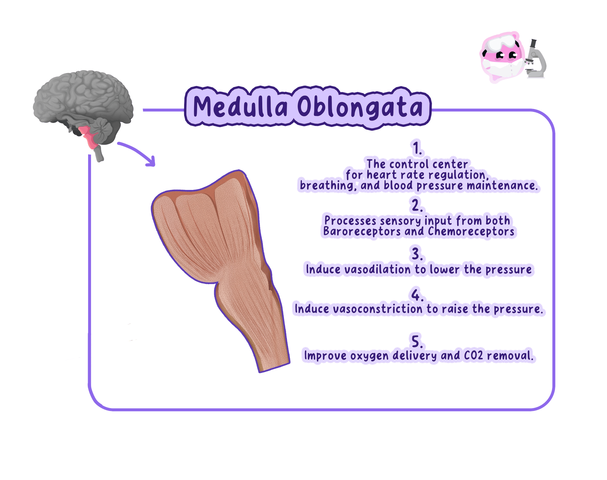
The medulla is the region at the base of the brain, joining the spinal cord, that controls unconscious activities such as heart rate and breathing rate.
- The medulla lies at the base of the brain, where it joins the top of the spinal cord.
- It controls unconscious activities that continue whether you are awake or asleep.
- It sets the heart rate by sending impulses to the heart, speeding it up during exercise and slowing it down at rest.
- It sets the breathing rate by sending impulses to the muscles of the ribs and diaphragm.
- It receives information from receptors that monitor the blood, so a rise in dissolved carbon dioxide makes it increase the breathing rate.
- It also controls blood pressure, swallowing, coughing and vomiting.
- Because these processes keep you alive from moment to moment, damage to the medulla is far more likely to be fatal than damage of the same size in the cerebrum.
- Do not swap the cerebellum and the medulla, because the cerebellum coordinates movement while the medulla controls heart rate and breathing rate.
- Do not write that the medulla controls all muscles, because the muscles you move on purpose are controlled by the cerebrum.
The hypothalamus and pituitary gland
Hypothalamus
The hypothalamus is the region of the brain that monitors internal conditions such as body temperature and water content and triggers the responses that keep them steady.
Pituitary gland
The pituitary gland is a small gland hanging below the hypothalamus that releases hormones which control other glands in the body.
- The hypothalamus is a small region near the base of the brain, just above the pituitary gland.
- It monitors the blood flowing through it, checking its temperature and its water content.
- When a reading moves away from normal it triggers the correcting response, so it acts as the body's thermoregulatory centre.
- It also helps control sleep, hunger and thirst, all of which depend on the state of the blood.
- The pituitary gland hangs just below it on a short stalk, and the hypothalamus controls what that gland releases.
- The pituitary gland releases hormones into the blood, and several of those hormones act on other glands and make them release hormones of their own.
- This pair is therefore the point where electrical control by the brain is turned into chemical control carried in the blood.
- That link explains how a change detected inside the brain can produce a slow, long-lasting response across the whole body.
Matching a region to an ability
- Questions often describe a patient's symptoms and ask which region is damaged, so read the symptom and work backwards to the job.
- Loss of memory, of speech or of conscious control of a limb points to the cerebrum.
- Unsteady walking, poor balance or shaky, badly aimed movements point to the cerebellum.
- An irregular heartbeat or difficulty breathing points to the medulla.
- A body temperature that drifts, or unusual thirst, points to the hypothalamus.
Answering on brain regions
- Name the region and its job in the same sentence, because a bare name rarely gets the explain mark.
- Use unconscious for the medulla and coordination of movement for the cerebellum, since those are the phrases the mark scheme credits.
- On a diagram, find the spinal cord first, because the region joined to it is the medulla and everything else can be placed from there.
- If a case study gives several symptoms, name the one region that explains all of them rather than a different region for each.
Why the regions work together
- A single action usually needs several regions at once, so they are connected by large bundles of neurones.
- Running upstairs starts as a decision in the cerebrum, is smoothed by the cerebellum, and is supported by the medulla raising your heart and breathing rates.
- Meanwhile the hypothalamus notices your blood warming and starts the responses that cool you down.
- Because the regions are so tightly linked, damage in one place often disturbs abilities that seem to belong to another.
- Name the five regions of the brain covered here and give one function of each.
- Explain why the cerebral cortex is folded.
- Describe the information the cerebellum uses to keep a movement smooth.
- Explain why damage to the medulla is especially dangerous.
- State what the hypothalamus monitors in the blood.
- Explain how the hypothalamus and pituitary gland link nervous control to chemical control.
3.1.7 Difficulties of investigating brain function
Why the brain is hard to investigate
Central nervous system
The central nervous system is the brain and spinal cord, which receive impulses from sensory neurones, coordinate a response and send impulses out along motor neurones.
- The brain is enclosed by the skull, so it cannot be seen or reached without surgery.
- Brain tissue is extremely delicate, so any attempt to reach one region risks destroying healthy tissue on the way in.
- It contains billions of neurones with an enormous number of connections between them, so the wiring is far too complex to map by looking at it.
- Regions do not work in isolation, because most abilities depend on several regions passing impulses to each other.
- Deliberately removing or damaging part of a healthy brain to see what changes would harm the person, so that experiment can never be done.
- Scientists therefore have to work with the damage that has already happened, or with methods that leave the brain intact.
Brain research is limited less by a lack of ideas than by the fact that the one experiment that would settle a question cannot ethically be carried out.
Studying patients with brain damage
- A case study is a detailed record of one patient whose brain has been damaged by an injury, a stroke, a tumour or an infection.
- Researchers test what the patient can and cannot do, then find out which region was damaged, either by scanning them or by examining the brain after death.
- If a lost ability lines up with a damaged region, that region is likely to be needed for the ability.
- Confidence grows only when many patients with damage in the same place show the same loss.
- Much of what is known about the roles of the cerebrum was built up this way over more than a century.
A patient who can no longer form new memories after an operation, but who still remembers their childhood clearly, shows that storing new memories and recalling old ones are handled by different parts of the brain.
Problems with obtaining case studies
- Suitable patients are rare, because damage confined to one small region hardly ever happens.
- Researchers cannot choose when a case appears, so the work is slow and depends on waiting for an injury to happen to someone.
- Patients are often seriously ill, so testing them may be impossible or may have to be kept very short.
- The sample size is tiny, sometimes a single person, so a result cannot safely be applied to everybody.
- The study cannot be repeated, because no second patient will ever have exactly the same damage.
- There is no control, because the same person cannot be studied both with and without the damage.
Problems with interpreting case studies
- Damage usually spreads across more than one region, so it is hard to tell which of them caused the lost ability.
- A damaged region may only be part of a longer pathway, so the loss may really be caused by the connection being cut rather than by that region doing the job.
- There is rarely a record of what the patient could do before the damage, so the size of the loss has to be estimated.
- The brain can partly reroute a task through other neurones over time, so testing months later may underestimate what was lost.
- Every brain differs slightly in the exact position of its regions, so the same map does not fit everyone.
- A case study therefore shows a correlation between a damaged region and a lost ability, which is weaker evidence than a controlled experiment.
- Do not write that a case study proves a region controls an ability, because a single unrepeatable observation cannot prove cause.
- Write instead that the evidence suggests a link, and say what further evidence would strengthen it.
Stimulating the brain electrically
- A weak electric current can be applied to a small area of the exposed brain during surgery, and the effect on the patient is recorded.
- The current starts impulses in the neurones of that area, so stimulating one spot may make a finger twitch and stimulating another may produce a sensation or a memory.
- The brain itself has no pain receptors, so the patient can stay awake and describe what they feel while the surgeon works.
- This method gives a very precise map, because the area being tested is being touched directly.
- Its limitation is that it can only be done on patients who already need brain surgery for another reason, so the sample is small and unusual.
- Stimulating a region also produces an artificial burst of impulses, which is not the same as watching that region work normally.
Scanning the brain
MRI scan
An MRI scan is a technique that uses strong magnetic fields and radio waves to produce a detailed image of the structure of the brain without surgery.
- A scan produces an image of the brain from outside the skull, so no surgery is needed and the person is unharmed.
- An MRI scan uses strong magnetic fields and radio waves to build a detailed image of the structure of the brain, showing exactly where a tumour or a damaged region lies.
- Other scans show activity instead, because active neurones respire faster and so draw more oxygen and glucose from the blood.
- A person can be scanned while reading, listening or moving, and the region that lights up is matched to the task.
- Scanning removed the biggest limitation of case studies, because healthy volunteers can be studied and the same person can be scanned again and again.
- Scanners are expensive and need trained staff, so studies use small numbers of volunteers.
- The person must lie completely still inside a noisy machine, which is difficult for young children and for anyone in pain.
- A bright patch on a scan shows only that a region is more active during a task, which is again a correlation rather than proof that the region produces the ability.
Ethical issues
- Patients must give informed consent, which means understanding what will be done and agreeing to it freely.
- Brain damage can affect memory, understanding and judgement, so the very patients researchers most want to study may be the least able to give that consent.
- Patients are in a vulnerable position, so they must not feel pressured into taking part in order to receive treatment.
- Any procedure must not increase the harm to the patient, so extra surgery purely for research is not allowed.
- Personal and medical details must be kept confidential, which is difficult when a case is so rare that the patient could be identified from the description.
- Using animals instead avoids some of these problems but raises others, because the animal cannot consent and its brain differs from a human one.
- Research is therefore reviewed by an ethics committee before it starts, and the expected benefit has to justify the risk.
Writing about the difficulties
- Separate the practical difficulties from the ethical ones, because a question asking for both wants a point from each side.
- Give a reason with every difficulty, so complex is not enough on its own and needs the number of neurones and connections behind it.
- Use the words small sample, no control and cannot be repeated when you evaluate a case study, since those are the marking points.
- For an evaluate question, finish with a judgement, such as saying that scanning gives the more reliable evidence because it can be repeated on healthy volunteers.
How the evidence builds up
- No single method is strong enough on its own, so scientists look for the same conclusion from several of them.
- When case studies, electrical stimulation and scanning all point to the same region, the conclusion becomes far more secure.
- Because of these limits our understanding of the brain is still incomplete, and it is being revised as scanning technology improves.
- Give three reasons why the brain is difficult to investigate.
- Describe how a case study of a damaged brain is used to work out what a region does.
- Give two reasons why suitable case studies are hard to obtain.
- Explain two reasons why the results of a case study are hard to interpret.
- State one advantage of scanning over studying patients with brain damage.
- Explain why obtaining informed consent can be difficult in brain research.
3.1.8 Limitations in treating nervous system damage
Why nervous tissue does not repair
Neurone
A neurone is a nerve cell adapted to carry electrical impulses rapidly from one part of the body to another.
- Most tissues heal because their cells divide to replace the ones that were lost, which is how a cut in the skin closes within days.
- Neurones in the brain and spinal cord do not divide once they are mature, so a neurone that dies is not replaced by a new one.
- A cut axon inside the central nervous system does not regrow along its old route, so the connection it made is lost even if the cell body survives.
- The gap fills instead with scar tissue, which physically blocks any regrowth and does not carry impulses.
- Even if an axon did regrow, it would have to find its original target among billions of others to restore the same function.
- The effects of the damage are therefore usually permanent, which is what makes this different from an injury to a muscle or a bone.
- Do not write that nervous tissue heals slowly, because the point is that neurones in the CNS are not replaced at all.
- Do not use a broken bone as your comparison, since bone repairs itself and nervous tissue does not.
Irreversible damage to surrounding tissue
Paralysis
Paralysis is the loss of movement in part of the body caused when impulses can no longer pass between the central nervous system and the muscles.
- Nervous tissue is soft and easily torn, so instruments passing through healthy tissue to reach a damaged region destroy neurones on the way.
- Because those neurones are not replaced, the harm done by the operation itself is permanent as well.
- Bleeding and swelling after an injury press on nearby tissue, and since the skull cannot expand that pressure kills further neurones.
- Squashed blood vessels then cut off the oxygen supply, so more cells die of starvation in the hours after the original damage.
- Neurones lie so close together that different functions are packed within millimetres, so a small area of damage can remove several abilities at once.
- If the motor pathways in the spinal cord are cut, impulses can no longer reach the muscles below that point and paralysis follows.
- Sensory pathways usually run in the same region, so feeling is often lost from the same part of the body.
Why the position of a spinal injury matters
- The spinal cord carries impulses between the brain and every part of the body below the head.
- A cut in the lower back stops impulses reaching the legs, so the legs are paralysed while the arms still work.
- A cut in the neck is above the point where the nerves to the arms leave, so the arms are affected as well as the legs.
- A cut high in the neck also blocks the impulses running to the muscles that control breathing.
- The higher the injury, the more of the body is cut off from the brain, so the effects are more severe.
- Reflexes below the injury can still work, because their arc is completed in the spinal cord without needing the brain.
Difficulty in reaching the nervous system
Spinal cord
The spinal cord is the column of nervous tissue running inside the backbone that carries impulses between the body and the brain and coordinates many reflexes.
- The brain is enclosed by the skull and the spinal cord runs inside the vertebrae, so bone has to be cut through before either can be reached.
- The protection that keeps the CNS safe from everyday knocks is the same protection that keeps a surgeon out.
- Many important structures lie deep inside the brain, so reaching them means passing through the regions above.
- A tumour often grows in among healthy neurones rather than as a separate lump, so it cannot be removed without taking healthy tissue too.
- Drugs are another route in, but the blood vessels of the brain have an unusually tight lining that stops most substances leaving the blood.
- That barrier protects the brain from poisons and pathogens, but it also blocks many medicines that would otherwise treat the damage.
- The nerves outside the CNS are spread throughout the body, so damage to them can be equally hard to locate and reach.
What treatment can achieve
- Most treatment manages the effects rather than repairing the tissue, which is the key limitation to state in an answer.
- Surgery can prevent further damage by removing a tumour, draining a bleed or relieving pressure, but it cannot restore neurones already lost.
- Drugs can control symptoms such as pain, seizures or muscle stiffness, and some slow the rate at which further neurones are lost.
- Physiotherapy helps a patient recover some function because repeated practice strengthens the connections in pathways that survived, so the task is rerouted rather than the damage repaired.
- Electrical implants can take over a particular job, such as stimulating a muscle directly or steadying the tremor of a movement disorder.
- Research into replacing lost neurones with stem cells is under way, and the difficulty is not only growing new neurones but getting them to connect correctly.
Recovery after a stroke happens because surviving neurones take over the task, which is why progress is slow, partial and depends heavily on how much practice the patient does.
Why prevention matters so much
- Since the damage cannot be reversed, avoiding it in the first place is far more effective than any treatment.
- A helmet spreads the force of an impact over a larger area and over a longer time, so less force reaches the skull and brain.
- Seatbelts, head restraints and airbags reduce the sudden movement of the head, which is what tears neurones inside the skull during a crash.
- Rules about head injuries in sport keep a concussed player off the pitch, because a second impact before recovery causes far worse damage.
- Not moving someone with a suspected spinal injury matters for the same reason, since moving them can turn a partly damaged cord into a completely severed one.
Explaining the limitations
- Give the three named limitations separately: nervous tissue cannot be repaired, surrounding tissue is damaged irreversibly, and parts of the nervous system are hard to access.
- Explain each one rather than naming it, so say that neurones do not divide and therefore are not replaced.
- When a question describes a patient, link the loss to the specific pathway that has been cut rather than answering in general terms.
- The mark most often dropped is the one for saying the effects are permanent, so make that consequence explicit.
Weighing up a treatment
- Deciding whether to operate means comparing the harm the damage is already doing with the harm the operation itself could cause.
- A tumour pressing on the medulla may be worth the risk of surgery, while one sitting deep among the pathways for speech may not be.
- The patient shares that decision, because only they can judge which abilities they are least willing to lose.
- Explain why a neurone lost from the brain is not replaced.
- Describe two ways in which tissue around an injury is damaged after the original injury has happened.
- Give two reasons why parts of the nervous system are difficult to reach.
- Explain why an injury high in the neck has more severe effects than one in the lower back.
- State what physiotherapy achieves after a stroke, and what it does not achieve.
- Explain how wearing a helmet reduces the chance of permanent brain damage.
