1.1.1 Viewing cells with light microscopes
Parts of a light microscope
Light microscope
An instrument that uses visible light and glass lenses to produce a magnified image of a specimen.
Specimen
The piece of biological material that is mounted on a slide and examined under a microscope.
- A light microscope shines visible light up through a thin specimen and uses curved glass lenses to bend that light outwards, so the image reaching your eye is far larger than the object on the slide.
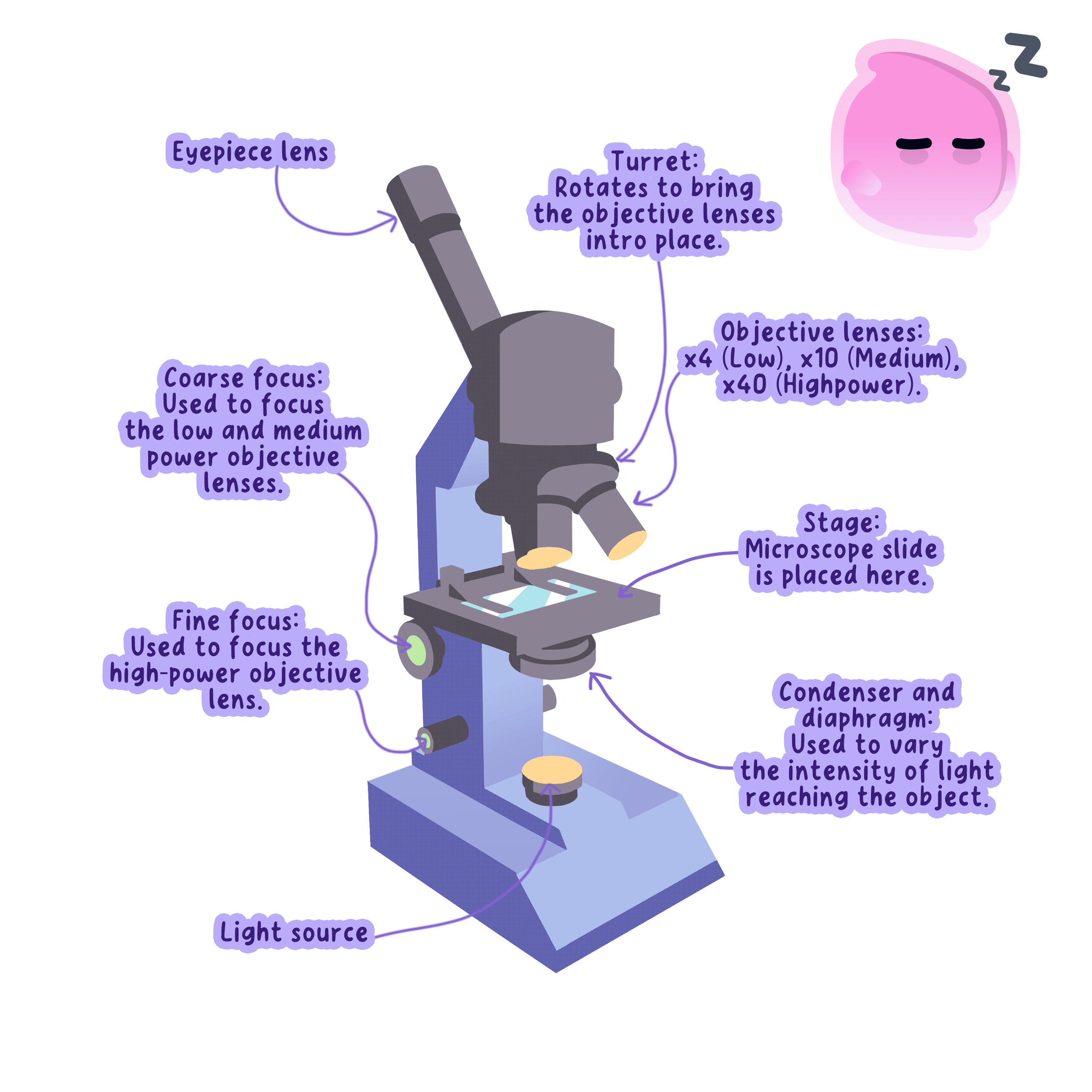
- The lamp sits in the base beneath the stage and supplies the beam of light that travels upwards through the specimen.
- The condenser is a lens under the stage that gathers light from the lamp and concentrates it onto the small area where the specimen lies.
- The iris diaphragm is an adjustable ring in the condenser that changes how much of that light passes through, which sets the contrast of the image.
- The stage is the flat platform the slide rests on, and it has a hole in the middle so light can pass straight through the specimen.
- Stage clips hold the slide firmly in place so the specimen does not slip out of view while you move it.
- The objective lenses are mounted on a rotating turret directly above the specimen, and a school microscope normally carries three, marked ×4\times4×4, ×10\times10×10 and ×40\times40×40.
- The eyepiece lens sits at the top of the tube and is the lens you look down, and it is usually ×10\times10×10.
- The coarse focus knob moves the stage or the tube through a large distance and brings the specimen roughly into view at low power.
- The fine focus knob moves it through a very small distance and sharpens the image once the specimen is almost in focus.
- Light therefore travels from the lamp, through the condenser, through the specimen, into the objective lens and then into the eyepiece lens, and each of the two lenses enlarges the image in turn.
- Always begin with the lowest-power objective lens in place, because the field of view is widest at low power and a small specimen is far easier to find.
- Lower the objective lens towards the slide while watching from the side, then focus by moving the lens away from the slide, so the lens can never be driven down into the glass.
Preparing and staining a slide
Stain
A coloured chemical that binds to particular structures in a specimen so that they can be seen under a microscope.
- Light has to pass through the specimen to reach your eye, so the specimen must be thin enough to be almost transparent, ideally a single layer of cells.
- The specimen is laid flat in the centre of a clean glass microscope slide, the rigid strip of glass that carries it under the lenses.
- A drop of water or stain is added on top, which stops the specimen drying out and shrivelling while you look at it.
- A cover slip is the thin square of glass placed over the specimen, and it flattens the specimen into an even layer and keeps the liquid away from the objective lens.
- The cover slip is held at roughly 45∘45^\circ45∘ on a mounted needle and lowered slowly, so the liquid spreads sideways and drives the air out ahead of it.
- Air trapped under the cover slip appears as a circle with a thick dark rim, and it is easy to mistake for a cell until you notice that nothing lies inside it.
- Most plant and animal cells are colourless and nearly transparent, so under plain light they show little more than faint grey outlines.
- A stain is a coloured chemical that binds strongly to some structures and weakly to others, so different parts of the cell take on different depths of colour and become visible.
- Iodine solution stains plant cytoplasm pale yellow and stains the nucleus a much darker orange-brown, which is why the nucleus of an onion cell stands out against the rest of the cell.
- Methylene blue stains the nucleus of an animal cell dark blue, which makes the nucleus easy to pick out in a cheek cell that is otherwise almost invisible.
- Crystal violet and eosin work in the same way and are chosen when a different structure or a different tissue needs to be picked out.
- Staining kills the cells, so a stained slide shows the structure of a cell but never shows it carrying out a living process.
- Do not flood the slide with stain, because the surplus runs out beyond the cover slip and darkens the whole field of view until nothing can be told apart.
- Do not drop the cover slip flat onto the specimen, because the air trapped underneath hides every cell beneath a bubble.
- Do not describe a stained specimen as living material, because the stain has killed it.
Observing plant and animal cells
- Aim: to prepare stained slides of onion epidermis and human cheek cells, view them with a light microscope, and record them as labelled biological drawings with a stated magnification.
- Apparatus: light microscope, glass microscope slides, cover slips, forceps, mounted needle, scalpel, white tile, a fleshy onion scale, sterile cotton buds, a beaker of disinfectant, iodine solution and methylene blue in dropping bottles, distilled water in a dropping pipette, filter paper, sharp pencil, ruler and eye protection.
- Method, the onion slide:
- Cut a square of about 1 cm1\ \text{cm}1 cm from a fleshy onion scale on a white tile, cutting away from your fingers.
- Grip the thin skin on the inner curved surface with forceps and peel it away in one piece. This epidermis is a single cell thick, which is what makes it transparent enough to view.
- Lay the peel flat in the centre of a slide and tease out any folds with the mounted needle, because a fold stacks cells on top of one another and blurs the image.
- Add a single drop of iodine solution directly onto the peel.
- Hold a cover slip at about 45∘45^\circ45∘ on the mounted needle, rest one edge on the slide beside the drop, and lower it slowly so the liquid spreads and the air is driven out.
- Touch the corner of a piece of filter paper to the edge of the cover slip to draw off the surplus stain.
- Method, the cheek cell slide:
- Rub the inside of your own cheek gently with a clean cotton bud to collect a few loose surface cells.
- Smear the bud across the centre of a slide in one thin sweep, then put the used bud straight into the disinfectant.
- Add one drop of methylene blue, lower a cover slip in the same way, and blot off the surplus.
- Method, viewing and recording:
- Clip the slide to the stage with the specimen centred over the hole, and open the iris diaphragm.
- Rotate the ×4\times4×4 objective into place, watch from the side while the coarse focus brings the lens close to the slide, then look down the eyepiece and focus upwards until the cells appear.
- Sharpen the image with the fine focus, then close the iris diaphragm slightly until the cell outlines look crisp rather than washed out.
- Move up to ×10\times10×10 and then ×40\times40×40, using the fine focus only, so the high-power lens cannot be driven into the cover slip.
- Draw a small group of cells with a sharp pencil, using single unbroken lines with no shading, no sketching and no arrow heads.
- Label the structures with straight ruled lines that do not cross one another, and write the eyepiece and objective magnifications beside the drawing.
- Results: onion epidermis cells appear as regular brick shapes packed in tidy rows, each with a straight wall and a stained nucleus. Cheek cells appear as separate, rounded, irregular flakes with a stained nucleus and no wall, and they lie much further apart.
- Maths: total magnification is the eyepiece magnification multiplied by the objective magnification, so ×10\times10×10 with ×40\times40×40 gives ×400\times400×400. Measure a drawn cell with a ruler and use A=IMA=\dfrac{I}{M}A=MI to find its real width, converting with 1 mm=1000 μm1\ \text{mm}=1000\ \mu\text{m}1 mm=1000 μm.
- Watch out: too much stain darkens the field, trapped bubbles hide cells, a folded peel gives a double layer, and focusing downwards at high power cracks the cover slip. Draw only what is on the slide, not the textbook cell you expect to see.
- Safety: wear eye protection, cut on a tile away from your fingers, treat iodine and methylene blue as staining chemicals that mark skin and clothing, use only your own cotton bud and place it in disinfectant straight afterwards, and carry the microscope with one hand under the base.
Calculating magnification
Magnification
The number of times larger an image is than the real object it shows.
Scale bar
A labelled line printed on an image that shows the real length the line stands for.
- Magnification compares the size of the image with the size of the real object, and it has no unit because it is one length divided by another.
- The equation is magnification=size of imagesize of real object\text{magnification}=\dfrac{\text{size of image}}{\text{size of real object}}magnification=size of real objectsize of image.
- In symbols this is M=IAM=\dfrac{I}{A}M=AI, where III is the measured size of the image and AAA is the actual size of the object.
- Rearranging gives I=M×AI=M\times AI=M×A for the image size and A=IMA=\dfrac{I}{M}A=MI for the real size.
- The total magnification of a microscope is the eyepiece magnification multiplied by the objective magnification, so a ×10\times10×10 eyepiece used with a ×40\times40×40 objective gives 10×40=×40010\times40=\times40010×40=×400.
- Both lengths must be converted into the same unit before dividing, because a mismatch of units changes the answer by a factor of a thousand or more.
- The conversions needed are 1 mm=1000 μm1\ \text{mm}=1000\ \mu\text{m}1 mm=1000 μm and 1 μm=1000 nm1\ \mu\text{m}=1000\ \text{nm}1 μm=1000 nm.
- Cells are normally measured in micrometres, so a typical animal cell is roughly 20 μm20\ \mu\text{m}20 μm across and a bacterial cell roughly 2 μm2\ \mu\text{m}2 μm long.
- Very small measurements are written in standard form to keep them manageable, so 0.002 mm0.002\ \text{mm}0.002 mm is written as 2×10⁻³ mm2\times10^{-3}\ \text{mm}2×10⁻³ mm.
- A scale bar printed beside a photograph is a line of known real length, so measuring the bar with a ruler and dividing by the length it stands for gives the magnification of that photograph.
- Once the magnification of a photograph is known, the real size of anything else in the same photograph is found by measuring it and dividing by that magnification.
Magnification from a photograph
- An onion cell measures 45 mm45\ \text{mm}45 mm across on a printed photograph, and the real cell is 300 μm300\ \mu\text{m}300 μm across.
- Convert the image size into the unit used for the object: 45 mm=45 000 μm45\ \text{mm}=45\,000\ \mu\text{m}45 mm=45000 μm.
- Substitute into M=IAM=\dfrac{I}{A}M=AI: M=45 000300M=\dfrac{45\,000}{300}M=30045000.
- The magnification is ×150\times150×150, written with no unit.
Real size from a scale bar
- A photograph carries a scale bar labelled 50 μm50\ \mu\text{m}50 μm that measures 10 mm10\ \text{mm}10 mm when checked with a ruler.
- Convert the measured length: 10 mm=10 000 μm10\ \text{mm}=10\,000\ \mu\text{m}10 mm=10000 μm.
- The magnification of the photograph is M=10 00050=×200M=\dfrac{10\,000}{50}=\times200M=5010000=×200.
- A chloroplast in the same photograph measures 1.2 mm1.2\ \text{mm}1.2 mm, which is 1200 μm1200\ \mu\text{m}1200 μm.
- Its real length is A=IM=1200200=6 μmA=\dfrac{I}{M}=\dfrac{1200}{200}=6\ \mu\text{m}A=MI=2001200=6 μm.
Answering magnification questions
- Write the equation out before substituting, because the equation line carries its own mark even when the arithmetic goes wrong.
- Convert both lengths into micrometres first and write the converted value down, since a missing factor of 100010001000 is the most common way marks are lost here.
- Give a magnification as a bare number with a multiplication sign in front, such as ×150\times150×150, and never attach a unit to it.
- Measure a scale bar or an image to the nearest millimetre with a ruler rather than estimating it, because a measured value falls inside the allowed tolerance and a guess often does not.
- Name the two lenses that magnify the image in a light microscope and state which one you look down.
- Explain why a specimen must be thin and why a stain is added before it is viewed.
- State the equation that links magnification, image size and real size.
- Calculate the total magnification when a ×10\times10×10 eyepiece is used with a ×40\times40×40 objective lens.
- A cell measures 30 mm30\ \text{mm}30 mm on a photograph taken at a magnification of ×1500\times1500×1500. Calculate its real width in micrometres.
1.1.2 Sub-cellular structures and functions
Eukaryotic and prokaryotic cells
Eukaryotic cell
A cell whose genetic material is enclosed inside a nucleus.
Prokaryotic cell
A cell whose genetic material lies free in the cytoplasm rather than inside a nucleus.
Organelle
A structure inside a cell that is specialised to carry out one particular function.
- Every living organism is built from cells, and all cells fall into one of two groups according to where the genetic material is kept.
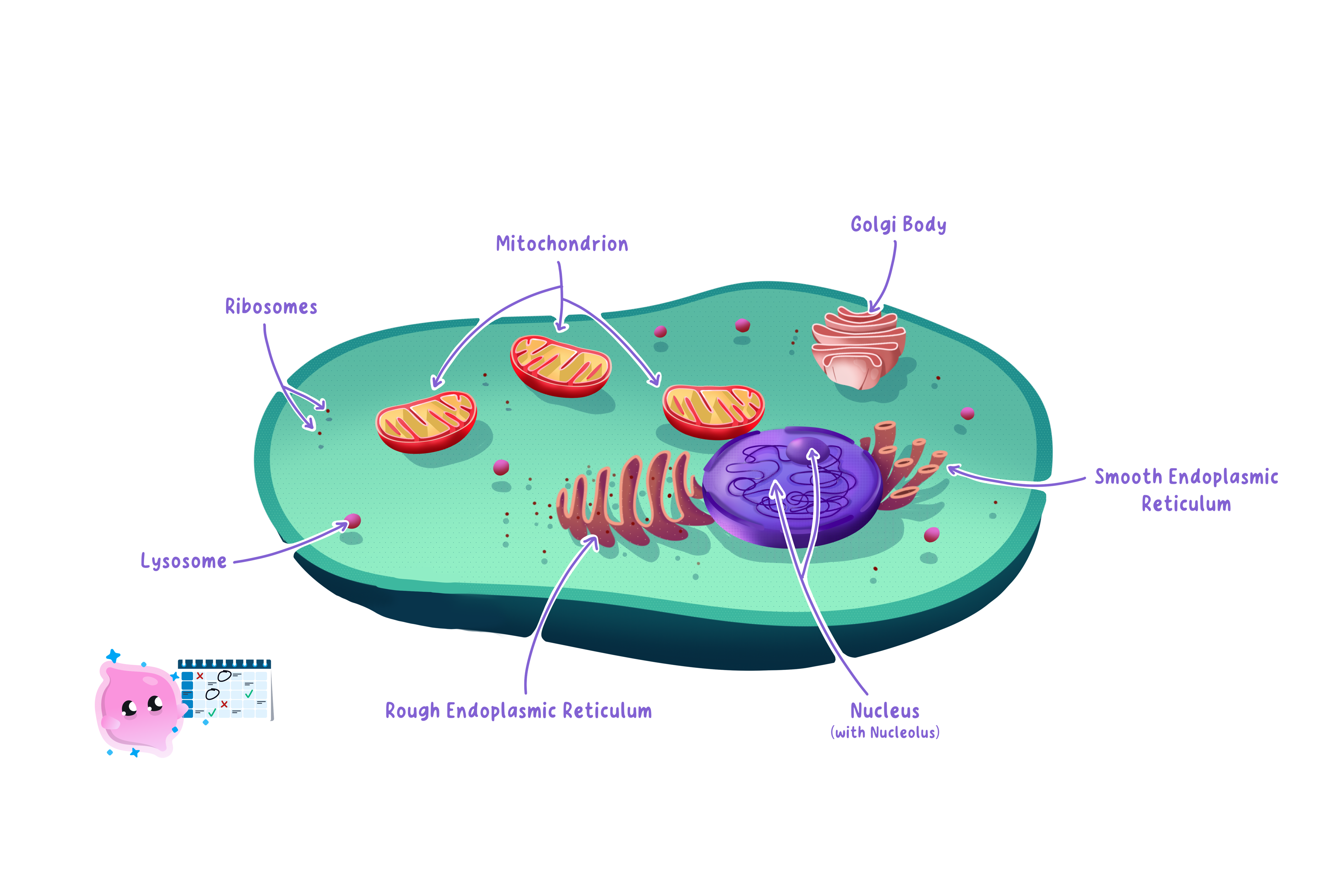
- A eukaryotic cell keeps its genetic material inside a nucleus, a compartment sealed off by its own membrane, and animals, plants and fungi are all built from eukaryotic cells.
- A prokaryotic cell has no nucleus, so its genetic material lies loose in the cytoplasm, and bacteria are prokaryotic.
- Eukaryotic cells are much the larger of the two, typically 10 μm10\ \mu\text{m}10 μm to 100 μm100\ \mu\text{m}100 μm across, while prokaryotic cells are typically 1 μm1\ \mu\text{m}1 μm to 5 μm5\ \mu\text{m}5 μm long.
- A eukaryotic cell contains many membrane-bound organelles, each a separate compartment specialised for one job.
- Dividing the cell into compartments lets different reactions run at the same time without interfering with one another, because each organelle holds the particular enzymes its own reaction needs.
- A prokaryotic cell has no membrane-bound organelles at all, so its reactions take place in the cytoplasm or at the cell membrane.
- Both types of cell have cytoplasm, a cell membrane and ribosomes, because every cell has to hold its contents together, control what crosses its surface and make its own proteins.
- A prokaryotic cell is a complete living cell in its own right, not a simplified piece of a eukaryotic one.
- The absence of a nucleus is the single feature that defines a prokaryotic cell, so start there whenever you are asked to tell the two types apart.
Where the genetic material is kept
Nucleus
The membrane-bound organelle that contains the chromosomes and controls the activities of the cell.
Chromosome
A long, coiled molecule of DNA that carries the genes of an organism.
Plasmid
A small circular loop of DNA in the cytoplasm of a prokaryotic cell, separate from its main chromosome.
- The nucleus of a eukaryotic cell is enclosed by its own membrane and is usually the largest structure that can be picked out under a light microscope.
- Inside the nucleus the DNA is coiled into long structures called chromosomes, and a human body cell holds 464646 of them arranged in 232323 pairs.
- Each chromosome carries many genes, and a gene is a length of DNA that codes for one protein.
- The nucleus controls the activities of the cell because the proteins built from those instructions include the enzymes that decide which reactions the cell can carry out.
- Enclosing the DNA in a membrane keeps it apart from the reactions going on in the surrounding cytoplasm, which protects it from damage.
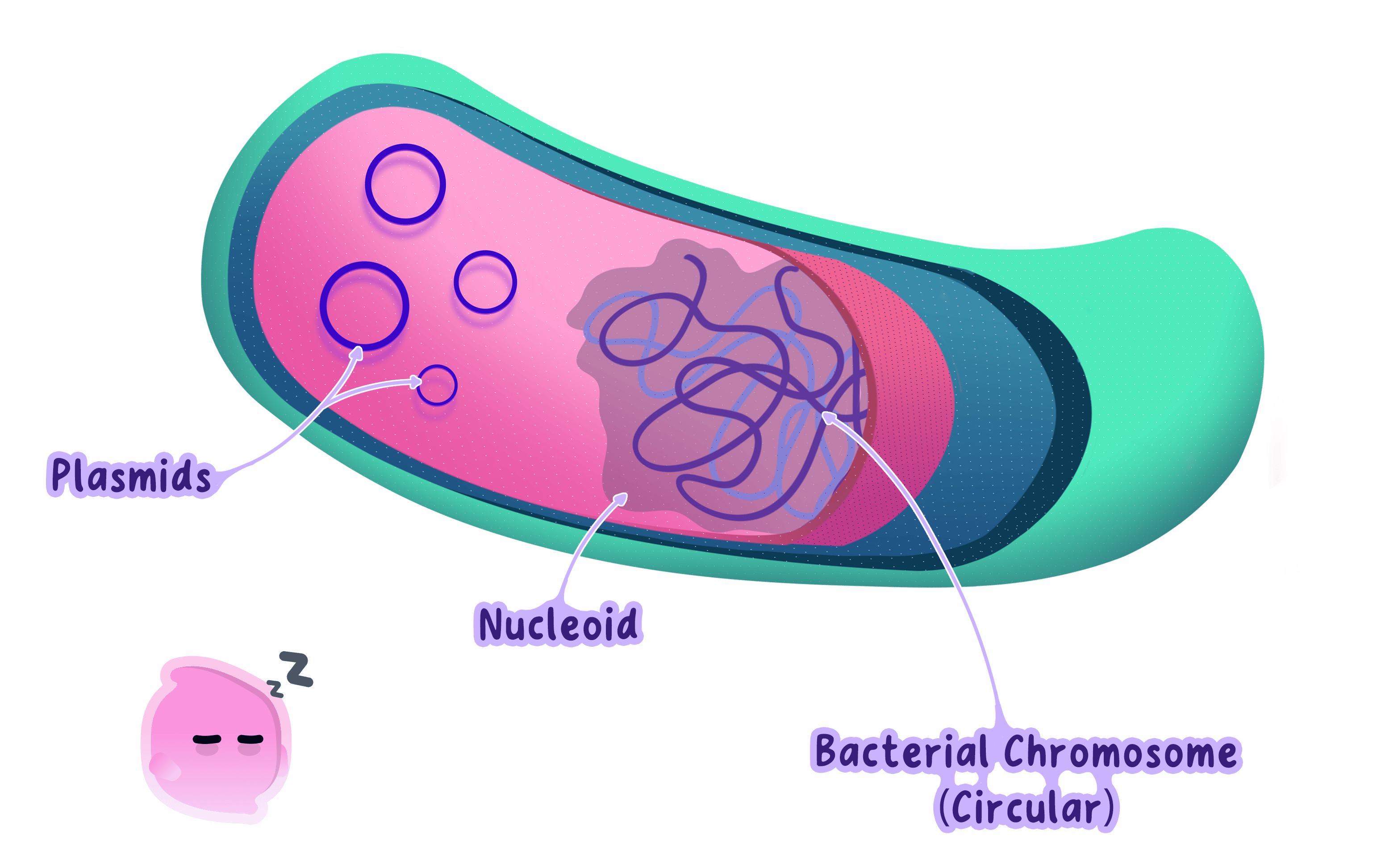
- A prokaryotic cell instead has a single circular loop of DNA lying free in the cytoplasm, and this loop does the job the chromosomes do in a eukaryotic cell.
- Many prokaryotic cells also carry plasmids, which are small extra rings of DNA kept separate from the main loop.
- A plasmid usually carries genes that are only useful in particular conditions, such as genes giving resistance to an antibiotic.
- Plasmids can be copied and passed from one bacterium to another, so a useful gene can spread through a population of bacteria quickly.
- Do not confuse a plasmid with a nucleus, because a plasmid is a small ring of DNA and a nucleus is a membrane-bound compartment.
- Do not write that a bacterium has no DNA, because it has a circular chromosome lying free in its cytoplasm.
- Do not say the nucleus makes the proteins, because it holds the instructions and the proteins are built elsewhere.
Mitochondria, chloroplasts and ribosomes
Mitochondrion
The organelle that contains the enzymes for aerobic respiration and is where most of a cell's energy is released.
Chloroplast
The organelle in plant and algal cells that contains chlorophyll and is the site of photosynthesis.
Chlorophyll
The green pigment held inside chloroplasts that absorbs the light energy used in photosynthesis.
Ribosome
The sub-cellular structure in the cytoplasm at which amino acids are joined together to make a protein.
- Mitochondria are rod-shaped organelles with a double membrane, and the inner one is deeply folded.
- They hold the enzymes that catalyse the reactions of aerobic respiration, which is the process that transfers energy from glucose for the cell to use.
- The folds of the inner membrane give a large surface area for those enzymes, so more respiration can take place inside the same small volume.
- Cells that use a great deal of energy contain far more mitochondria, so a muscle cell holds thousands while a fat-storage cell holds very few.
- The number of mitochondria in a cell is therefore a direct clue to how much energy that cell demands.
- Chloroplasts are found in plant and algal cells and are the site of photosynthesis.
- Each chloroplast contains chlorophyll, the green pigment that absorbs the light energy used to build glucose from carbon dioxide and water.
- The chlorophyll is held on stacks of internal membranes, which spreads it out so that as much light as possible is absorbed.
- Chloroplasts are found only in the plant cells that receive light, so leaf cells are packed with them and root cells have none.
- Ribosomes are the smallest of these organelles and are the site of protein synthesis.
- At a ribosome, amino acids are joined together in the order coded by a gene, and the chain that results is a protein.
- Ribosomes are found in every cell, prokaryotic cells included, because every cell has to build its own enzymes and other proteins.
- Some ribosomes float free in the cytoplasm while others are attached to membranes, and a cell that secretes a lot of protein carries very large numbers of them.
Reading a cell's job from its organelles
- A heart muscle cell contracts without resting and needs a constant supply of energy, so it is packed with mitochondria.
- A palisade cell sits just below the upper surface of a leaf where the light is strongest, so it is packed with chloroplasts.
- A gland cell in the pancreas releases large amounts of digestive enzyme, so it carries an unusually high number of ribosomes.
- A root hair cell absorbs water and mineral ions but never receives light, so it has many mitochondria and no chloroplasts.
The cell membrane and its receptors
Cell membrane
The partially permeable layer around every cell that controls which substances enter and leave it.
Partially permeable
Describing a membrane that lets some substances pass through it while holding others back.
Receptor molecule
A protein in the cell membrane that binds to one specific substance and triggers a response inside the cell.
- The cell membrane surrounds every cell, holding the cytoplasm and organelles inside and separating them from the surroundings.
- It is built from a double layer of lipid molecules with proteins set into it, which is why it is flexible rather than rigid.
- The membrane acts as a selective barrier because it is partially permeable, letting some substances through while holding others back.
- Small molecules such as oxygen, carbon dioxide and water cross it easily, while large molecules such as starch and proteins, and charged ions, do not cross freely.
- Controlling what crosses the surface lets the cell hold its internal conditions steady even when the surroundings change, and steady conditions are what keep its enzymes working.
- The membrane also carries receptor molecules, which are proteins that project from its outer surface.
- Each receptor has a shape that matches one particular molecule, so only that molecule can bind to it.
- When the matching molecule binds, the receptor sets off a change inside the cell, which is how a cell detects a chemical signal and responds to it.
- Different cells carry different receptors, so a hormone carried in the blood past every cell in the body changes the behaviour of only the cells whose receptors match it.
- Do not describe the cell membrane as fully permeable, because it lets some substances through and blocks others.
- Do not swap the roles of the membrane and the wall, because the membrane controls what enters and leaves while the wall supports the cell.
Structures found only in plant cells
Cytoplasm
The jelly-like material inside a cell in which most of the cell's chemical reactions take place.
Cell wall
The rigid layer of cellulose outside the cell membrane of a plant cell that supports and strengthens it.
Permanent vacuole
The large fluid-filled sac in a plant cell that contains cell sap and helps keep the cell firm.
- Cytoplasm fills both plant and animal cells, holds the organelles in place and is where most of the cell's chemical reactions happen.
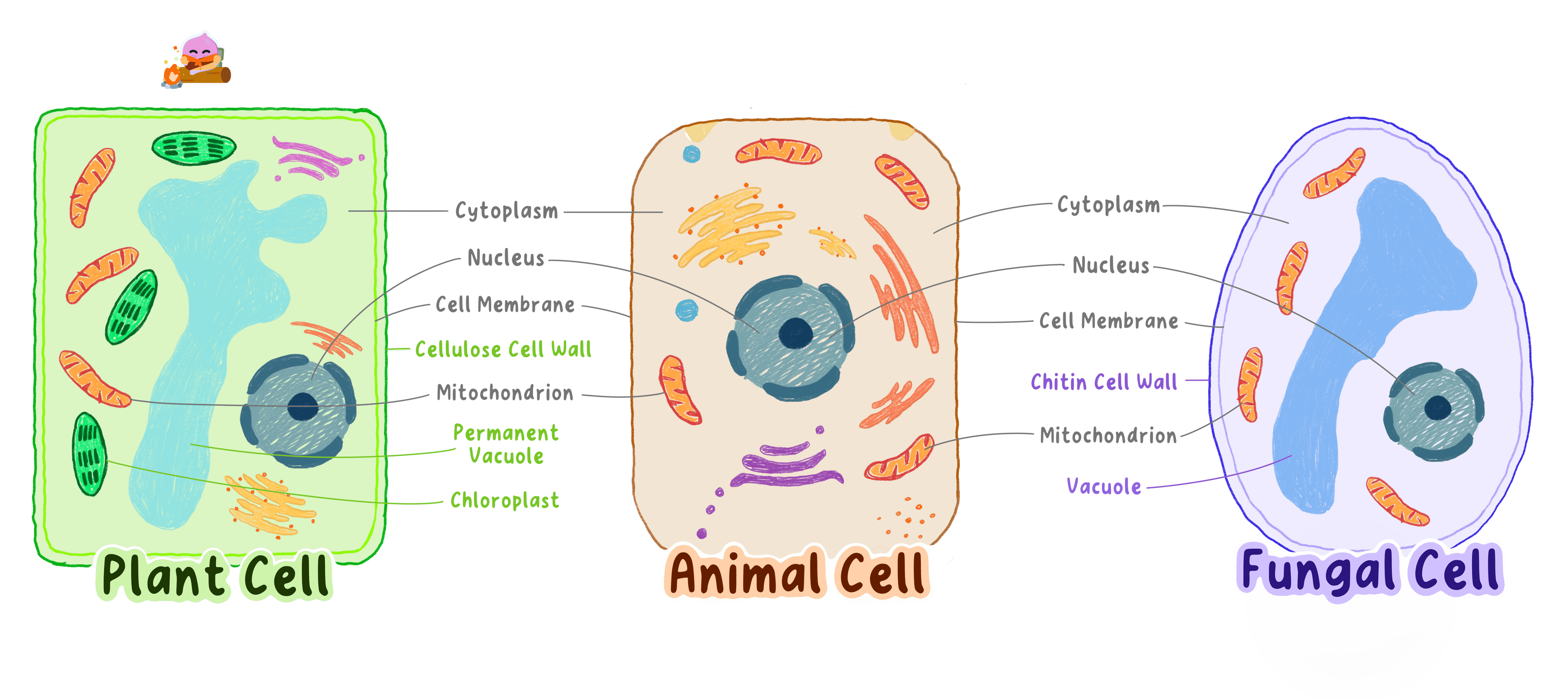
- A plant cell has three structures that an animal cell never has: a cell wall, a permanent vacuole and chloroplasts.
- The cell wall lies outside the cell membrane and is made of cellulose, a tough carbohydrate laid down as criss-crossing fibres.
- Those crossed fibres make the wall strong in every direction, so it stops the cell bursting when water enters and holds the plant upright.
- The wall is fully permeable, so it plays no part in choosing what enters the cell, and that job is left entirely to the membrane beneath it.
- The permanent vacuole is a large sac filled with cell sap, a solution of sugars and mineral ions.
- A full vacuole pushes the cytoplasm outwards against the wall and keeps the cell firm, and a plant whose cells lose that pressure wilts.
- The vacuole also stores the sugars and ions the cell is not using at that moment.
- Fungal cells also have a cell wall, but it is made of chitin rather than cellulose, and a fungal cell has no chloroplasts because it does not photosynthesise.
- A cell wall is fully permeable while a cell membrane is partially permeable, so the wall gives support and the membrane does the controlling.
- A root hair cell is a plant cell with no chloroplasts, so a cell without chloroplasts is not automatically an animal cell.
- State the one feature that decides whether a cell is prokaryotic or eukaryotic.
- Describe where the genetic material is found in a bacterial cell and name the extra rings of DNA it may also carry.
- Explain why a muscle cell contains far more mitochondria than a fat-storage cell.
- Name the organelle that is the site of protein synthesis and explain why it is found in prokaryotic cells as well as eukaryotic ones.
- Explain how receptor molecules in the cell membrane allow a hormone to affect some cells and not others.
- Give one function of the cell wall and one function of the permanent vacuole in a plant cell.
1.1.3 Electron microscopy and resolution
Resolution and the limit of light
Resolution
The smallest distance between two points that can still be seen as two separate points.
- Resolution is the smallest distance between two points at which they can still be seen as two separate points instead of merging into one blur.
- Resolution is measured as a distance, so a smaller number means a better resolution and finer detail.
- Magnification and resolution do different jobs: magnification sets how large the image is, and resolution sets how much detail that image can hold.
- Once two structures lie closer together than the resolution of the instrument, no extra magnification will separate them and the image simply becomes a larger blur.
- The resolution of a light microscope is limited by the wavelength of visible light, because two points closer together than about half a wavelength cannot be told apart.
- Visible light has wavelengths of roughly 400 nm400\ \text{nm}400 nm to 700 nm700\ \text{nm}700 nm, which fixes the best resolution of a light microscope at about 200 nm200\ \text{nm}200 nm.
- That limit also caps the useful magnification, which is why a light microscope is not taken far beyond about ×1500\times1500×1500.
- A structure smaller than 200 nm200\ \text{nm}200 nm, such as a ribosome at roughly 20 nm20\ \text{nm}20 nm across, is therefore invisible to light no matter how the microscope is set up.
- Do not treat a larger magnification as more detail, because the detail available is fixed by the resolution.
- Do not describe a good resolution as a large number, because resolution is a distance and the smaller that distance is the finer the detail.
How an electron microscope works
Electron microscope
An instrument that uses a beam of electrons instead of light to produce a magnified image of a specimen.
Transmission electron microscope
An electron microscope in which electrons pass through a very thin specimen to reveal the detail inside it.
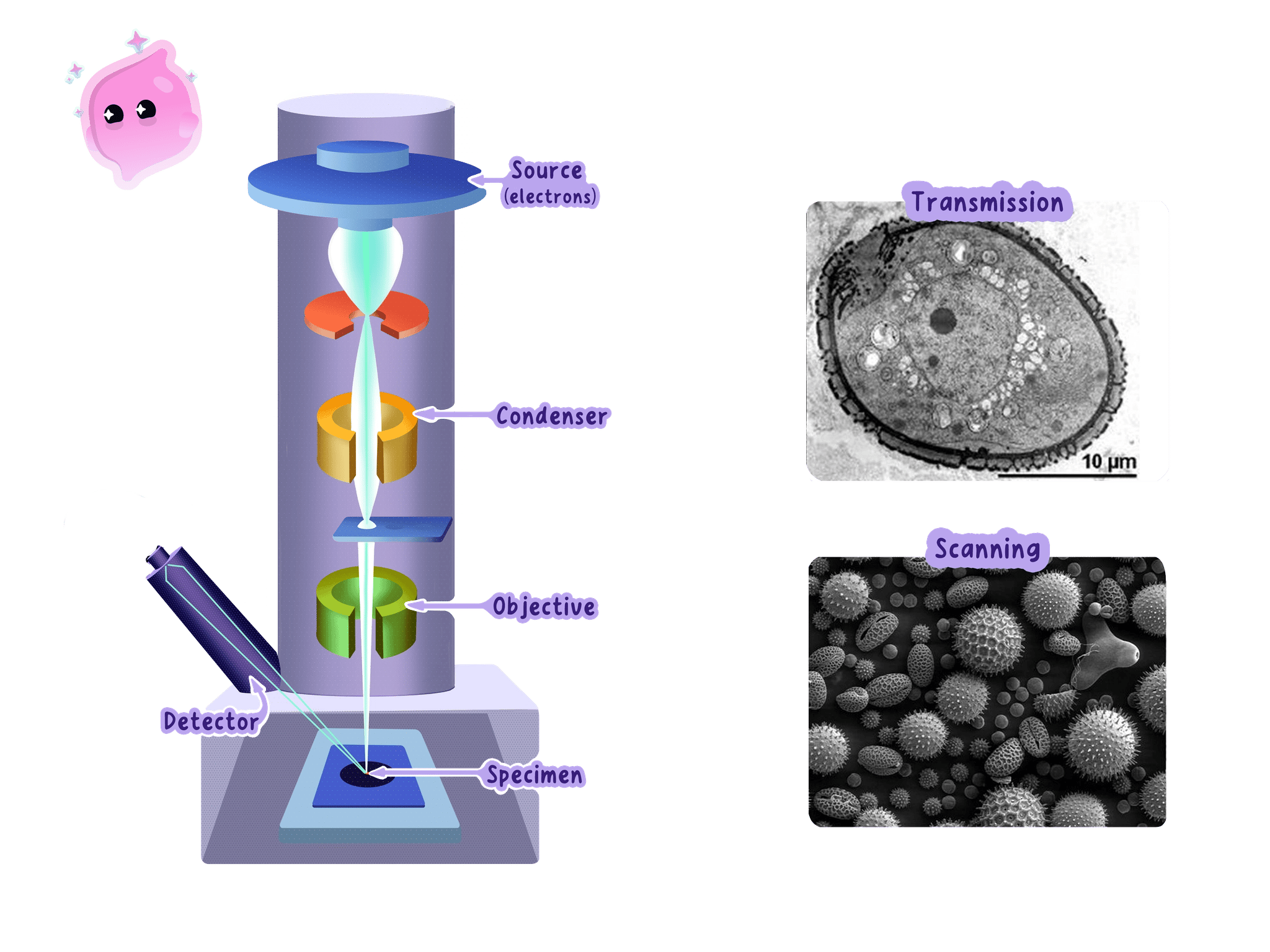
- An electron microscope replaces the beam of light with a beam of electrons.
- The electrons are released from a heated filament called the electron gun at the top of the column and are accelerated downwards by a high voltage.
- Electrons carry a negative charge, so the beam is focused by electromagnets rather than glass lenses, and changing the current through a magnet changes how strongly the beam is bent.
- The whole column is held under a vacuum, because electrons collide with air particles and would be scattered off course long before reaching the specimen.
- In a transmission electron microscope, the electrons pass straight through a specimen that has been cut into an extremely thin slice.
- The specimen is treated with heavy metal salts, which gather on particular structures and scatter electrons strongly, in the same way that a stain builds contrast on a light microscope slide.
- Dense parts of the specimen scatter more electrons and appear dark, while thinner or less dense parts let more electrons through and appear pale, and this difference is what forms the image.
- The electrons that get through strike a fluorescent screen or an electronic detector, producing an image in shades of grey because electrons carry no colour information.
- A beam of electrons behaves as though it has a far shorter wavelength than visible light, and that shorter wavelength is the reason the resolution is so much better.
- A transmission electron microscope resolves detail down to about 0.2 nm0.2\ \text{nm}0.2 nm and magnifies usefully to well over ×1 000 000\times1\,000\,000×1000000.
- A scanning electron microscope instead sweeps the beam back and forth across the surface of a whole specimen and builds a three-dimensional picture of that surface, at a lower resolution than a transmission instrument reaches.
Comparing what each microscope can separate
- A light microscope resolves down to about 200 nm200\ \text{nm}200 nm and a transmission electron microscope down to about 0.2 nm0.2\ \text{nm}0.2 nm.
- Dividing one limit by the other gives 2000.2=1000\dfrac{200}{0.2}=10000.2200=1000.
- The electron microscope therefore separates points that are a thousand times closer together.
- A ribosome is about 20 nm20\ \text{nm}20 nm across, which is ten times smaller than the light microscope's limit but a hundred times larger than the electron microscope's, so only the electron microscope can show it.
What electron microscopy revealed
Ultrastructure
The fine internal detail of a cell that can only be seen using an electron microscope.
- Under a light microscope a cell showed a nucleus, cytoplasm, a membrane, chloroplasts and the outlines of mitochondria, and nothing finer than that.
- Mitochondria appeared as featureless rods, so their internal arrangement could only be guessed at.
- The far higher resolution of the electron microscope revealed the ultrastructure of the cell, the fine internal detail that light could never separate.
- It showed that a mitochondrion has two membranes and that the inner one is folded into shelves, which explained how the organelle packs a large surface area of respiration enzymes into a tiny volume.
- It revealed ribosomes for the first time, as tiny dense granules lying free in the cytoplasm and attached to membranes, which showed where proteins are assembled.
- It showed that a chloroplast contains stacks of flattened membrane sacs carrying the chlorophyll, which explained how the pigment is spread out so that as much light as possible is absorbed.
- It showed the cell membrane as two distinct dark lines rather than one, which supported the idea of a membrane built from a double layer of lipid.
- It revealed folded internal membrane systems that light had never shown at all, so the cytoplasm turned out to be organised rather than a featureless jelly.
- Each of these discoveries let biologists match a structure to the job it does, and it is that link between structure and function that transformed how cells were understood.
- A specimen for an electron microscope has to be dead, dried out and held in a vacuum, so the instrument can never be used to watch a living process.
- The preparation can create artefacts, which are structures that appear in the image but were not present in the living cell.
- A newly seen structure is therefore only accepted once it shows up again after a different preparation method.
- Electron microscopes are also large, expensive and need trained operators, so a light microscope is still the practical choice for routine work.
Writing about what the electron microscope changed
- Answer in terms of higher resolution, because credit goes to the ability to separate close points and not to magnification on its own.
- Name a specific structure it revealed, such as ribosomes or the folded inner membrane of a mitochondrion, since a vague reference to seeing more detail rarely scores.
- Follow the structure with the job it does when the command word is explain, for example that folds give a large surface area for respiration enzymes.
- State a limitation as a consequence rather than a bare fact, for example that the specimen is dead so no living process can be observed.
- State what is meant by the resolution of a microscope.
- Explain why raising the magnification of a light microscope beyond a certain point reveals no extra detail.
- Describe how a transmission electron microscope produces an image of a specimen.
- Explain why the column of an electron microscope is kept under a vacuum.
- Name two structures the electron microscope revealed and state what each one told biologists about how a cell works.
