Question 5
HA scientist used PET scanning to investigate brain activity.
Before each scan, the person was injected with radioactive glucose.
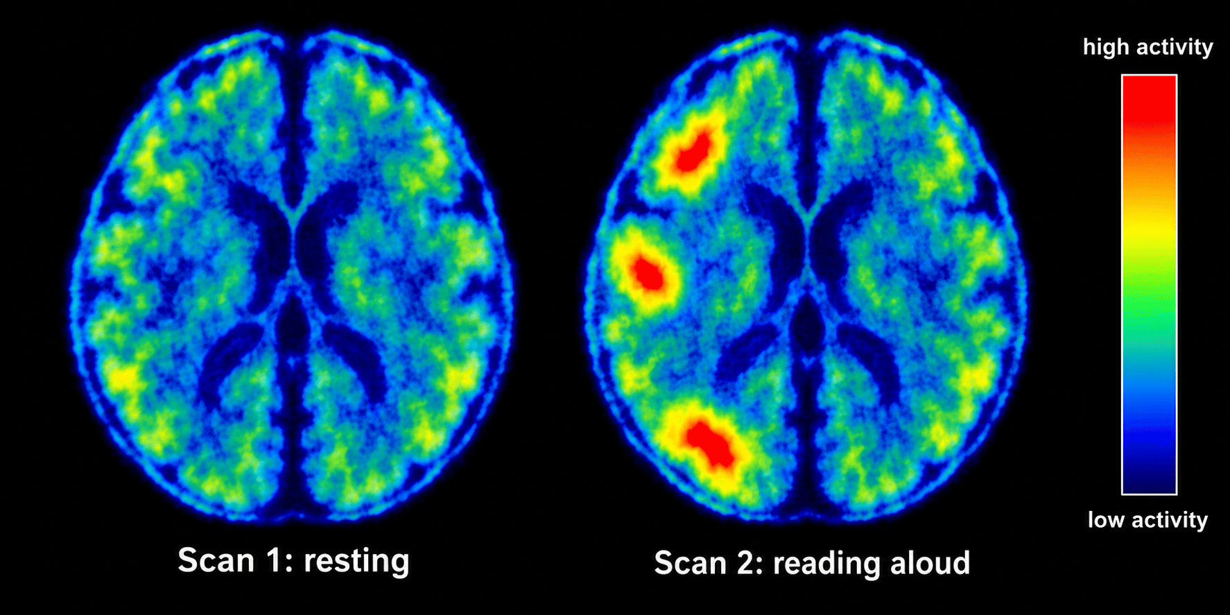
The Figure shows two PET scans of the same person's brain. Scan 1 was taken while the person was resting. Scan 2 was taken while the person was reading aloud.
Figure
a.[3]
Explain why active areas of the brain show up brightly on a PET scan.
b.[2]
Describe how the activity of the brain in Scan 2 is different from the activity in Scan 1.
c.[2]
Explain why a CT scan could not be used to identify the areas of the brain used for reading aloud.
d.[1]
Suggest why doctors do not carry out PET scans on the same person many times.
2.3 The nervous system and brain Questions
43 exam-style questions on Edexcel GCSE Biology 2.3 The nervous system and brain, covering 2.3.1 Structures and functions of the brain, 2.3.2 Imaging brain function with scanning, 2.3.3 Limitations in treating nervous system damage, 2.3.4 Neurones and impulse transmission, and 2.3.5 Structure and function of a reflex arc. Each one has a worked solution and a mark scheme showing where the marks go.