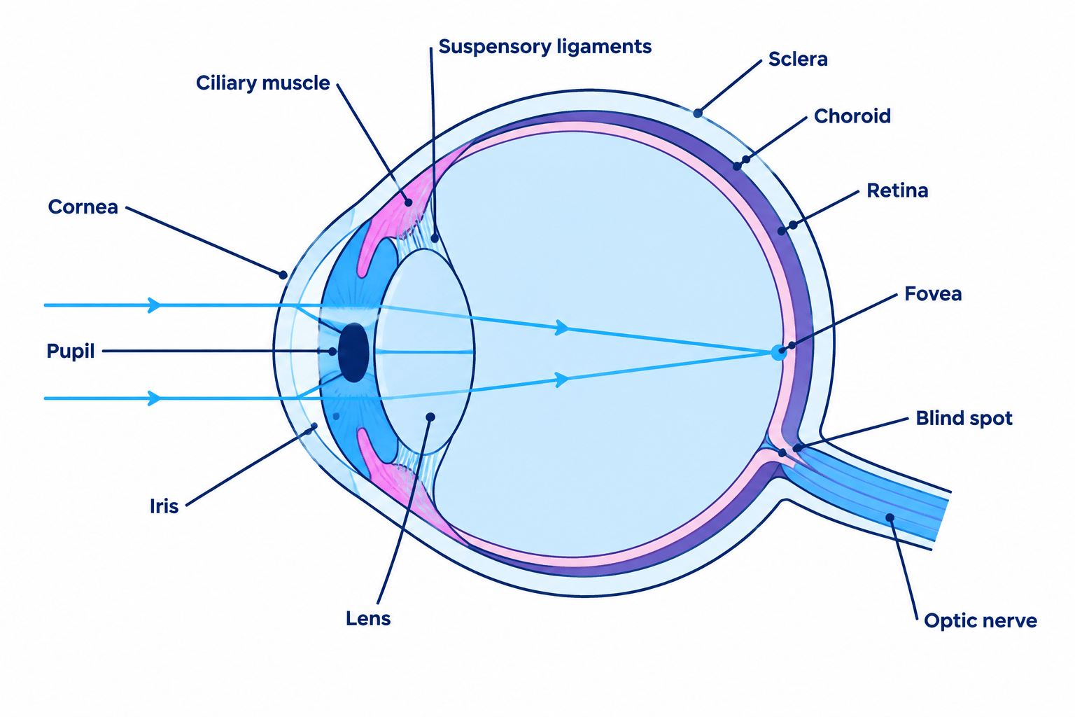
The Eye Is a Sense Organ
Sense organ
An organ containing receptor cells that detect one particular type of stimulus, such as the eye detecting light.
- The eye is the sense organ that detects light, which is the stimulus it responds to.
- Its job is to focus light onto the retina and to convert that light into electrical impulses.
- The impulses travel along the optic nerve to the brain, where they are interpreted as an image.
- The sclera is the tough white outer coat that protects the eyeball and keeps its shape, and the choroid beneath it is a dark layer that absorbs stray light and supplies blood.
The Cornea
Cornea
The transparent front layer of the eye that refracts light entering the eye and does most of the focusing.
- The cornea is the transparent layer at the front of the eye, curved outwards like a dome.
- It refracts the light entering the eye, bending the rays inwards so that they converge.
- It does most of the focusing, because the change in speed as light passes from air into the cornea is large.
- Its curvature is fixed, so it cannot adjust for objects at different distances.
- It has no blood vessels, which keeps it transparent, and it takes oxygen directly from the air instead.
The Lens and Accommodation
Accommodation
The change in shape of the lens that allows the eye to focus on objects at different distances.
- The lens sits behind the pupil, and it is transparent and flexible so it can change shape.
- It carries out the fine focusing that the fixed cornea cannot do, and this change of shape is called accommodation.
- The lens is held in place by suspensory ligaments, which are attached to a ring of ciliary muscles.
- Focusing on a near object. The ciliary muscles contract, the ring gets smaller, the suspensory ligaments slacken, and the lens becomes thicker and more curved so it refracts light more strongly.
- Focusing on a distant object. The ciliary muscles relax, the ring gets wider, the suspensory ligaments are pulled tight, and the lens is stretched thin so it refracts light less.
- Light from a distant object arrives almost parallel and needs little extra bending, while light from a near object is spreading out and needs much more.
The ciliary muscles contract to view a near object, even though the ligaments slacken. Contracting a ring of muscle makes the ring smaller, which is what releases the tension on the ligaments.
The Iris and the Pupil Reflex
Iris
The ring of muscle that controls the size of the pupil and so the amount of light reaching the retina.
- The pupil is the hole in the middle of the iris that lets light into the eye, and it is not a structure in itself.
- The iris is the coloured ring of muscle that changes the size of the pupil, controlling how much light reaches the retina.
- It contains two sets of muscle: circular muscles that run around the pupil and radial muscles that run outwards like the spokes of a wheel.
- In bright light the circular muscles contract and the radial muscles relax, so the pupil gets smaller and less light enters.
- In dim light the radial muscles contract and the circular muscles relax, so the pupil gets larger and more light enters.
- This is a reflex action coordinated by the brain, and its function is to protect the retina from being damaged by bright light.
The two sets of muscle are antagonistic, so whenever one contracts the other relaxes. Write both halves, because a full answer names what happens to each set.
The Retina
Retina
The light-sensitive layer at the back of the eye, containing the rod and cone receptor cells.
- The retina is the light-sensitive layer lining the back of the eye.
- It contains the receptor cells, which are the rods and the cones.
- When light hits a receptor cell it is converted into an electrical impulse, and the impulses leave along the optic nerve.
- The image formed on the retina is upside down and back to front, and the brain interprets it the right way up.
Rod Cells
Rod cells
Light receptor cells in the retina that are sensitive in dim light but do not detect colour.
- Rod cells are sensitive to low light intensity, so they allow vision in dim conditions.
- They cannot detect colour, which is why everything looks grey at night.
- They are spread across most of the retina but are absent from the fovea.
- They give less detailed vision than cones, because many rods share a single neurone to the brain.
- This sharing is what makes them sensitive, since the small responses of many rods are added together to trigger one impulse.
Cone Cells
Cone cells
Light receptor cells in the retina that detect colour and work best in bright light.
- Cone cells detect colour and work best in bright light.
- There are three types, sensitive to red, green and blue light.
- The brain compares the responses of the three types to work out the colour being viewed.
- Cones give highly detailed vision, because each cone tends to have its own neurone to the brain.
- They need bright light to respond, which is why colours cannot be seen in a dark room.
The Fovea and the Blind Spot
Fovea
The small pit at the centre of the retina, packed with cone cells, where the sharpest and most detailed image is formed.
- The fovea is a small pit at the centre of the retina, directly opposite the pupil.
- It is packed with cone cells and contains almost no rods, so it gives the sharpest and most detailed vision.
- When you look straight at something you are moving your eyes so the image falls on the fovea.
- The blind spot is the point where the optic nerve leaves the retina, and it contains no rods or cones.
- No light is detected there, so a small part of the field of view is missing, and the brain fills the gap using information from the other eye and the surroundings.
Cones are concentrated at the fovea, so in dim light an object is easier to see slightly off centre, where the rods are. Astronomers use this deliberately when looking for a faint star.
Investigating Responses of the Eye
Investigation: The Pupil Reflex and the Blind Spot
- Aim: To observe the pupil reflex and to locate the blind spot. Edexcel lists investigating human responses to external stimuli as a suggested practical for Topic 2, and the eye is the clearest example.
- Apparatus: A torch, a ruler with a millimetre scale, a mirror, a partner, a plain card marked with a cross on the left and a filled circle on the right about 8 cm apart, a dimly lit room.
- Method: the pupil reflex:
- Sit your partner in a dimly lit room for two minutes so their pupils widen fully, then measure the pupil diameter in millimetres against a ruler held beside the eye.
- Shine a torch towards the eye from about 20 cm away, at an angle rather than straight into it, and hold it there for 5 seconds.
- Measure the pupil diameter again while the light is still on.
- Switch the torch off, wait 30 seconds, and measure once more.
- Repeat three times on the same eye and calculate a mean for each condition.
- Method: finding the blind spot:
- Hold the card at arm's length with the cross in front of your left eye.
- Close your right eye and look steadily at the cross with the left.
- Move the card slowly towards your face while continuing to stare at the cross.
- At about 25 cm the filled circle vanishes, because its image has fallen on the blind spot where the optic nerve leaves the retina.
- Keep moving the card closer and the circle reappears once the image moves off the blind spot.
- Variables: For the pupil reflex the independent variable is the light intensity and the dependent variable is the pupil diameter. Keep the same person, the same eye, the same torch and the same distance from the eye, and allow the same time to adjust before each measurement.
- Results: The pupil constricts within about a second of the torch being switched on, typically from around 6 mm to around 2 mm, and widens again once it is switched off. In the blind spot test the circle disappears completely rather than fading gradually.
- Maths: Express the change as a percentage so that people with different starting pupil sizes can be compared. percentage change in diameter=final diameter−initial diameterinitial diameter×100\text{percentage change in diameter} = \frac{\text{final diameter} - \text{initial diameter}}{\text{initial diameter}} \times 100percentage change in diameter=initial diameterfinal diameter−initial diameter×100
- Watch out: Never shine the torch directly into the eye, and never use a laser or a phone torch at close range, because the reflex protects the retina but does not make bright light safe. Measuring a moving pupil against a ruler is imprecise, so photographing the eye and measuring the image gives a better result.
- Safety: Use a low power torch held at an angle and limit each exposure to a few seconds. Stop at once if your partner finds it uncomfortable.
- For an accommodation question, name the ciliary muscles, the suspensory ligaments and the shape of the lens in the same answer, because one mark is available for each.
- State what the change achieves as well as what happens, so finish with more refraction for a near object or less refraction for a distant one.
- Keep the cornea and the lens apart in your answer: the cornea does most of the refraction and cannot change, while the lens fine tunes the focus.
- When comparing rods and cones, contrast the same features in turn, which are light intensity, colour, detail and position in the retina.
- Which part of the eye does most of the refraction of light?
- Describe what happens to the ciliary muscles, the suspensory ligaments and the lens when you focus on a near object.
- Explain what happens to the iris in bright light and why.
- Give two differences between rod cells and cone cells.
- Why is no image detected at the blind spot?