Magnification and resolution
Resolution
The smallest distance between two points that can still be seen as two separate points.
- A microscope has to do two separate jobs, and they are easily confused.
- Magnification is how many times bigger the image is than the real object.
- Resolution is the smallest gap between two points that still appears as two points rather than one blur.
- Once the magnification goes past what the resolution can support, the image gets bigger but no clearer.
- Resolution is therefore what decides which structures can be seen at all, and it is the reason new microscope technology mattered.
Higher resolution means finer detail, and every advance in what scientists knew about organelles followed an advance in resolution.
The light microscope
Light microscope
A microscope that uses a beam of light focused by glass lenses to produce a magnified image of a specimen.
- A light microscope passes a beam of light through a thin specimen and focuses it with glass lenses.
- Its useful magnification stops at roughly ×1500\times 1500×1500, and its resolution is about 200 nm200\,\text{nm}200nm.
- That limit exists because two points closer together than about half the wavelength of light cannot be separated, however good the lenses are.
- A light microscope shows the nucleus, cytoplasm, cell membrane, cell wall, chloroplasts and the vacuole.
- It cannot show ribosomes, plasmids, the inside of a mitochondrion or any detail of the membrane itself.
- It is still used every day because it is cheap, portable, simple to use, and it can show living cells in colour.
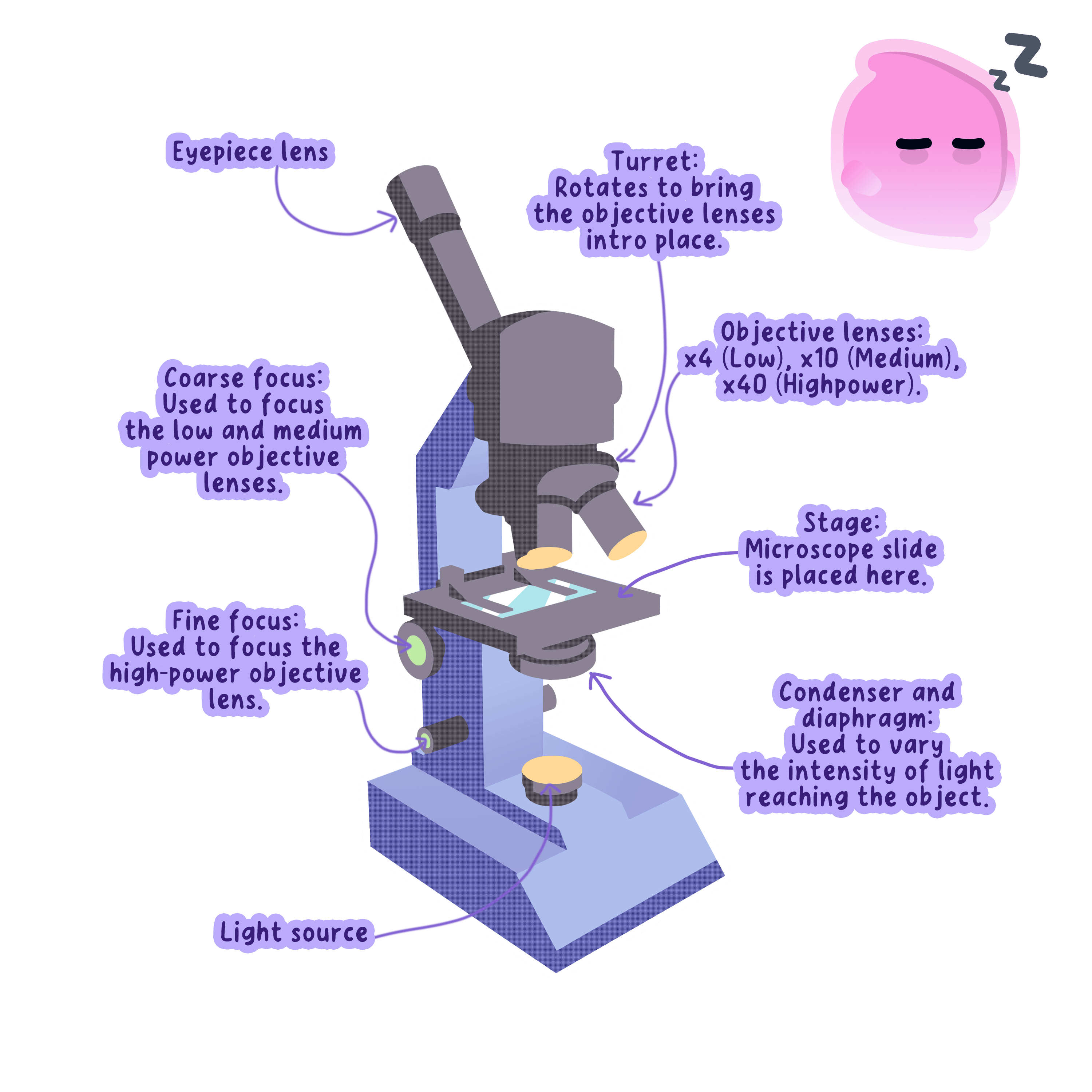
Total magnification is the eyepiece lens multiplied by the objective lens, so a ×10\times 10×10 eyepiece with a ×40\times 40×40 objective gives ×400\times 400×400.
The electron microscope
Electron microscope
A microscope that uses a beam of electrons rather than light, giving far higher magnification and resolution than a light microscope.
- Electron microscopes were developed in the 1930s and use a beam of electrons in place of light.
- An electron beam has a far shorter wavelength than light, so the resolution improves to about 0.2 nm0.2\,\text{nm}0.2nm.
- That is roughly a thousand times better than a light microscope, and magnifications above ×1 000 000\times 1\,000\,000×1000000 become useful.
- Specimens must be dead, chemically fixed and placed in a vacuum, because air would scatter the electron beam.
- The instruments are large, expensive and need trained operators, and the images they produce are black and white until colour is added artificially.
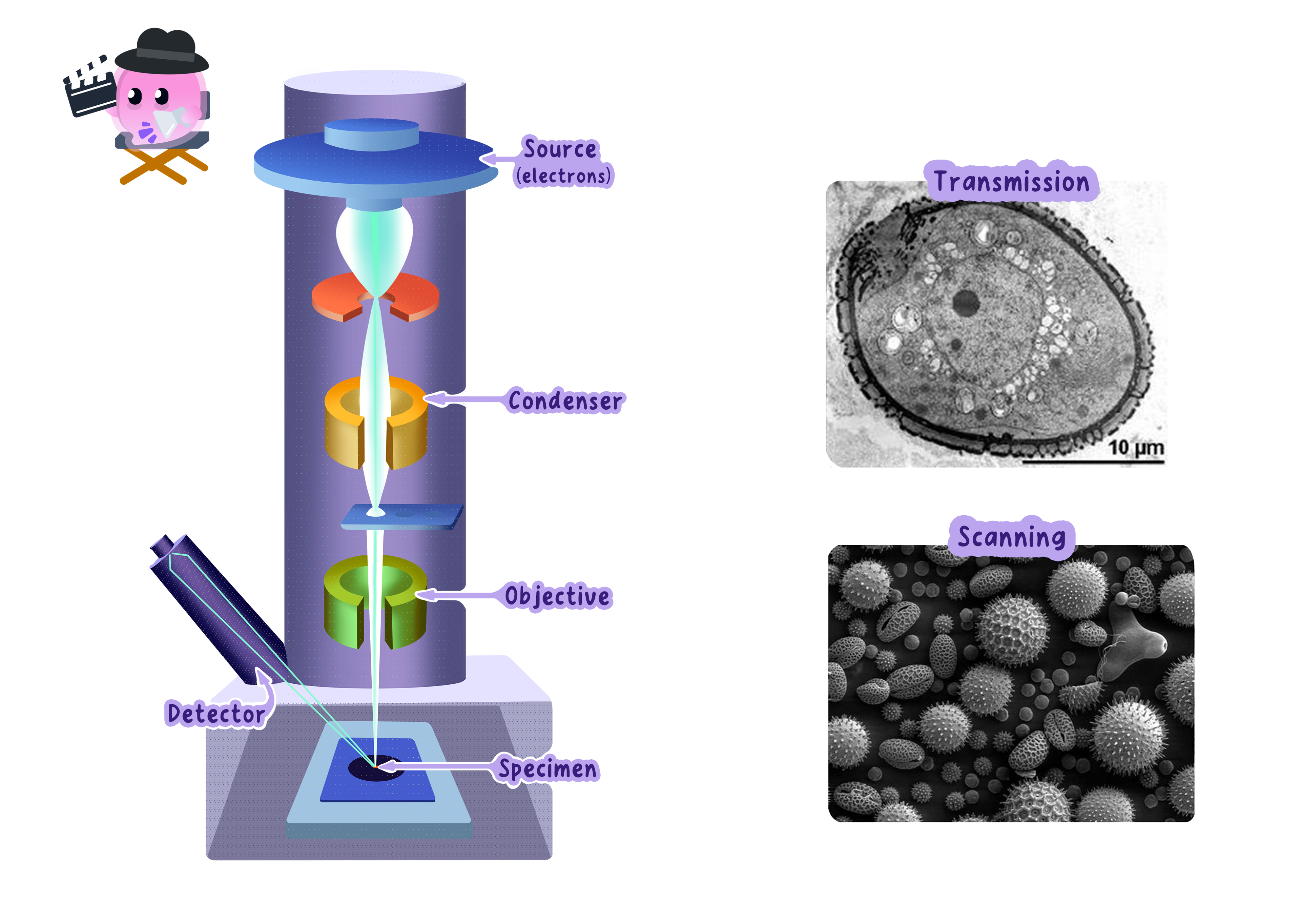
Saying only that an electron microscope has a higher magnification is not enough for the marks, because the reason more can be seen is the higher resolution.
What better microscopes revealed
- Under a light microscope a mitochondrion was no more than a dot, and its job was unknown.
- Electron microscopy showed a double membrane with the inner one deeply folded, and that folded surface was then linked to the reactions of aerobic respiration.
- Ribosomes were invisible before electron microscopy, and only once they could be seen was protein synthesis traced to them.
- Chloroplasts turned out to contain stacks of internal membranes, which explained how they hold enough chlorophyll to absorb light efficiently.
- The cell membrane was resolved as a distinct double layer, which supported the model of a partially permeable barrier.
- Plasmids and viruses became visible for the first time, and both later became central to medicine and genetic engineering.
- The pattern is the one Edexcel wants you to describe: better technology gave better evidence, and better evidence changed the accepted explanation.
- A question asking how microscope technology changed our understanding wants a named organelle, what was newly seen inside it, and the function that was then worked out.
- Use both words correctly in the same answer, because credit is usually split between a magnification point and a resolution point.
- If asked to evaluate the two microscopes, give a strength of the light microscope as well, since cost, portability and living specimens are creditable answers.
Looking at Cells
- Aim: to prepare slides of onion epidermis and cheek cells, observe them under a light microscope, draw them, and calculate magnification and actual size.
- Apparatus: light microscope and lamp, glass slides, coverslips, mounted needle, forceps, pipette, iodine solution, methylene blue, an onion, a sterile wooden spatula, paper towels, a beaker of 1% Virkon, gloves and eye protection.
- Method, plant slide:
- Peel one thin layer of epidermis from the inside of an onion scale with forceps.
- Lay it flat in a drop of water on the slide with no folds, because folded tissue is too thick for light to pass through and will never come into focus.
- Add one drop of iodine solution to stain it, which raises the contrast so the nucleus and cell wall stand out.
- Rest one edge of the coverslip on the slide and lower it slowly at about 45∘45^\circ45∘ with a mounted needle, so air is pushed out ahead of it instead of being trapped as bubbles.
- Blot the excess stain with a paper towel.
- Method, cheek cells:
- Rub a sterile wooden spatula gently along the inside of your cheek.
- Smear it thinly onto a clean slide.
- Add a drop of methylene blue and lower a coverslip in the same way.
- Drop the used spatula straight into 1% Virkon.
- Method, focusing:
- Clip the slide onto the stage.
- Start on the lowest-power objective.
- Watching from the side, turn the coarse focus to bring the objective close to the slide.
- Now look down the eyepiece and turn the coarse focus the other way, moving the lens away until the cells come into view.
- Switch to a higher-power objective and sharpen using the fine focus only.
- Results: onion cells appear as neat rectangular boxes with a clear cell wall and a stained nucleus, while cheek cells are irregular blobs with a nucleus, cytoplasm and membrane and no wall.
- Maths: total magnification = eyepiece ×\times× objective, and actual size = image size ÷\div÷ magnification. To estimate real cell size, lay a transparent ruler across the low-power field of view, measure the field diameter in mm, convert to μm\mu\text{m}μm by multiplying by 100010001000, then judge what fraction of that diameter one cell takes up.
- Watch out: trapped air bubbles have thick dark outlines and are regularly mistaken for cells, the specimen has to be a single cell layer thick, and the coarse focus must never be used at high power because the objective can crack the slide.
- Safety: wear eye protection because iodine and methylene blue stain skin and clothes, never re-use a spatula, put anything that touched a mouth into 1% Virkon, and handle coverslips carefully because they snap easily.
Scientific drawings
- Use a sharp pencil and draw clean single lines, with no shading and no sketchy repeated strokes.
- Make the drawing large enough to fill the space you are given.
- Draw label lines straight, without arrowheads, and do not let them cross one another.
- Give the drawing a title and state the magnification you used.
- Draw only what is actually on your slide, not the textbook version of the cell.
- Give the difference between magnification and resolution.
- State the approximate resolution of a light microscope and of an electron microscope.
- Name two structures visible with an electron microscope but not with a light microscope.
- Explain why the coverslip is lowered at an angle when preparing a slide.
- Describe one way electron microscopy changed what scientists knew about an organelle.