The image below shows two micrographs of animal cells. Micrograph A was taken using a light microscope, while Micrograph B was taken using a transmission electron microscope (TEM).
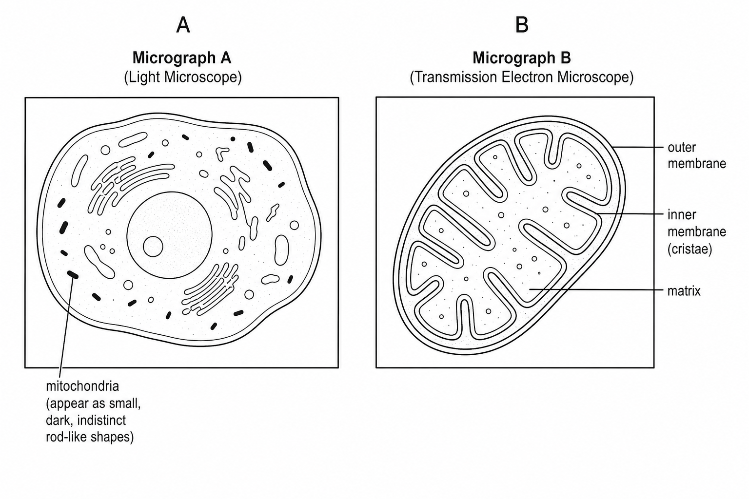
Explain how the transmission electron microscope image (Micrograph B) helps us to understand more about the structure and function of mitochondria compared to the light microscope image (Micrograph A).