Magnification and Resolution Are Not the Same Thing
Resolution
The shortest distance between two points that can still be seen as separate; higher resolution shows finer detail.
- A microscope makes small objects, such as cells, appear much larger.
- Magnification is how many times larger the image is than the real object.
- Resolution is the shortest distance between two points that can still be seen as separate.
- Higher resolution means more fine detail can be seen, not just a bigger image.
Increasing magnification on its own does not reveal more detail; a blurry image just becomes a bigger blurry image.
The Light Microscope
Light microscope
A microscope that uses light and glass lenses to magnify a specimen, with lower magnification and resolution than an electron microscope.
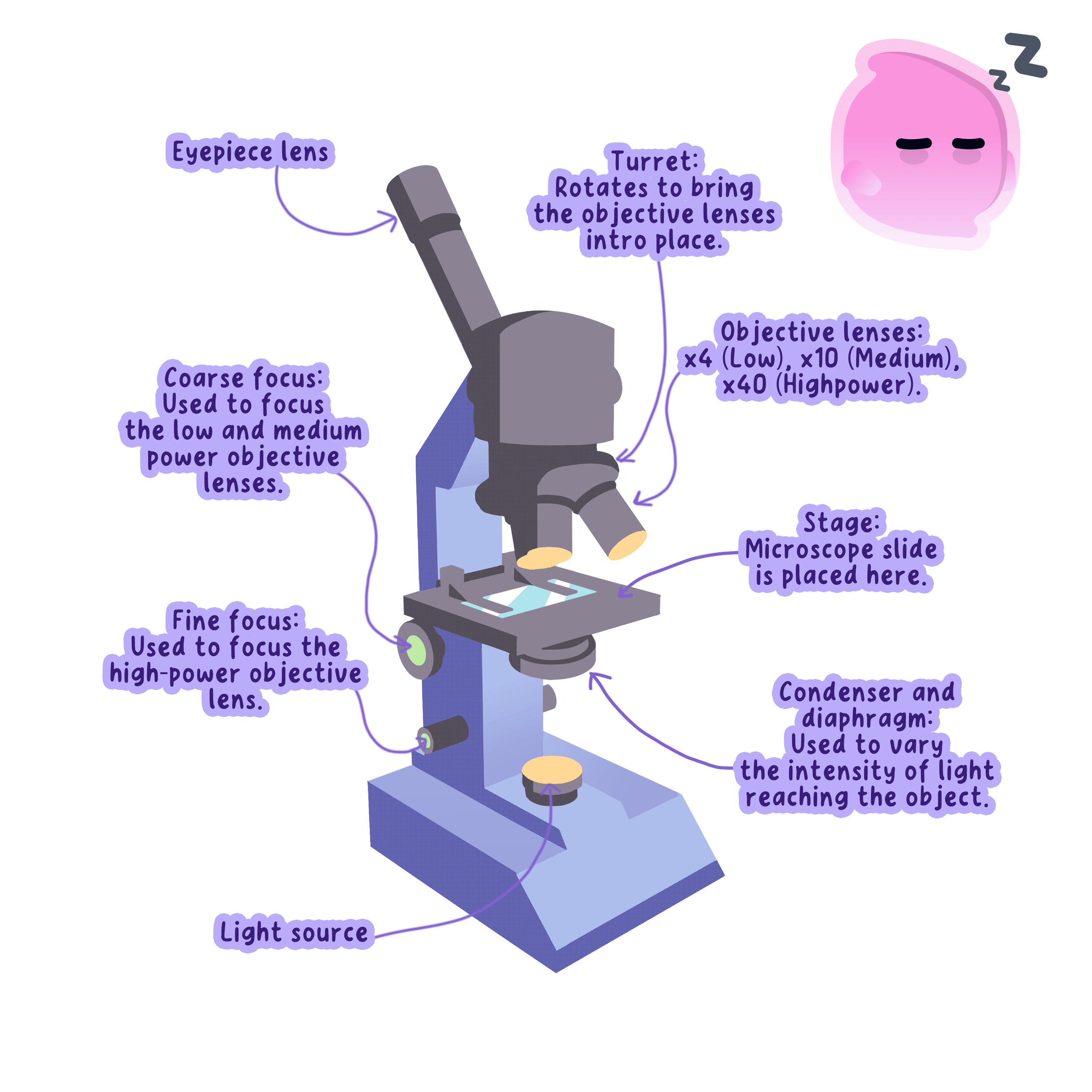
- A light microscope uses light and glass lenses to magnify a specimen.
- It can magnify up to about 1500 to 2000 times.
- It shows cells and their larger structures, such as the nucleus, cytoplasm and chloroplasts.
- Light microscopes are cheap, portable and can be used to view living cells.
A stained slide of onion or cheek cells under a light microscope shows the cell walls, nuclei and chloroplasts.
The Electron Microscope
Electron microscope
A microscope that uses a beam of electrons instead of light, giving much higher magnification and resolution so sub-cellular structures can be seen.
- An electron microscope uses a beam of electrons instead of light, focused by electromagnets in place of glass lenses.
- It has a much higher magnification and resolution, so it shows far finer detail.
- Its image is in greyscale, and the inside is kept under a vacuum so the electrons are not scattered by air.
- It is large and expensive and cannot be used to view living cells.
A light microscope resolves detail down to about 200 nm200\ \text{nm}200 nm, while an electron microscope resolves detail smaller than 1 nm1\ \text{nm}1 nm.
How Microscopy Developed and Revealed Sub-cellular Structures
- Light microscopes were developed first, and electron microscopes came much later, in the twentieth century.
- The higher resolving power of the electron microscope let scientists see fine detail inside cells for the first time.
- It revealed sub-cellular structures such as the internal structure of mitochondria and small structures like ribosomes.
- This greatly increased biologists' understanding of how cells are built and how they work.
The electron microscope's advantage is its higher resolution, not simply higher magnification.
Working Out Magnification
Magnification
How many times larger an image is than the real object, found by dividing image size by real size; it has no units.
- Magnification is worked out with a single formula.
- magnification=size of imagesize of real object\text{magnification} = \dfrac{\text{size of image}}{\text{size of real object}}magnification=size of real objectsize of image
- Rearranging it gives real size=image sizemagnification\text{real size} = \dfrac{\text{image size}}{\text{magnification}}real size=magnificationimage size, and image size=magnification×real size\text{image size} = \text{magnification} \times \text{real size}image size=magnification×real size.
- Magnification has no units and is written as, for example, ×100\times 100×100 or ×3000\times 3000×3000.
- The image size and real size must be in the same unit before you divide, so convert one of them if needed.
- An image 30 mm across at ×3000\times 3000×3000 gives a real size of 30÷3000=0.01 mm30 \div 3000 = 0.01\ \text{mm}30÷3000=0.01 mm, which is 10 μm10\ \mu\text{m}10 μm.
- A leaf 2000 μm2000\ \mu\text{m}2000 μm thick drawn 50 mm thick becomes 2 mm after converting, so 50÷2=×2550 \div 2 = \times 2550÷2=×25.
Light microscope
A microscope that uses light and glass lenses to magnify a specimen, with lower magnification and resolution than an electron microscope.
Using a Light Microscope
- Aim: to use a light microscope to observe, draw and label plant and animal cells, with a magnification scale.
- Prepare a thin specimen on a slide, such as onion epidermis for plant cells or cheek cells for animal cells.
- Add a stain to make the structures show up, using iodine for onion cells and methylene blue for cheek cells.
- Lower a coverslip on carefully to avoid trapping air bubbles.
- Start with the lowest-power objective lens to find the cells, then focus with a higher power.
- Make a clear, labelled biological drawing with sharp lines and add a scale.
- What is the difference between magnification and resolution?
- Give two advantages of a light microscope.
- Why does an electron microscope have a higher resolution than a light microscope?
- Write down the formula for magnification.
- Which stain is used for onion cells, and which for cheek cells?
