

Welcome to one of the most powerful and exciting areas of organic chemistry! In this topic, we transition from simple chemical test-tube reactions to using sophisticated physical techniques to solve chemical structures. You will learn to decipher spectroscopic puzzles and determine the exact structure of an unknown organic compound.
What you'll learn:
- How Nuclear Magnetic Resonance (NMR) spectroscopy works and how to interpret both 13C^{13}\text{C}13C and 1H^1\text{H}1H spectra.
- How to predict NMR spectra from a given organic structure.
- The role of tetramethylsilane (TMS), deuterated solvents like CDCl3\text{CDCl}_3CDCl3, and deuterium oxide (D2O\text{D}_2\text{O}D2O).
- How to combine elemental analysis, mass spectrometry, IR spectroscopy, and NMR to identify unknown molecules.
Introduction to NMR Spectroscopy
Nuclear Magnetic Resonance (NMR) is an analytical technique used to determine the detailed carbon-hydrogen framework of organic molecules. It relies on the magnetic properties of certain atomic nuclei.
Some nuclei have a property called nuclear spin. Any nucleus with an odd number of nucleons (protons + neutrons) has a non-zero spin, which generates a tiny magnetic field.
The two most important nuclei with spin in organic chemistry are:
- Hydrogen-1 (1H^1\text{H}1H): representing abundance >99%>99\%>99% of natural hydrogen.
- Carbon-13 (13C^{13}\text{C}13C): representing about 1.1%1.1\%1.1% of natural carbon (the dominant isotope, 12C^{12}\text{C}12C, has an even number of nucleons and does not have spin).
Nuclear Magnetic Resonance (NMR)
Nuclear Magnetic Resonance (NMR) is an analytical technique that measures the absorption of radiofrequency electromagnetic radiation by spinning nuclei placed in a strong external magnetic field.
When these spinning nuclei are placed into a strong external magnetic field, they align either with the external field (lower energy state) or against it (higher energy state). By applying electromagnetic radiation in the radiofrequency range, the nuclei can absorb energy and "flip" from the lower to the higher energy state. This transition is called resonance.
Chemical Shift
The exact frequency of radiation absorbed depends on the local environment around the nucleus. Electronegative atoms (like oxygen, nitrogen, or halogens) pull electron density away from adjacent nuclei, "deshielding" them. This changes the energy gap required for resonance.
We measure this magnetic environment relative to a standard, expressing it as chemical shift, denoted by the Greek letter δ\deltaδ (delta), with the unit of parts per million (ppm).
Chemical Shift (δ)
Chemical shift (δ\deltaδ) is the shift in the NMR absorption frequency of a nucleus relative to a reference standard (TMS), measured in parts per million (ppm).
Sample Preparation: Standards, Solvents, and Exchange
Before running an NMR spectrum, the sample must be carefully prepared. In your exam, you are expected to understand three critical aspects of sample preparation:
1. Tetramethylsilane (TMS) as a Standard
To calibrate the chemical shift scale, we need a reference standard. We use tetramethylsilane, Si(CH3)4\text{Si}(\text{CH}_3)_4Si(CH3)4 (TMS).
Why TMS?
TMS is used as the universal reference standard because:
- It contains 12 equivalent protons (1H^1\text{H}1H) and 4 equivalent carbons (13C^{13}\text{C}13C), giving a single, highly intense, sharp absorption peak.
- Silicon is less electronegative than carbon, meaning the protons and carbons in TMS are highly shielded. Its peak appears far to the right of almost all other organic signals. We define its position exactly as δ=0 ppm\delta = 0\ \text{ppm}δ=0 ppm.
- It is chemically inert, non-toxic, and volatile (meaning it can be easily evaporated off to recover the sample after analysis).
2. The Need for Deuterated Solvents
Organic samples must be dissolved in a solvent to run an NMR spectrum. However, common organic solvents (such as ethanol, acetone, or water) are packed with carbon and hydrogen atoms, which would overwhelm the spectrum with massive solvent peaks.
To prevent this, we use deuterated solvents. In these solvents, the normal hydrogen-1 (1H^1\text{H}1H) atoms are replaced with deuterium (2H^2\text{H}2H or D\text{D}D), an isotope of hydrogen with one proton and one neutron.
Because deuterium has an even number of nucleons, it does not produce a signal in the range where 1H^1\text{H}1H absorbs. A common deuterated solvent used in NMR is deuterated chloroform, CDCl3\text{CDCl}_3CDCl3.
Carbon-13 solvent peaks
While CDCl3\text{CDCl}_3CDCl3 does not produce a proton signal, it does contain a carbon-13 atom. When running a 13C^{13}\text{C}13C NMR spectrum, CDCl3\text{CDCl}_3CDCl3 produces a small triplet peak around 77 ppm77\ \text{ppm}77 ppm. Computer software automatically filters this solvent peak out, or it is ignored during interpretation.
3. Identifying O–H and N–H Protons by D2O\text{D}_2\text{O}D2O Exchange
Protons attached to oxygen (-OH\text{-OH}-OH) and nitrogen (-NH-\text{-NH-}-NH-) are called labile protons. They do not have fixed chemical shifts; their peaks can appear broad and vary wildly in position depending on the temperature, solvent, and concentration. This makes them tricky to identify.
To prove whether a peak is due to an -OH\text{-OH}-OH or -NH-\text{-NH-}-NH- group, we use a technique called deuterium exchange:
- Run a standard 1H^1\text{H}1H NMR spectrum of the sample.
- Add a few drops of heavy water, D2O\text{D}_2\text{O}D2O, shake the mixture, and re-run the spectrum.
- The deuterium atoms in D2O\text{D}_2\text{O}D2O rapidly exchange with the labile protons in the sample:
- Because the -OH\text{-OH}-OH or -NH-\text{-NH-}-NH- protons have been replaced by deuterium (-OD\text{-OD}-OD or -ND-\text{-ND-}-ND-), their corresponding peak disappears from the second spectrum.
Carbon-13 (13C^{13}\text{C}13C) NMR Spectroscopy
Carbon-13 NMR is the simpler of the two types of NMR. Every unique carbon environment in a molecule produces a single peak (as all 13C^{13}\text{C}13C spectra assessed at A-Level are proton-decoupled, meaning they show singlets only).
When analyzing a 13C^{13}\text{C}13C NMR spectrum, look for two things:
- The number of peaks = the number of unique carbon environments.
- The chemical shift (δ\deltaδ) of the peaks = the type of carbon environment (refer to your OCR Data Sheet).
Determining Carbon Environments
Carbon atoms that are symmetrically identical belong to the same environment and will contribute to the same peak.
Determining Carbon Environments
Predict the number of peaks in the 13C^{13}\text{C}13C NMR spectra of:
- Propan-2-ol
- Butanone
Step-by-step resolution:
- Draw the structure of propan-2-ol: CH3-CH(OH)-CH3\text{CH}_3\text{-CH(OH)-CH}_3CH3-CH(OH)-CH3. Identify symmetry. The two methyl (-CH3\text{-CH}_3-CH3) carbons are chemically identical because they are both bonded directly to the central -CH(OH)-\text{-CH(OH)-}-CH(OH)- carbon. The central carbon is in its own unique environment.
- Draw the structure of butanone: CH3-CO-CH2-CH3\text{CH}_3\text{-CO-CH}_2\text{-CH}_3CH3-CO-CH2-CH3. Check for symmetry. There is no plane of symmetry in this molecule. Each carbon is in a different chemical environment:
- C1\text{C}_1C1 (-CH3\text{-CH}_3-CH3 adjacent to carbonyl)
- C2\text{C}_2C2 (C=O\text{C=O}C=O carbonyl carbon)
- C3\text{C}_3C3 (-CH2−\text{-CH}_2--CH2− carbon)
- C4\text{C}_4C4 (-CH3\text{-CH}_3-CH3 adjacent to methyl carbon)
High-Resolution Proton (1H^1\text{H}1H) NMR Spectroscopy
Proton (1H^1\text{H}1H) NMR is significantly more powerful because it provides four distinct pieces of information:
| Feature | What it tells you |
|---|---|
| Number of peaks (signals) | The number of unique proton (hydrogen) environments. |
| Chemical shift (δ\deltaδ) | The chemical environment of the protons (using the Data Sheet). |
| Relative peak area | The relative number of protons in that environment (given by an integration trace or ratio). |
| Spin-spin splitting pattern | The number of non-equivalent protons on the adjacent carbon atoms, using the n+1n + 1n+1 rule. |
The n+1n + 1n+1 Rule for Spin-Spin Splitting
Splitting is caused by the magnetic fields of protons on adjacent carbon atoms interacting (coupling) with the magnetic field of the protons being measured.
The n + 1 Rule
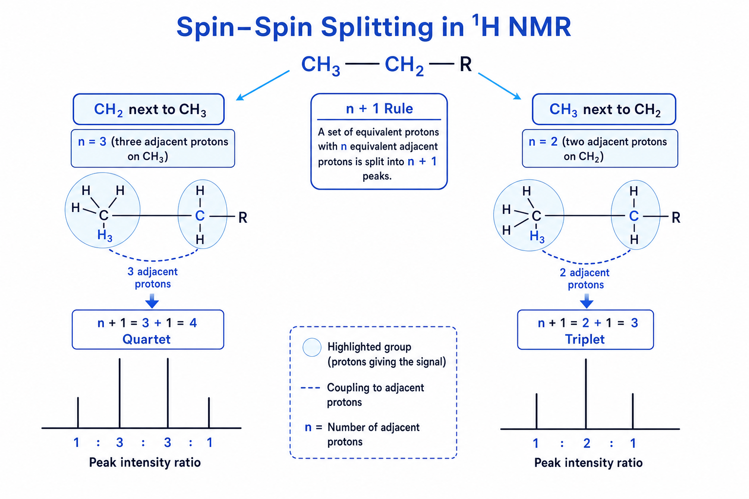
If a proton environment has nnn non-equivalent protons on adjacent carbon atoms, its NMR peak will split into n+1n + 1n+1 sub-peaks.
The intensity of the split peaks follows the mathematical ratios of Pascal's Triangle:
| Adjacent Protons (nnn) | Splitting Pattern (n+1n+1n+1) | Relative Intensity Ratio |
|---|---|---|
| 0 | Singlet | 1 |
| 1 | Doublet | 1:1 |
| 2 | Triplet | 1:2:1 |
| 3 | Quartet | 1:3:3:1 |
| Multi | Multiplet | Complex |
Splitting across heteroatoms
Spin-spin coupling does not typically occur across oxygen or nitrogen atoms. Protons in -OH\text{-OH}-OH or -NH-\text{-NH-}-NH- groups almost always appear as singlets, and they do not cause splitting in the protons on adjacent carbons.
The classic ethyl group triplet-quartet pair
If you see a triplet integrating to 3H3\text{H}3H and a quartet integrating to 2H2\text{H}2H in a spectrum, it is almost certainly an ethyl group (-CH2-CH3\text{-CH}_2\text{-CH}_3-CH2-CH3). The methyl protons (3H3\text{H}3H) are split by the two adjacent protons (n=2→n=2 \ton=2→ triplet). The methylene protons (2H2\text{H}2H) are split by the three adjacent protons (n=3→n=3 \ton=3→ quartet).
Combined Analytical Techniques
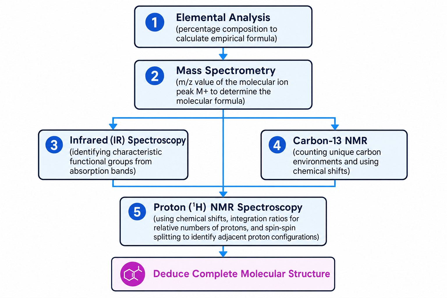
In the A-Level exam, you will rarely get an NMR question in isolation. You will be expected to synthesize multiple streams of analytical data to deduce the structural formula of an unknown compound.
The systematic approach is summarized in the diagnostic flowchart below:
Let's look at how to put these pieces together.
Deducing Structure from Combined Data
An unknown organic compound X is analyzed and found to have the following data:
- Elemental Analysis: C=66.7%\text{C} = 66.7\%C=66.7%, H=11.1%\text{H} = 11.1\%H=11.1%, O=22.2%\text{O} = 22.2\%O=22.2% by mass.
- Mass Spectrum: Molecular ion peak (M+M^+M+) at m/z=72.0m/z = 72.0m/z=72.0.
- Infrared Spectrum: Strong, sharp peak at 1715 cm−11715\ \text{cm}^{-1}1715 cm−1. No broad absorption above 3200 cm−13200\ \text{cm}^{-1}3200 cm−1.
- 1H^1\text{H}1H NMR Spectrum:
- Triplet at δ=1.0 ppm\delta = 1.0\ \text{ppm}δ=1.0 ppm (relative area = 3)
- Singlet at δ=2.1 ppm\delta = 2.1\ \text{ppm}δ=2.1 ppm (relative area = 3)
- Quartet at δ=2.4 ppm\delta = 2.4\ \text{ppm}δ=2.4 ppm (relative area = 2)
Deduce the structure of compound X.
Step-by-step resolution:
- Find the empirical formula. Divide each percentage by the relative atomic mass (ArA_rAr) of the element:
Divide by the smallest value (1.391.391.39):
Ratio for C=5.561.39=4 \text{Ratio for C} = \frac{5.56}{1.39} = 4 Ratio for C=1.395.56=4 Ratio for H=11.11.39=8 \text{Ratio for H} = \frac{11.1}{1.39} = 8 Ratio for H=1.3911.1=8 Ratio for O=1.391.39=1 \text{Ratio for O} = \frac{1.39}{1.39} = 1 Ratio for O=1.391.39=1The empirical formula is C4H8O\text{C}_4\text{H}_8\text{O}C4H8O.
- Determine the molecular formula. Calculate the empirical formula mass:
Since the M+M^+M+ peak is at m/z=72.0m/z = 72.0m/z=72.0, the empirical formula is identical to the molecular formula: C4H8O\text{C}_4\text{H}_8\text{O}C4H8O.
-
Identify functional groups from the IR spectrum.
- The strong, sharp peak at 1715 cm−11715\ \text{cm}^{-1}1715 cm−1 is characteristic of a carbonyl group (C=O\text{C=O}C=O).
- The absence of a broad peak above 3200 cm−13200\ \text{cm}^{-1}3200 cm−1 confirms there is no O-H\text{O-H}O-H group (ruling out an alcohol). This indicates compound X is either an aldehyde or a ketone.
-
Interpret the proton NMR environments. Let's analyze each signal:
- Singlet at δ=2.1 ppm\delta = 2.1\ \text{ppm}δ=2.1 ppm (3H3\text{H}3H): This represents a methyl group (-CH3\text{-CH}_3-CH3). Since it is a singlet, there are no protons on the adjacent carbon (n=0n=0n=0). Looking at chemical shifts on the Data Sheet, a proton adjacent to a carbonyl (H-C-C=O\text{H-C-C=O}H-C-C=O) absorbs in the 2.1−2.6 ppm2.1 - 2.6\ \text{ppm}2.1−2.6 ppm range. This suggests a CH3-CO-\text{CH}_3\text{-CO-}CH3-CO- group.
- Triplet at δ=1.0 ppm\delta = 1.0\ \text{ppm}δ=1.0 ppm (3H3\text{H}3H): A methyl group (-CH3\text{-CH}_3-CH3) adjacent to a carbon with 2 protons (n=2→n=2 \ton=2→ triplet). This suggests a -CH2-CH3\text{-CH}_2\text{-CH}_3-CH2-CH3 environment.
- Quartet at δ=2.4 ppm\delta = 2.4\ \text{ppm}δ=2.4 ppm (2H2\text{H}2H): A methylene group (-CH2−\text{-CH}_2--CH2−) adjacent to a carbon with 3 protons (n=3→n=3 \ton=3→ quartet). Its chemical shift (δ=2.4 ppm\delta = 2.4\ \text{ppm}δ=2.4 ppm) also indicates it is directly bonded to the carbonyl group (O=C-CH2-\text{O=C-CH}_2\text{-}O=C-CH2-).
-
Assemble the pieces. Combining the fragment CH3-CO-\text{CH}_3\text{-CO-}CH3-CO- with the ethyl group -CH2-CH3\text{-CH}_2\text{-CH}_3-CH2-CH3 yields butanone:
Let's double check: it has molecular formula C4H8O\text{C}_4\text{H}_8\text{O}C4H8O, a carbonyl group, and the correct splitting patterns. The structure is correct!
In the exam
When tackling combined spectroscopy structure-determination questions:
- Structure your response clearly. Use subheadings for each technique (e.g., Mass Spectrometry, IR Spectroscopy, NMR Spectroscopy). Examiners love structured answers and will easily award identification marks even if you make a mistake in your final structure.
- State peak assignments precisely. Don't just write "peak at 1700 is carbonyl". Write: "IR absorption peak at 1715 cm−11715\ \text{cm}^{-1}1715 cm−1 indicates the presence of a C=O\text{C=O}C=O bond."
- Account for all protons. Sum the integration ratios of your 1H^1\text{H}1H NMR peaks to check if they match the total number of hydrogens in your molecular formula. If they don't, you need to find the scaling factor (e.g. if the ratio is 1:3 but your molecular formula has 8 hydrogens, the actual proton numbers are 2 and 6).
- Explain splitting logically. Always explicitly state: "The quartet indicates an adjacent -CH3\text{-CH}_3-CH3 group because of the n+1n+1n+1 rule (3+1=43+1 = 43+1=4)."
Check yourself
- Why does deuterated chloroform (CDCl3\text{CDCl}_3CDCl3) need to be used as a solvent in 1H^1\text{H}1H NMR instead of normal chloroform (CHCl3\text{CHCl}_3CHCl3)?
- A compound has the molecular formula C3H6O\text{C}_3\text{H}_6\text{O}C3H6O. Its 13C^{13}\text{C}13C NMR spectrum has only 2 peaks. Deduce the name of the compound.
- Explain how you would use D2O\text{D}_2\text{O}D2O to confirm the presence of an alcohol (-OH\text{-OH}-OH) group in an unknown sample when analyzing its proton NMR spectrum.
