

Welcome to your study notes on Cell structure. Cells are the fundamental building blocks of all living organisms. While you may have met basic cell diagrams at GCSE, A-Level Biology takes a much deeper look into the cell's ultructure—the detailed structure of a cell that can only be resolved using high-magnification electron microscopes.
What you'll learn
- How to compare the principles, advantages, and limitations of light and electron microscopes.
- How to prepare slide specimens and use calibration techniques to measure cells under a microscope.
- The functions and interactions of eukaryotic organelles in protein synthesis and secretion.
- The role of the cytoskeleton, and how eukaryotic cells differ fundamentally from prokaryotic cells.
1. Magnification vs. Resolution
When we look at biological specimens, we need microscopes to make small structures visible to our eyes. To understand how they work, we must distinguish between two vital optical terms: magnification and resolution.
Magnification
Magnification is how many times larger an image appears compared to the actual size of the real object.
Resolution
Resolution is the ability to distinguish between two separate points that are close together. It is a measure of image clarity; the higher the resolution, the more detail can be seen.
If a microscope has high magnification but low resolution, the image will just look like a giant, blurry blob. Resolution is fundamentally limited by the wavelength of the radiation used to view the specimen:
- Light microscopes use visible light (wavelength between 400 nm400\ \text{nm}400 nm and 700 nm700\ \text{nm}700 nm), limiting their maximum resolution to about 200 nm200\ \text{nm}200 nm.
- Electron microscopes use a beam of electrons (which have a much shorter wavelength than light, around 1 nm1\ \text{nm}1 nm or less), allowing a maximum resolution of up to 0.2 nm0.2\ \text{nm}0.2 nm for Transmission Electron Microscopes (TEM).
2. Types of Microscopes
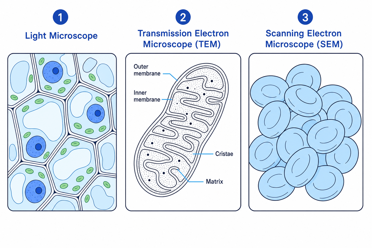
You need to appreciate the different images produced by, and features of, three main types of microscopes:
Light Microscope
- Radiation used: Light (photons).
- Focusing mechanism: Glass lenses.
- Maximum Magnification: Approximately ×1500\times 1500×1500.
- Maximum Resolution: 200 nm200\ \text{nm}200 nm.
- Specimen state: Can view living or dead specimens.
- Image produced: 2D, in color, showing whole cells or larger organelles (like nuclei, chloroplasts, and cell walls).
Transmission Electron Microscope (TEM)
- Radiation used: Electromagnetically focused electron beam passed through a thin specimen.
- Focusing mechanism: Electromagnets.
- Maximum Magnification: Over ×500,000\times 500,000×500,000.
- Maximum Resolution: 0.2 nm0.2\ \text{nm}0.2 nm (very high).
- Specimen state: Must be dead (placed in a vacuum; dehydrated and stained with heavy metals).
- Image produced: 2D, black-and-white, highly detailed cross-sections showing internal organelle ultrastructure.
Scanning Electron Microscope (SEM)
- Radiation used: Electron beam bounced off the surface of a specimen.
- Focusing mechanism: Electromagnets.
- Maximum Magnification: Usually up to ×100,000\times 100,000×100,000.
- Maximum Resolution: 3–10 nm3\text{–}10\ \text{nm}3–10 nm (lower than TEM, but higher than light microscopy).
- Specimen state: Must be dead (in a vacuum).
- Image produced: 3D, black-and-white (often false-colored later), showing detailed surface topography.
3. The Magnification Formula
You must be able to calculate magnification, image size, and actual size of specimens using the standard formula:
Magnification=Size of ImageSize of Real Object \text{Magnification} = \frac{\text{Size of Image}}{\text{Size of Real Object}} Magnification=Size of Real ObjectSize of ImageThis is often remembered using the I AM triangle:
I=A×M I = A \times M I=A×Mwhere:
- III is the measured size of the image.
- AAA is the actual size of the biological specimen.
- MMM is the magnification.
Units must match!
Before performing any calculation, ensure the units of the image size (III) and the actual size (AAA) are identical. Usually, you will measure the image size using a physical ruler in millimetres (mm\text{mm}mm), but cells and organelles are measured in micrometres (μm\mu\text{m}μm) or nanometres (nm\text{nm}nm). Always convert measurements first!
- To convert mm→μm\text{mm} \to \mu\text{m}mm→μm: multiply by 1,0001,0001,000.
- To convert μm→nm\mu\text{m} \to \text{nm}μm→nm: multiply by 1,0001,0001,000.
Calculating actual cell size
A micrograph of a mitochondrion shows its image length to be 48 mm48\text{ mm}48 mm on paper. The magnification of the image is stated as ×8,000\times 8,000×8,000. Find the actual size of the mitochondrion in micrometres (μm\mu\text{m}μm).
- Rearrange the magnification formula to solve for actual size (AAA):
- Convert the measured image size (III) from millimetres into micrometres (μm\mu\text{m}μm) so that it matches biological units:
- Substitute the values of image size (I=48,000 μmI = 48,000\ \mu\text{m}I=48,000 μm) and magnification (M=8,000M = 8,000M=8,000) into the rearranged equation:
- Solve the division to find the final actual size of the mitochondrion:
4. Slide Preparation, Staining, and Calibration
Specimen Preparation
To examine slides under a light microscope (PAG 1), you can prepare them in two main ways:
- Wet Mount: The specimen is suspended in a liquid drop (such as water or stain) and a cover slip is angled down carefully to avoid trapping air bubbles. This is ideal for aquatic organisms or living cheek cells.
- Dry Mount: Thinly sliced specimens (cut using a sectioning razor) are placed directly onto the dry slide with a coverslip placed over them (e.g., pollen grains, hair).
The Use of Staining
Most biological specimens are naturally transparent, making it extremely difficult to distinguish between individual organelles.
Staining works by binding to specific molecules within the cell. This provides contrast between different structures. Differential staining is a technique where multiple stains are used to bind selectively to different cellular components:
- Methylene blue: An alkaline stain that binds negatively charged molecules (like DNA/RNA), staining nuclei blue in animal cells.
- Acetic orcein: Binds strongly to DNA and stains chromosomes a deep red.
- Iodine in potassium iodide solution (KI): Binds to starch granules in plant cells, staining them blue-black.
- Eosin: Stains cytoplasm and extracellular proteins pink.
Microscope Calibration
An eyepiece graticule is a small glass disc with an arbitrary scale (from 1 to 100) fitted inside the microscope eyepiece. It does not have real units (like millimetres) because its apparent size changes when you change objectives.
To find the physical size of one eyepiece unit (epu) at a specific magnification, you must calibrate it against a stage micrometer—a special microscope slide with an incredibly precise scale of known length engraved on it (typically 1 mm1\text{ mm}1 mm divided into 100 divisions, meaning each division is exactly 10 μm10\ \mu\text{m}10 μm wide).
The Golden Rule of Calibration
You must calibrate your eyepiece graticule separately for every objective lens magnification you use.
Calibrating an eyepiece graticule
Under a specific high-power objective lens (×400\times 400×400 total magnification), you align the scales of the eyepiece graticule and the stage micrometer. You observe that exactly 40 eyepiece units (epu) align with 15 divisions of the stage micrometer. Each division on the stage micrometer is known to be 10 μm10\ \mu\text{m}10 μm long. Calculate the actual physical value of 1 eyepiece unit under this objective.
- Determine the total actual distance represented by the aligned stage micrometer divisions by multiplying the number of divisions by the value of each division:
- Equate the total eyepiece units (epu) to this actual distance, showing that 40 epu corresponds to the calculated micrometer distance:
- Calculate the length of a single eyepiece unit by dividing the total actual distance by the number of aligned eyepiece units:
5. Representing Cell Structure (Scientific Drawing)
When drawing structures observed under a light microscope, you must follow strict scientific drawing guidelines to earn marks:
- Use a sharp pencil and draw clean, single, continuous lines. No sketching, feathering, or shading.
- Draw what you actually see, not what you think should be there. Keep proportions accurate.
- Label lines must be drawn with a ruler, touch the structure directly, and never cross each other. Do not use arrowheads on label lines.
- State the magnification or include a clear scale bar.
- Include a title stating what the specimen is and whether it is a whole mount or a cross-section.
6. Eukaryotic Cell Ultrastructure
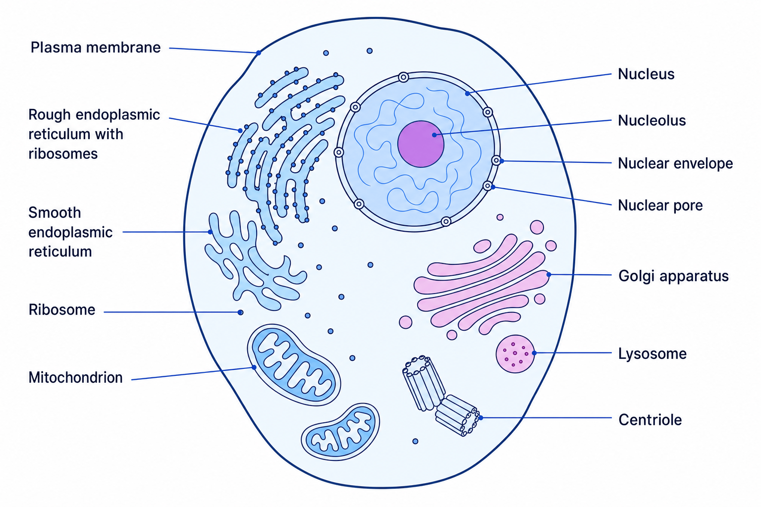
Eukaryotic cells make up plants, animals, fungi, and protoctists. They contain a variety of membrane-bound organelles that partition the cell into distinct compartments, allowing different chemical environments to exist simultaneously.
Core Organelles and Their Functions
- Nucleus: Surrounded by a double membrane called the nuclear envelope, which contains nuclear pores to allow molecules (like mRNA) to enter and leave. It contains chromatin (DNA coiled around histone proteins), which contains the genetic code for protein synthesis.
- Nucleolus: A dense region within the nucleus that does not have a membrane. It is responsible for synthesizing ribosomal RNA (rRNA) and assembling ribosomes.
- Mitochondrion: The site of aerobic respiration, producing ATP. It is enclosed by a double membrane; the inner membrane is highly folded into cristae to increase surface area, enclosing a central fluid-filled matrix.
- Chloroplasts (plants only): The site of photosynthesis. Surrounded by a double membrane, containing fluid stroma and membrane-bound sacks called thylakoids stacked into grana, which contain chlorophyll.
- Ribosomes: Tiny organelles composed of rRNA and proteins, not bound by a membrane. Found free in the cytoplasm or bound to the RER. They are the site of protein translation (80S80\text{S}80S in eukaryotes, 70S70\text{S}70S in prokaryotes).
- Rough Endoplasmic Reticulum (RER): A system of fluid-filled, membrane-bound flattened sacs (cisternae) coated with ribosomes. It folds and packages proteins synthesized by the ribosomes into transport vesicles.
- Smooth Endoplasmic Reticulum (SER): A system of tubular cisternae without ribosomes. It is responsible for the synthesis, storage, and transport of lipids and carbohydrates.
- Golgi Apparatus: A stack of flattened, curved membrane-bound sacs (cisternae) that receives proteins from the RER. It modifies proteins (e.g., adding carbohydrate chains to make glycoproteins) and packages them into secretory vesicles.
- Lysosomes: Specialized vesicles containing hydrolytic (digestive) enzymes. They break down waste materials, worn-out organelles, and pathogens.
- Centrioles: Small tubes made of microtubules (composed of tubulin). They occur in pairs near the nucleus and form the spindle fibres that organize and separate chromosomes during cell division (mitosis/meiosis).
- Plasma Membrane: A phospholipid bilayer containing proteins, controlling the passage of substances in and out of the cell.
- Cell Wall (plants, fungi, algae): A rigid structure providing mechanical strength and preventing the cell from bursting. Made of cellulose in plants and chitin in fungi.
- Flagella and Cilia: Hair-like projections extending from the cell surface. They contain a 9+29+29+2 arrangement of microtubules. Flagella are longer and enable cell movement (e.g., sperm), while cilia are shorter and beat in a coordinated fashion to move fluids over a cell surface (e.g., in the trachea).
7. Organelle Interrelationship: Protein Secretion
One of the most important concepts to master for OCR is how organelles work cooperatively to produce and secrete proteins.
The Protein Secretion Pathway
Transcription in Nucleus →\to→ Translation on RER →\to→ Transport via Vesicle →\to→ Modification in Golgi →\to→ Secretion via Exocytosis.
  [Nucleus] (DNA transcribed to mRNA)
     │
     ▼
  (mRNA leaves through nuclear pore)
     │
     ▼
  [Ribosomes on RER] (mRNA translated; protein folds in RER lumen)
     │
     ▼
  [Transport Vesicles] (pinch off RER, move along cytoskeleton)
     │
     ▼
  [Golgi Apparatus] (vesicles fuse; proteins modified & sorted)
     │
     ▼
  [Secretory Vesicles] (pinch off Golgi, move to plasma membrane)
     │
     ▼
  [Plasma Membrane] (vesicles fuse, releasing protein via exocytosis)
Don't forget the mitochondria!
Mitochondria are crucial throughout this process because they undergo aerobic respiration to synthesize ATP. This active transport/secretion (including vesicle movement along the cytoskeleton and exocytosis) requires energy!
8. The Cytoskeleton
The cytoskeleton is a dynamic network of protein fibres extending throughout the cytoplasm. It is not rigid; it constantly reorganizes to support cell functions.
It consists of three main components:
- Microfilaments (contractile fibres made of the protein actin): Provide mechanical strength, maintain cell shape, and are responsible for cell movement (e.g., amoeboid movement and cytokinesis).
- Microtubules (hollow cylinders made of the protein tubulin): Form scaffold-like structures. They act as "tracks" along which motor proteins (like dynein and kinesin) move organelles and vesicles. They also form spindle fibres, centrioles, cilia, and flagella.
- Intermediate filaments: Provide mechanical strength to help cells withstand physical pulling forces and anchor organelles (like the nucleus) in place.
Monorail of the cell
Think of microtubules as train tracks and vesicles as carriages. Motor proteins are the locomotives that use ATP to walk along these microtubule tracks, carrying materials from one side of the cell to the other.
9. Prokaryotic vs. Eukaryotic Cells
Prokaryotes (bacteria and archaea) are structurally simpler and much smaller than eukaryotes.
| Feature | Prokaryotic Cells | Eukaryotic Cells |
|---|---|---|
| Size | Extremely small (0.5–5 μm0.5\text{–}5\ \mu\text{m}0.5–5 μm) | Larger (10–100 μm10\text{–}100\ \mu\text{m}10–100 μm) |
| Genetic Material | Circular DNA; "naked" (no histone proteins); free in cytoplasm (nucleoid); may have plasmids | Linear DNA; associated with histone proteins; enclosed inside a nuclear envelope |
| Organelles | No membrane-bound organelles | Has membrane-bound organelles (mitochondria, RER, Golgi, etc.) |
| Ribosomes | Smaller, 70S70\text{S}70S ribosomes | Larger, 80S80\text{S}80S ribosomes (70S70\text{S}70S inside mitochondria/chloroplasts) |
| Cell Wall | Made of peptidoglycan (murein) | Made of cellulose (plants) or chitin (fungi) |
| Cell Division | Binary fission (no spindle fibres) | Mitosis or meiosis (uses spindle fibres) |
In the exam
- Double-check units before using I=A×MI = A \times MI=A×M. Examiners love to give the image size in mm and ask for the actual size in μm\mu\text{m}μm.
- Be specific about membranes: If describing mitochondria or chloroplasts, always state they have a double membrane.
- Remember the Golgi roles: Do not just write "packages proteins". You must write "modifies and packages proteins" to get full marks.
- Link the cytoskeleton to ATP: If asked about vesicle transport, always explain that motor proteins move vesicles along microtubules and that this process requires ATP produced by mitochondria.
Check yourself
- Why is the maximum resolution of a light microscope lower than that of an electron microscope?
- Outline the path taken by an enzyme from its synthesis to its secretion outside the cell, naming every organelle and vesicle involved.
- Under a microscope, 20 eyepiece units align with 4 divisions of a stage micrometer. If each division is 10 μm10\ \mu\text{m}10 μm, what is the value of 1 eyepiece unit?
