

Welcome to the foundation of all living systems! Biochemistry is the study of the chemical processes and substances that occur within all living organisms—from the smallest prokaryotic bacterium to the largest eukaryotic blue whale. By mastering the structure of these molecules, you will unlock the key to understanding how they function.
What you'll learn
- How the unique chemical properties of water support life as a solvent, transport medium, coolant, and habitat.
- How monomers link via condensation reactions to form vital biological polymers (carbohydrates, lipids, and proteins) and how they are broken down by hydrolysis.
- How to perform and interpret biochemical tests, chromatography, and quantitative colorimetry.
1. The Elements of Life and the Power of Water
To understand biochemistry, we must first look at the elements that build macromolecules.
Macromolecule
A very large molecule, such as a protein, polymer, or nucleic acid, built up from the covalent bonding of smaller subunits.
Different classes of biological molecules are constructed from specific combinations of elements:
- Carbohydrates: Carbon (C), Hydrogen (H), and Oxygen (O) only.
- Lipids: Carbon (C), Hydrogen (H), and Oxygen (O) only (except phospholipids, which also contain Phosphorus).
- Proteins: Carbon (C), Hydrogen (H), Oxygen (O), Nitrogen (N), and sulfur (S) (found in the R-groups of some amino acids like cysteine).
- Nucleic Acids: Carbon (C), Hydrogen (H), Oxygen (O), Nitrogen (N), and Phosphorus (P).
The Structure and Polarity of Water
Water (H2OH_2OH2O) is the medium in which all metabolic reactions take place. To understand its properties, we must look at its bonding.
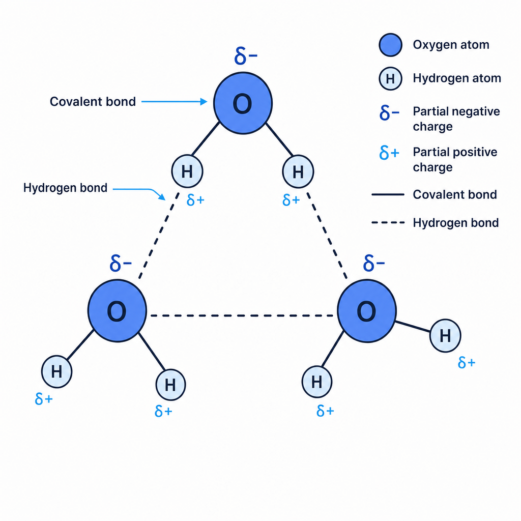
A water molecule consists of one oxygen atom covalently bonded to two hydrogen atoms. Oxygen is highly electronegative, meaning it has a much stronger pull on the shared electrons in the covalent bonds than hydrogen does. This creates an uneven distribution of charge:
- The oxygen atom pulls the electrons closer and gains a partial negative charge (δ−\delta^-δ−).
- The hydrogen atoms are left with a partial positive charge (δ+\delta^+δ+).
This separation of charge makes water a polar molecule (a dipole). The opposite charges on neighbouring water molecules attract one another.
Hydrogen Bond
A weak electrostatic bond formed when a partially positive hydrogen atom (δ+\delta^+δ+) in one polar molecule is attracted to a partially negative electronegative atom (δ−\delta^-δ−) (usually oxygen or nitrogen) in another molecule.
Properties of Water and Their Biological Roles
While individual hydrogen bonds are weak, the cumulative effect of billions of them in a small volume of water gives it extraordinary properties:
- As a Solvent: Because water is polar, the positive and negative parts of the water molecules cluster around solute ions or polar molecules (like glucose). This surrounds them, separating them and keeping them in solution. This allows metabolic reactions to occur in the cytoplasm of prokaryotes and eukaryotes alike.
- As a Transport Medium: Water is highly cohesive (water molecules stick together due to hydrogen bonding) and adhesive (water molecules stick to polar surfaces, such as xylem walls). This allows columns of water to be pulled up the xylem vessels in plants (transpiration stream) and allows blood to transport dissolved nutrients in mammals.
- As a Coolant: Water has a high latent heat of vaporisation. This means it requires a large amount of thermal energy to break the hydrogen bonds and turn liquid water into water vapour (gas). This makes evaporation an incredibly effective cooling mechanism (e.g., sweating in humans, transpiration cooling leaf surfaces in plants).
- Thermal Stability: Water has a high specific heat capacity because a lot of energy is required to break the hydrogen bonds and increase the kinetic energy of the molecules. This prevents rapid temperature fluctuations, providing a stable aquatic habitat and keeping internal cellular temperatures stable so that enzymes can function at their optimum rate.
- As a Habitat: Unlike most substances, solid ice is less dense than liquid water because, below 4 °C, the hydrogen bonds fix the water molecules into a rigid, open crystalline lattice. This causes ice to float, forming an insulating layer on top of ponds and lakes. This prevents the water below from freezing solid, allowing aquatic organisms to survive beneath the surface during winter.
2. Monomers, Polymers, and Carbohydrates
All living organisms rely on giant macromolecules. Many of these are polymers built from repeating chemical subunits called monomers.
Monomer vs Polymer
- Monomer: A small, simple molecule that can bind chemically to other monomers to form a polymer (e.g., monosaccharides, amino acids).
- Polymer: A large molecule made up of many repeating monomer subunits bonded together (e.g., starch, proteins).
The formation and breakdown of these macromolecules rely on two universal chemical reactions:
Condensation vs Hydrolysis
- Condensation: A reaction that joins two molecules together with the formation of a chemical bond and the elimination (release) of a water molecule.
- Hydrolysis: A reaction that splits a chemical bond between two molecules using the addition of a water molecule.
Monosaccharides: Glucose and Ribose
Monosaccharides are the single-sugar monomers. You need to know two main types:
- Pentose sugars: Have 5 carbon atoms (e.g., ribose, C5H10O5C_5H_{10}O_5C5H10O5, which forms the backbone of RNA).
- Hexose sugars: Have 6 carbon atoms (e.g., glucose, C6H12O6C_6H_{12}O_6C6H12O6, the primary respiratory substrate).
The Two Isomers of Glucose
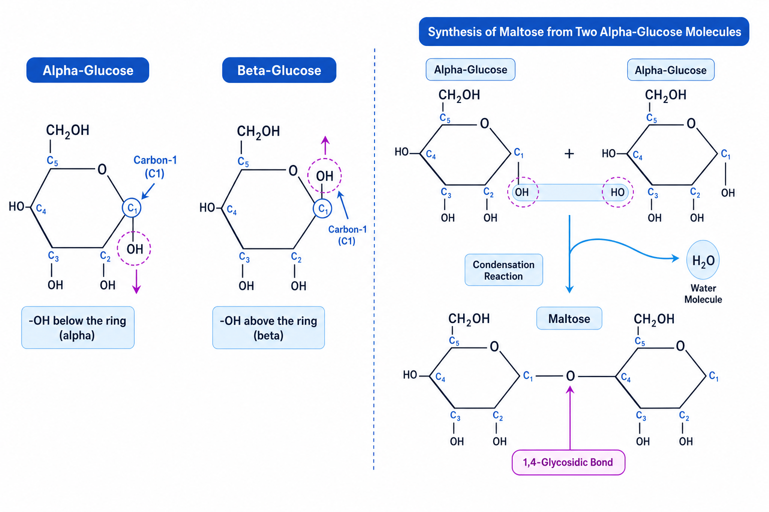
Glucose exists in two structural forms (isomers): α\alphaα-glucose and β\betaβ-glucose. The only difference between them lies in the orientation of the hydroxyl group (-OH) on Carbon-1 (C1):
- α\alphaα-glucose: The -OH group on C1 is located below the carbon ring.
- β\betaβ-glucose: The -OH group on C1 is located above the carbon ring.
How to remember the glucose isomers
Remember ABBA: Alpha = Below, Beta = Above.
Synthesis and Breakdown of Disaccharides
When two monosaccharides undergo a condensation reaction, they form a disaccharide held together by a covalent glycosidic bond. You must know these three key disaccharides:
- Maltose: α\alphaα-glucose + α\alphaα-glucose (joined by a 1,4-glycosidic bond).
- Sucrose: glucose + fructose.
- Lactose: glucose + galactose.
During a condensation reaction between two alpha-glucose molecules, the hydroxyl group (-OH) on C1 of one glucose aligns with the -OH group on C4 of the adjacent glucose. One molecule of H2OH_2OH2O is removed, leaving an oxygen bridge (a 1,4-glycosidic bond) to link the two rings together.
To break this bond during digestion, a water molecule is added back in a hydrolysis reaction, splitting maltose back into two separate glucose molecules.
Polysaccharides: Starch, Glycogen, and Cellulose
Polysaccharides are polymers of monosaccharides. Their structural differences determine their biological functions:
| Polysaccharide | Monomer Unit | Glycosidic Bonds | Structure | Function & Properties |
|---|---|---|---|---|
| Starch: Amylose | α\alphaα-glucose | 1,4-only | Unbranched, tightly coiled helix. | Energy storage in plants. Highly compact, meaning lots of glucose can be stored in a small space. Insoluble so it does not affect the water potential of the cell. |
| Starch: Amylopectin | α\alphaα-glucose | 1,4- and 1,6- | Branched (approx. every 24–30 glucose units). | Energy storage in plants. Multiple branches mean it has many terminal ends, allowing rapid hydrolysis by amylase enzymes to release glucose for respiration. Insoluble. |
| Glycogen | α\alphaα-glucose | 1,4- and 1,6- | Highly branched (approx. every 8–12 glucose units). | Energy storage in animals and fungi. It is even more branched than amylopectin, providing a massive number of free ends. This is vital for animals with high metabolic rates, as they can rapidly mobilise glucose when needed. Insoluble. |
| Cellulose | β\betaβ-glucose | 1,4-only | Straight, unbranched, parallel chains. | Structural component of plant cell walls. Because β\betaβ-glucose has its -OH on C1 pointing up and its -OH on C4 pointing down, every alternate β\betaβ-glucose molecule must be rotated 180∘180^\circ180∘ relative to its neighbour to form a bond. This rotation prevents coiling and results in straight, flat chains. |
How Cellulose gains its immense strength
The straight, parallel cellulose chains run side-by-side. Hydrogen bonds form cross-links between the hydroxyl groups of adjacent chains. Thousands of these hydrogen bonds grouped together form microfibrils, which bundle into macrofibrils. These run in different directions across the plant cell wall, providing high tensile strength to prevent the plant cell from bursting when turgid.
3. Lipids (Triglycerides, Phospholipids, and Cholesterol)
Lipids are a diverse group of non-polar, hydrophobic macromolecules. Unlike carbohydrates and proteins, they are not true polymers because they are not made of repeating monomer subunits.
Saturated vs Unsaturated Fatty Acids
Fatty acids consist of a carboxyl group (-COOH) attached to a long hydrocarbon tail:
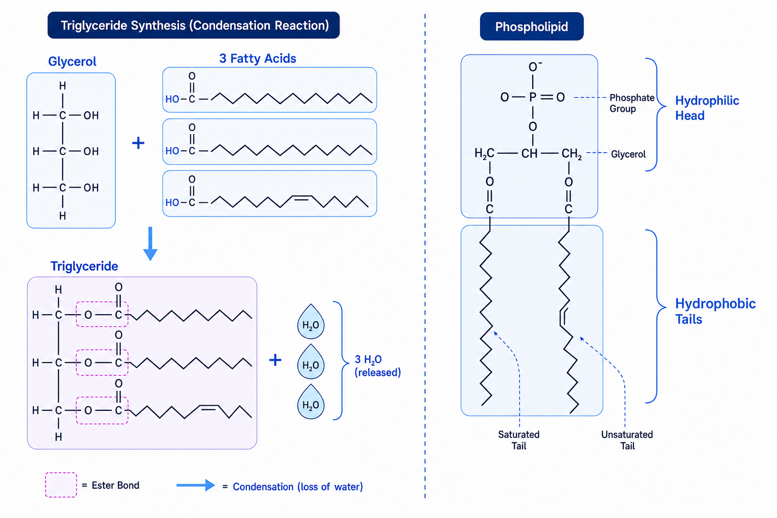
- Saturated fatty acids: Contain no carbon-carbon double bonds (C=CC=CC=C) in the hydrocarbon tail. The chain is straight, allowing molecules to pack closely together. They are typically solids at room temperature (e.g., animal fats).
- Unsaturated fatty acids: Contain one or more carbon-carbon double bonds (C=CC=CC=C) in the hydrocarbon tail. This double bond introduces a kink (bend) in the chain, preventing the molecules from packing tightly. They are typically liquids at room temperature (e.g., plant oils).
Triglycerides
A triglyceride is composed of one glycerol molecule bonded to three fatty acids.
During synthesis, the hydroxyl groups (-OH) of the glycerol react with the carboxyl groups (-COOH) of the fatty acids. This forms three ester bonds via three separate condensation reactions, releasing three water molecules. Hydrolysis breaks these ester bonds by adding three water molecules.
- Function: Triglycerides are used primarily for energy storage. The long hydrocarbon tails contain a very high ratio of carbon-hydrogen bonds to carbon atoms, making them highly reduced. As a result, lipids store about double the chemical energy per unit mass compared to carbohydrates. Being hydrophobic, they pack closely together without attracting water, making them lightweight and efficient for mobile organisms.
Phospholipids
In a phospholipid, one of the three fatty acids is replaced by a phosphate group.
- Properties: The phosphate group is highly polar and negatively charged, making the "head" of the molecule hydrophilic (water-attracting). The two fatty acid "tails" remain non-polar and hydrophobic (water-repelling). This dual nature is called being amphipathic.
- Function: When placed in water, phospholipids naturally self-assemble into a bilayer. The hydrophilic heads face outwards, interacting with the aqueous cytoplasm and extracellular fluid. The hydrophobic tails point inwards, away from water, forming a hydrophobic core. This forms the basis of all cell membranes, acting as a barrier that controls what enters and exits the cell.
Cholesterol
Cholesterol is a small lipid molecule with a four-carbon ring structure.
- Function: It is highly hydrophobic and fits directly between the fatty acid tails of phospholipids within the cell membrane. It regulates membrane fluidity: at high temperatures, it stabilizes the membrane to prevent it from becoming too fluid; at low temperatures, it prevents the tails from packing too tightly and freezing.
4. Proteins (Structure and Diversity)
Proteins are polymers made of monomers called amino acids. They carry out almost every functional role in living cells, including catalysis, structural support, transport, and cell signalling.
The General Structure of an Amino Acid
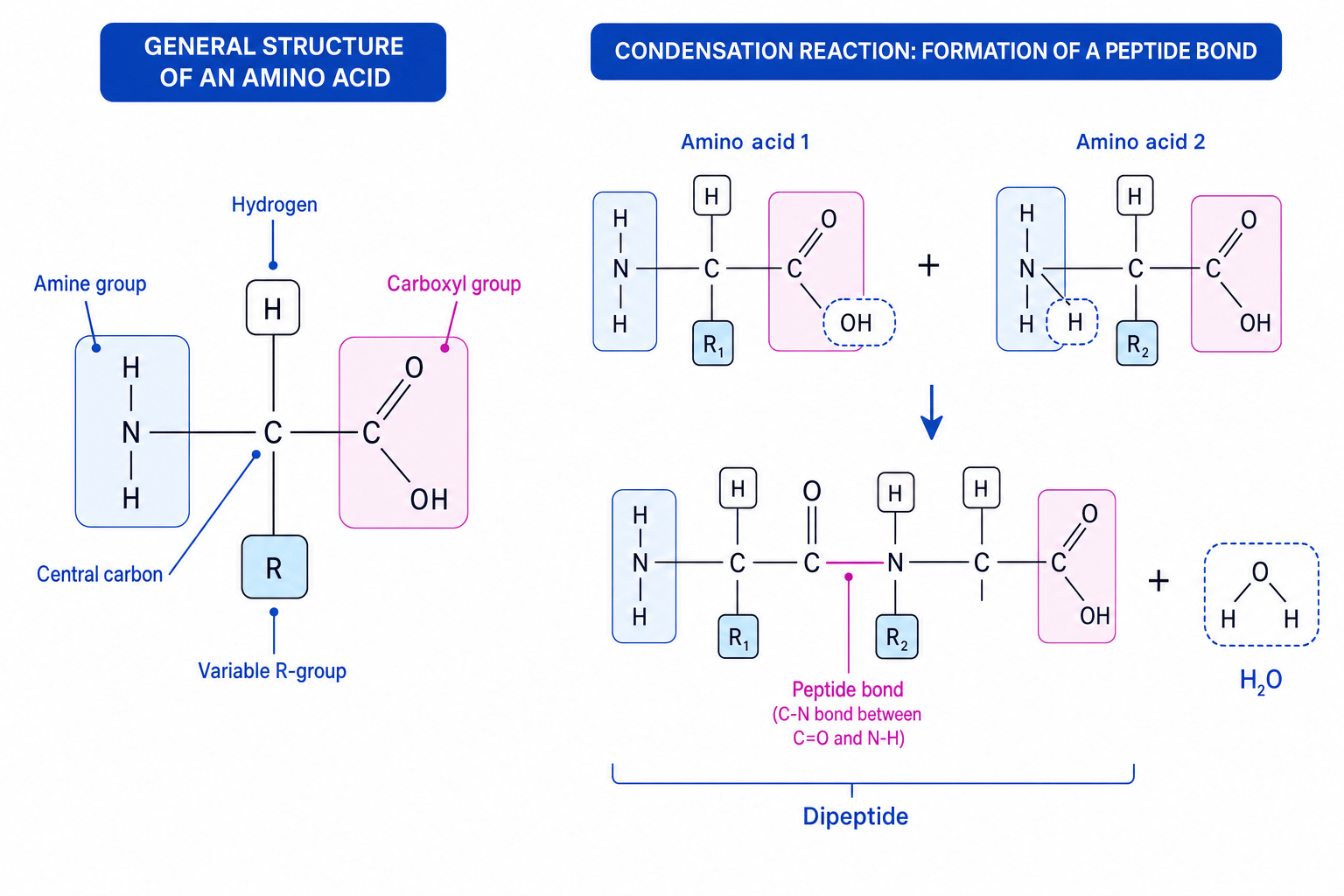
All amino acids share the same basic structure centered around a central carbon atom (often called the alpha-carbon):
- An amine group (−NH2-NH_2−NH2)
- A carboxyl group (−COOH-COOH−COOH)
- A hydrogen atom (−H-H−H)
- A variable R-group (side chain), which is unique to each of the 20 amino acids and determines its chemical properties (polar, non-polar, acidic, basic).
Synthesis of Dipeptides and Polypeptides
Two amino acids join together via a condensation reaction to form a dipeptide. The reaction occurs between the carboxyl group of one amino acid and the amine group of the next, forming a covalent peptide bond and releasing a molecule of water.
A chain of many amino acids joined by peptide bonds is called a polypeptide. This chain is broken down back into amino acids via hydrolysis.
The Four Levels of Protein Structure
To become a functional protein, a polypeptide chain must fold into a precise 3D shape. This folding is described in four levels:
Primary Structure (1∘1^\circ1∘)
The unique sequence of amino acids in the polypeptide chain. This sequence is determined by DNA.
- Bonds involved: Covalent peptide bonds only.
Secondary Structure (2∘2^\circ2∘)
The folding or coiling of the primary polypeptide chain.
- Bonds involved: Hydrogen bonds form between the δ−\delta^-δ− oxygen of the carboxyl group (C=OC=OC=O) and the δ+\delta^+δ+ hydrogen of the amine group (N−HN-HN−H) along the polypeptide backbone.
- Key shapes: This regular bonding forms either an α\alphaα-helix (a tight spiral) or a β\betaβ-pleated sheet (flat, parallel sheets).
Tertiary Structure (3∘3^\circ3∘)
The overall 3D folding of the entire polypeptide chain. This is determined by the interactions between the variable R-groups of the amino acids.
- Bonds involved:
- Hydrogen bonds: Form between polar R-groups. They are weak and easily broken by high temperatures or pH changes.
- Ionic bonds: Form between positively and negatively charged R-groups. Easily broken by pH changes.
- Disulfide bonds (bridges): Strong covalent bonds that form between the sulfur atoms of two cysteine amino acids. These are highly stable and resist heat.
- Hydrophobic and Hydrophilic interactions: Non-polar (hydrophobic) R-groups clump together in the center of the protein to shield themselves from water, while polar (hydrophilic) R-groups point outwards into the aqueous environment.
Confusing the location of bonds
Students often lose marks by not specifying where bonds form. In secondary structure, hydrogen bonds form between the peptide backbone groups (C=OC=OC=O and N−HN-HN−H). In tertiary structure, all bonds (hydrogen, ionic, disulfide, hydrophobic) form between the variable R-groups.
Quaternary Structure (4∘4^\circ4∘)
The structure formed when two or more polypeptide chains (called subunits) work together as a single functional protein. It can also involve non-protein groups (known as prosthetic groups).
- Bonds involved: All the same bond types found in tertiary structure, holding the subunits together.
Globular vs Fibrous Proteins
Proteins generally fall into one of two structural categories:
                  ┌───────────────────────┐
                  │   PROTEIN STRUCTURE   │
                  └───────────┬───────────┘
                              │
              ┌───────────────┴───────────────┐
              ▼                               ▼
    ┌───────────────────┐           ┌───────────────────┐
    │ Globular Proteins │           │  Fibrous Proteins │
    ├───────────────────┤           ├───────────────────┤
    │ • Spherical       │           │ • Long strands    │
    │ • Soluble         │           │ • Insoluble       │
    │ • Metabolic roles │           │ • Structural roles│
    └───────────────────┘           └───────────────────┘
Globular Proteins
Globular proteins fold so that hydrophobic R-groups are kept on the inside while hydrophilic R-groups face the outside. This makes them highly soluble in water. They have precise, round shapes that allow them to bind specifically to other molecules.
- Insulin: A hormone that regulates blood glucose. It is a small globular protein composed of two polypeptide chains held together by disulfide bonds. Its precise shape allows it to bind specifically to complementary glycoprotein receptors on cell surface membranes.
- Amylase: An enzyme that hydrolyses starch into maltose. Its globular shape forms a highly specific active site with a shape complementary to its substrate, starch.
- Haemoglobin (Conjugated Protein): A conjugated protein is a globular protein that contains a non-protein prosthetic group. Haemoglobin has a quaternary structure consisting of four polypeptide subunits (two α\alphaα-chains and two β\betaβ-chains). Each subunit contains a prosthetic haem group containing an Fe2+Fe^{2+}Fe2+ ion. Each Fe2+Fe^{2+}Fe2+ ion can bind reversibly to one oxygen molecule (O2O_2O2), allowing a single haemoglobin molecule to transport up to four oxygen molecules.
Fibrous Proteins
Fibrous proteins are made of long, parallel polypeptide chains. They have simple, repetitive primary structures, are insoluble in water, and have high tensile strength. Their function is almost entirely structural.
- Collagen: Found in skin, bone, tendons, and cartilage. It consists of three polypeptide chains wrapped around each other to form a tight, triple-helix "rope" with covalent cross-links between the chains, providing extreme tensile strength.
- Keratin: Found in hair, nails, and the outer layer of skin. It is rich in cysteine amino acids, forming many strong disulfide bridges, which makes it exceptionally tough and impermeable to water.
- Elastin: Found in walls of large blood vessels (like arteries) and alveoli. It contains cross-linked molecules that can stretch and recoil when deformed, allowing tissue to regain its original shape.
5. Key Inorganic Ions
Living organisms also require inorganic ions to function. You must recognise and know the biological roles of the following ions:
Cations (Positively Charged)
- Calcium (Ca2+Ca^{2+}Ca2+): Essential for bone and teeth structure, muscle contraction, and triggering neurotransmitter release at synapses. In plants, it forms calcium pectate, which glues plant cell walls together.
- Sodium (Na+Na^+Na+): Used in the co-transport of glucose and amino acids across cell membranes (e.g., in the ileum or kidney nephron) and in generating action potentials during nerve impulses.
- Potassium (K+K^+K+): Used to generate action potentials in neurons and active transport of K+K^+K+ into guard cells causes stomata to open.
- Hydrogen (H+H^+H+): The concentration of H+H^+H+ determines the pH of a solution. It is also vital in chemiosmosis during aerobic respiration and photosynthesis to generate ATP.
- Ammonium (NH4+NH_4^+NH4+): A key source of nitrogen that plants absorb from the soil to synthesise amino acids and nucleic acids.
Anions (Negatively Charged)
- Nitrate (NO3−NO_3^-NO3−): The primary source of nitrogen absorbed by plants from the soil to build proteins and DNA.
- Hydrogencarbonate (HCO3−HCO_3^-HCO3−): Acts as a chemical buffer in the blood, maintaining blood pH within a narrow range, and plays a major role in transporting carbon dioxide from respiring tissues to the lungs.
- Chloride (Cl−Cl^-Cl−): Acts as an inorganic cofactor for the enzyme amylase and is involved in maintaining water potential in cells.
- Phosphate (PO43−PO_4^{3-}PO43−): A structural component of DNA, RNA, ATP, and phospholipids in cell membranes.
- Hydroxide (OH−OH^-OH−): Involved in maintaining pH and acid-base balance in aqueous solutions.
6. Practical Skills: Testing and Separating Molecules
You need to know how to perform and interpret qualitative chemical tests, use a colorimeter to make quantitative measurements, and separate biological mixtures using chromatography.
Qualitative Biochemical Tests
| Nutrient | Reagent used | Method | Negative Result | Positive Result |
|---|---|---|---|---|
| Starch | Iodine in potassium iodide | Add a few drops of iodine solution directly to the sample. | Yellow-brown | Blue-black |
| Reducing Sugars (e.g. glucose, maltose) | Benedict's reagent | Add Benedict's, heat in a water bath at 80 °C for 5 minutes. | Blue | Green →\rightarrow→ Yellow →\rightarrow→ Orange →\rightarrow→ Brick-red precipitate |
| Non-Reducing Sugars (e.g. sucrose) | Hydrochloric acid + Benedict's reagent | Heat sample with dilute HCl (to hydrolyse glycosidic bonds). Neutralise with sodium hydrogencarbonate, then perform the standard Benedict's test. | Blue | Green →\rightarrow→ Yellow →\rightarrow→ Orange →\rightarrow→ Brick-red precipitate |
| Proteins | Biuret reagent | Add sodium hydroxide, then copper(II) sulfate (or premixed Biuret reagent). | Blue | Purple / Violet |
| Lipids | Ethanol (Emulsion test) | Mix sample with ethanol (to dissolve lipids), shake, then pour the mixture into water. | Clear | Cloudy, white emulsion |
Quantitative Methods: Colorimetry
Benedict's test is semi-quantitative: the closer the final colour is to brick-red, the higher the initial concentration of reducing sugar. To make this truly quantitative, we use a colorimeter to measure the concentration.
Colorimeter
An instrument that measures the light absorbance or light transmission of a solution at a specific wavelength.
Steps to determine an unknown glucose concentration:
- Create standard solutions: Use a serial dilution to prepare a range of known glucose concentrations (e.g., 0.0, 0.5, 1.0, 1.5, 2.0 mol dm⁻³).
- Perform Benedict's test: Add equal volumes of Benedict's reagent to each standard and heat.
- Centrifuge the samples: Spin the tubes to force the insoluble brick-red precipitate to the bottom, leaving a clear, blue liquid (the supernatant).
- Calibrate the colorimeter: Set the colorimeter to zero absorbance using a cuvette of distilled water (often using a red filter to maximise the detection of blue colour differences).
- Measure absorbance/transmission: Measure the light absorbance of the supernatant for each of your standard solutions.
- High glucose concentration →\rightarrow→ more precipitate forms →\rightarrow→ less blue copper(II) ions remain in the supernatant →\rightarrow→ lower absorbance (or higher transmission) of red light.
- Plot a calibration curve: Plot a graph of glucose concentration (x-axis) against absorbance (y-axis).
- Find the unknown: Perform the test on your unknown sample, measure its absorbance, and use your calibration curve to read across and find its exact concentration.
Separation: Paper and Thin-Layer Chromatography (TLC)
Chromatography is used to separate and identify individual biological molecules (such as amino acids, carbohydrates, or pigments) from a mixture.
How it works:
- Stationary Phase: A solid sheet (chromatography paper, or a thin layer of silica on a plastic sheet for TLC).
- Mobile Phase: A liquid solvent that moves up the stationary phase by capillary action.
- Molecules in the mixture separate because they have different solubilities in the mobile phase and different adsorption to the stationary phase. Highly soluble molecules spend more time in the solvent and travel further up the sheet.
The Chromatographic Method:
- Draw a pencil starting line (the origin) near the bottom of the chromatography paper (do not use ink, as ink contains pigments that will separate and ruin the results).
- Spot the concentrated sample mixture onto the line using a capillary tube. Let it dry, and repeat to build up a small, concentrated spot.
- Place the bottom of the paper into the solvent. Ensure the solvent level is below the pencil line, otherwise, the spots will wash off into the solvent bath.
- Cover the beaker with a lid to saturate the air with solvent vapour and prevent evaporation.
- Remove the paper just before the solvent reaches the top. Immediately mark the position of the solvent front with a pencil.
- If separating colourless molecules like amino acids, spray the paper with ninhydrin spray and heat. The amino acids will appear as purple or brown spots.
- Calculate the RfR_fRf (Retention Factor) value for each spot and compare it to known databases to identify the molecule.
Calculating retention factor (R_f)
A student separates a mixture of amino acids using TLC. They measure the following distances from the origin line:
- Solvent front distance = 8.4 cm8.4\text{ cm}8.4 cm
- Spot A (unknown amino acid) distance = 3.7 cm3.7\text{ cm}3.7 cm
Calculate the RfR_fRf value of amino acid A.
- State the formula clearly:
- Substitute the values with consistent units (both in cm, or converted to mm):
- Calculate the value to an appropriate number of decimal places (usually two):
(Note: RfR_fRf values are always dimensionless ratios and must be less than 1.00)
Units and measuring errors in chromatography
Always measure the distance from the pencil origin line to the centre of the spot, not the edge of the spot. Ensure you measure to the same point on every spot to keep your measurements valid.
In the exam
- Be specific with bonds: Always name the exact bond involved. Do not just say "carbohydrates have bonds"—say glycosidic bonds. For proteins, refer to peptide, hydrogen, ionic, or disulfide bonds depending on the structural level you are discussing.
- Describe cellulose rotation: If asked about the structure of cellulose, you must state that alternate beta-glucose molecules are rotated 180∘180^\circ180∘ to allow the 1,4-glycosidic bonds to form. Mentioning this rotation is a common mark-scheme requirement.
- Use the Benedict's non-reducing test protocol correctly: If asked how to test for non-reducing sugars, you must write down all three steps in order: (1) add hydrochloric acid and heat, (2) neutralise with sodium hydrogencarbonate, and (3) add Benedict's and heat. Skipping the neutralisation step will result in zero marks because Benedict's reagent will not work in acidic conditions!
- Be precise with chromatography calculations: When calculating RfR_fRf, show your working. Ensure your division is the right way around: solute distance (smaller number) divided by solvent distance (larger number).
Check yourself
- Why is it biologically significant that water has a high specific heat capacity? Contrast how this property supports both a single eukaryotic cell and an entire aquatic ecosystem.
- Draw the structural difference between α\alphaα-glucose and β\betaβ-glucose. Which of these monomers forms starch, and which forms cellulose?
- Outline the step-by-step procedure required to quantitatively determine the concentration of reducing sugar in an unknown fruit juice sample using a colorimeter.
- Explain how the structures of glycogen and amylose are suited to their roles as storage molecules.
