2.3.1 Structures and functions of the brain
The Brain and the Nervous System
Central nervous system
The brain and spinal cord, which receive impulses from receptors, coordinate a response and send impulses to effectors.
- The nervous system is divided into the central nervous system and the nerves that run to and from it.
- The central nervous system, usually shortened to the CNS, is made up of the brain and the spinal cord.
- The CNS receives impulses from receptors, coordinates a response, and sends impulses out to effectors.
- The human brain contains billions of interconnected neurones, and different regions of it carry out different jobs.
- The brain is protected by the bones of the skull, by three membranes called the meninges, and by a cushioning layer of fluid.
The Cerebral Hemispheres
Cerebral hemispheres
The two large folded halves at the top of the brain, responsible for conscious thought, memory, language, intelligence and voluntary movement.
- The cerebral hemispheres are the two large halves that make up the top and front of the brain, and together they are the largest part of it.
- Their outer layer is deeply folded, which packs a far larger surface area of nerve cells into the skull.
- They control conscious thought, memory, language, intelligence and voluntary movement.
- They also process the information arriving from the sense organs, so seeing, hearing and feeling pain all reach conscious awareness here.
- Damage to a cerebral hemisphere, for example after a stroke, can cause loss of speech, memory or movement on one side of the body.
The Cerebellum
Cerebellum
The region at the back and base of the brain that coordinates muscular activity, balance and posture.
- The cerebellum lies at the back and base of the brain, underneath the cerebral hemispheres.
- It coordinates muscular activity, so that the muscles of a movement contract in the right order and with the right force.
- It controls balance and posture, using information from the ears and from receptors in the muscles.
- It makes movement smooth and precise rather than jerky, and it is involved in learning a physical skill.
- Damage to the cerebellum causes unsteady walking, a loss of balance and clumsy, poorly aimed movements.
Riding a bicycle depends on the cerebellum, which constantly compares the intended movement with information from the muscles and the ears, then adjusts the muscle contractions to keep you upright.
The Medulla Oblongata
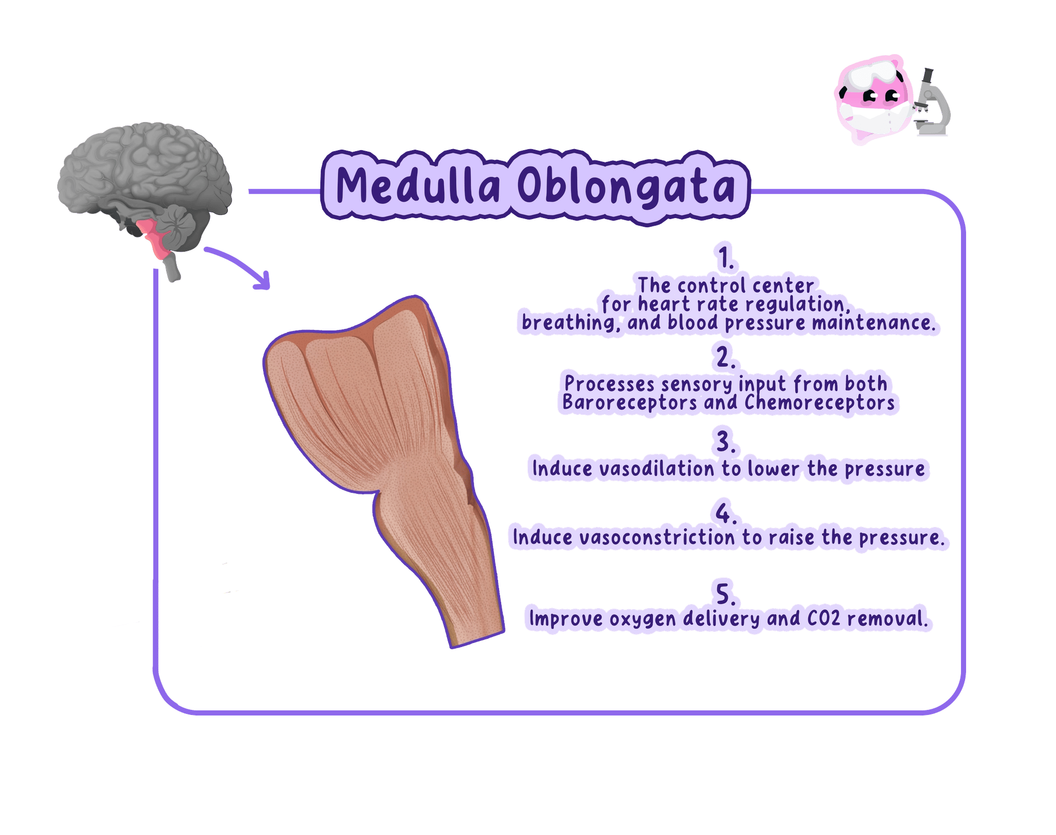
Medulla oblongata
The region at the base of the brain, joining the spinal cord, that controls unconscious activities such as heart rate and breathing rate.
- The medulla oblongata sits at the base of the brain, where it joins the spinal cord.
- It controls unconscious, or involuntary, activities that continue without any thought.
- It regulates heart rate, breathing rate and blood pressure.
- It also controls reflexes such as swallowing, coughing, sneezing and vomiting.
- Because it controls the processes that keep a person alive, damage to the medulla oblongata is usually fatal.
Do not confuse the cerebellum with the cerebral hemispheres. The cerebellum coordinates movement and balance, while the cerebral hemispheres handle conscious thought and decide on the movement in the first place.
Conscious and Unconscious Control
- Conscious control means you decide to act, and the cerebral hemispheres send the instruction.
- Unconscious control happens automatically, and the medulla oblongata handles the processes that must continue while you sleep.
- Some actions involve both, and breathing is the clearest case because it is normally unconscious but can be taken over deliberately.
- The cerebellum sits between the two, refining a consciously chosen movement without any conscious effort.
How the Regions Were Identified
- Much of what is known about the brain came from studying patients with damage to a particular region.
- Comparing the position of the damage with the ability the patient had lost suggested what that region normally does.
- Electrical stimulation during brain surgery gave more direct evidence, because stimulating a small area produces a specific response.
- Scanning techniques now allow the living brain to be studied without surgery, and these are covered in the next section.
- Label questions on a brain diagram are common, and the distractors are always the other three named regions, so learn the position of each as well as its job.
- Give a function that matches the region precisely, because coordination of movement is credited for the cerebellum but not for the cerebral hemispheres.
- When a question describes a patient's symptoms and asks which region is damaged, work backwards from the lost ability to the region that controls it.
- Use the full name medulla oblongata at least once, then the short form if you prefer.
- Name the two organs that make up the central nervous system.
- State three functions of the cerebral hemispheres.
- Which region of the brain coordinates balance and posture?
- Give two unconscious activities controlled by the medulla oblongata.
- A patient has become very unsteady on their feet but can speak and think normally. Which region of the brain is most likely to be damaged?
2.3.2 Imaging brain function with scanning
Why the Brain Is Difficult to Study
Central nervous system
The brain and spinal cord, which receive impulses from receptors, coordinate a response and send impulses to effectors.
- The brain is enclosed by the skull, so it cannot be seen or reached without surgery.
- Brain tissue is extremely delicate, and any attempt to reach it risks permanent damage to healthy regions.
- The brain contains billions of neurones with enormous numbers of connections, so its structure is very complex.
- Regions do not work in isolation, so removing or damaging one region to see what it does is neither ethical nor reliable.
- Studying patients with brain damage gives useful evidence but is slow and opportunistic, because researchers must wait for a suitable case.
Scanning Solves the Access Problem
- A scan produces an image of the brain from outside the skull, so no surgery is needed.
- The patient is conscious and unharmed, so the same person can be scanned repeatedly over time.
- The two techniques named in the specification are the CT scan and the PET scan.
- They answer different questions, because one shows what the brain looks like and the other shows what it is doing.
CT Scanning
CT scan
A scanning technique that uses many X-rays taken from different angles to build a detailed image of the structure of the brain.
- A CT scan uses a beam of X-rays taken from many different angles around the head.
- Dense tissue absorbs more X-rays than soft tissue, so the amount reaching the detector varies with what the beam passed through.
- A computer combines the readings into a detailed cross-sectional image of the brain.
- The image shows structure, so a tumour, a bleed, a blood clot or a region killed by a stroke can be located precisely.
- Brain function is investigated by linking the damaged region to the ability the patient has lost, which shows what that region normally does.
A patient who cannot form new memories after a head injury is given a CT scan, and the damaged area shown on the image tells researchers which region is needed for memory formation.
PET Scanning
PET scan
A scanning technique that uses a radioactive tracer to show which regions of the brain are most active while a task is being carried out.
- A PET scan requires the patient to be injected with a radioactive tracer, usually a form of glucose.
- The tracer travels in the blood and collects in the most active regions, because active neurones respire faster and take up more glucose.
- The tracer emits radiation as it decays, and detectors around the head record where it is coming from.
- A computer builds a colour coded image in which the most active regions stand out clearly.
- A PET scan therefore shows function in real time, and the patient can be scanned while reading, listening or moving so the active region can be matched to the task.
- Comparing a patient's scan with a healthy scan can reveal a region of unusually low activity, which is used in diagnosing conditions such as Alzheimer's disease.
Comparing the Two Techniques
- What they show. A CT scan shows structure, while a PET scan shows activity.
- How they work. A CT scan uses X-rays passed through the head, while a PET scan detects radiation emitted by a tracer inside the body.
- Detail. A CT scan gives sharper structural detail, while a PET scan gives poorer resolution but real time information.
- Speed and cost. A CT scan is quicker and cheaper, while a PET scan needs a tracer made in a nearby facility because it decays rapidly.
- The two are often used together, so the activity shown by the PET scan can be placed accurately on the structure shown by the CT scan.
Do not write that a CT scan shows which parts of the brain are working. A CT scan shows structure, and function is worked out indirectly by matching a damaged region to a lost ability.
Limitations and Risks of Scanning
- Both techniques expose the patient to ionising radiation, which slightly increases the long term risk of cancer.
- Repeated scans increase the total dose, so scans are only carried out when they are clinically justified.
- The equipment is expensive and requires trained staff, so scanners are concentrated in large hospitals.
- Patients must stay completely still, which is difficult for young children and for people in pain.
- A scan shows a correlation between a region and a task, and does not by itself prove that the region causes the ability.
- The command word here is usually explain how, so link the method to the problem it solves: the skull blocks access, and a scan produces an image from outside it.
- Name the technique and its physical basis in the same sentence, for example X-rays from many angles for CT and a radioactive tracer for PET.
- Finish the chain for CT by saying how a structural image is used to work out function, because the mark is for the link and not for the image.
- If asked to compare, use paired statements about the same feature rather than describing each scan separately.
- Give three reasons why the brain is difficult to investigate.
- Describe how a CT scan produces an image of the brain.
- Explain why active regions of the brain show up on a PET scan.
- State one advantage of a PET scan over a CT scan for studying brain function.
- Give one risk associated with having a brain scan.
2.3.3 Limitations in treating nervous system damage
Nervous Tissue Does Not Repair Itself
Central nervous system
The brain and spinal cord, which receive impulses from receptors, coordinate a response and send impulses to effectors.
- Most neurones in the central nervous system do not divide once they are mature, so lost neurones are not replaced.
- A cut axon in the CNS does not regrow, because scar tissue and chemical signals at the injury site block it.
- Skin and bone heal because the cells there divide by mitosis, and nervous tissue simply cannot do the same.
- Damage to the CNS is therefore usually permanent, and treatment aims to limit further damage and manage the symptoms.
The Problem of Access
- The brain is enclosed by the skull and the spinal cord by the vertebral column, so surgery means cutting through bone.
- Neurones are densely packed and interconnected, so an instrument reaching a deep structure must pass through healthy tissue.
- Damaging healthy tissue on the way in can cost the patient an ability the operation was never meant to affect.
- Some structures lie so deep that they are considered inoperable, and the risk of surgery outweighs the benefit.
The Blood Brain Barrier
- The capillaries supplying the brain have tightly joined walls, forming a blood brain barrier.
- This barrier protects the brain by preventing many substances in the blood from reaching the neurones.
- It also stops many drugs from reaching the brain, including some chemotherapy drugs and antibiotics.
- A drug that works well elsewhere in the body may therefore be useless against a brain tumour or a brain infection.
The features that protect the brain, the skull and the blood brain barrier, are the same features that make it hard to treat.
Treating Brain Tumours
Tumour
A mass of cells produced by uncontrolled cell division.
- A tumour deep inside the brain may be impossible to reach without destroying healthy tissue.
- A tumour may have no clear boundary, so a surgeon cannot tell exactly where the tumour ends and healthy brain begins.
- Radiotherapy damages the surrounding healthy neurones as well as the tumour, and those neurones will not be replaced.
- Chemotherapy is limited by the blood brain barrier, so the drug may not reach the tumour in a high enough concentration.
- Even a benign brain tumour is dangerous, because the skull cannot expand and a growing mass raises the pressure inside it.
Treating Spinal Injuries
- The spinal cord carries the neurones connecting the brain to the rest of the body, so damage cuts that connection.
- The severed axons do not regrow, so impulses can no longer pass between the brain and the body below the injury.
- The result is a loss of sensation and of voluntary movement below the level of the injury, which is paralysis.
- The higher up the spine the injury is, the more of the body is affected, and an injury in the neck can affect breathing.
- Surgery can stabilise the spine and relieve pressure on the cord, but it cannot reconnect the cut neurones.
Do not write that surgery repairs a severed spinal cord. Surgery prevents further damage, and the neurones themselves are not reconnected.
The Consequences for Patients
- Damage is usually permanent, so patients live with the effects for the rest of their lives.
- Physiotherapy helps patients make the most of the function they still have, and other regions of the brain sometimes take over part of a lost role.
- Patients may need long term care, adaptations to their home, and support for the mental health effects of losing an ability.
- Stem cell research offers a possible future treatment, and trials are testing whether transplanted cells can replace damaged neurones.
- Explain some of the limitations is a plural command, so give at least two distinct problems and develop each one rather than listing many briefly.
- Pair the limitation with its consequence, for example that the axons do not regrow so movement below the injury is lost permanently.
- Use the spinal injury and the brain tumour as your two named examples, because those are the cases the specification lists.
- If the question offers marks for a possible future treatment, name stem cells and say what the transplanted cells would need to differentiate into.
- Explain why damage to the central nervous system is usually permanent.
- How does the blood brain barrier limit the treatment of a brain tumour?
- Give two reasons why surgery on a brain tumour is risky.
- Explain why a spinal injury causes paralysis below the level of the injury.
- Why is a benign tumour inside the skull still dangerous?
2.3.4 Neurones and impulse transmission
The Nervous System Carries Electrical Impulses
Neurone
A specialised cell adapted to carry electrical impulses rapidly from one part of the body to another.
- A neurone is a cell specialised to carry an electrical impulse rapidly from one part of the body to another.
- Bundles of neurones are wrapped in connective tissue to form the nerves that run through the body.
- The pathway of any nervous response follows the same order: stimulus, receptor, sensory neurone, CNS, motor neurone, effector, response.
- Nervous responses are fast and short lived, which is what distinguishes them from the slower, longer lasting effects of hormones.
Receptors Detect the Stimulus
Receptor
A cell or group of cells that detects a stimulus and converts it into an electrical impulse.
- A receptor detects a stimulus and converts that stimulus into an electrical impulse.
- Receptors are usually grouped inside a sense organ, so light receptors sit in the retina of the eye and sound receptors sit in the cochlea of the ear.
- Each type of receptor responds to only one type of stimulus, which is why the eye cannot detect sound.
- The skin contains separate receptors for touch, pressure, temperature and pain.
- Receptors inside the body monitor conditions such as blood glucose concentration, blood pressure and the position of the muscles.
Sensory Neurones
Sensory neurone
A neurone that carries electrical impulses from a receptor to the central nervous system.
- A sensory neurone carries the impulse from a receptor to the central nervous system.
- It has a long dendron that carries the impulse towards the cell body, and a shorter axon that carries it onwards into the CNS.
- The cell body sits partway along the side of the neurone rather than at one end, which is the easiest way to recognise it in a diagram.
Relay Neurones
Relay neurone
A neurone inside the central nervous system that carries electrical impulses from a sensory neurone to a motor neurone.
- A relay neurone lies entirely within the central nervous system, in the brain or the spinal cord.
- It carries the impulse from a sensory neurone to a motor neurone.
- It is short, with a cell body in the middle and many short branches at both ends.
- Those branches connect to large numbers of other neurones, which is how the CNS links incoming information to a coordinated response.
Motor Neurones
Motor neurone
A neurone that carries electrical impulses from the central nervous system to an effector.
- A motor neurone carries the impulse from the central nervous system to an effector.
- Its cell body sits at one end, inside the CNS, with many short dendrites branching from it.
- A single very long axon then runs all the way to the effector, and in an adult a motor neurone reaching the foot can be about a metre long.
- The axon ends in branches that spread across the muscle so that many muscle fibres are stimulated at once.
The Axon and the Dendron
Axon
The long fibre of a neurone that carries the electrical impulse away from the cell body.
- The axon carries the impulse away from the cell body, and the dendron carries it towards the cell body.
- Both are long and thin, which allows a single cell to span a large distance without the impulse being passed between cells.
- Passing an impulse along one continuous fibre is far faster than passing it from cell to cell, because every junction adds a delay.
- A sensory neurone has both a dendron and an axon, while a motor neurone has dendrites and one long axon.
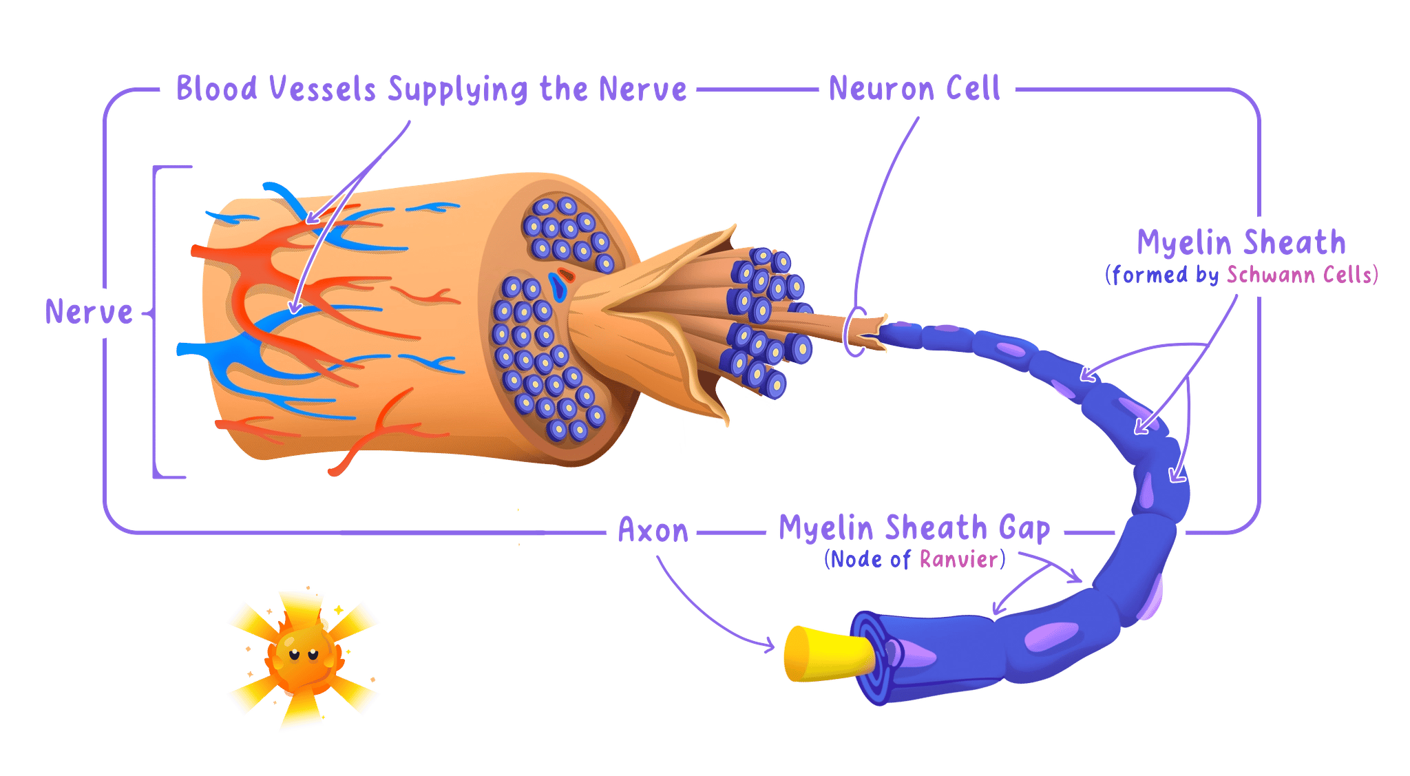
The Myelin Sheath
Myelin sheath
A fatty layer that insulates the axon of a neurone and increases the speed at which the electrical impulse travels.
- Many axons are wrapped in a myelin sheath, a fatty layer made by cells that coil around the fibre.
- The sheath acts as an electrical insulator, so the impulse cannot leak out through the membrane.
- The sheath is broken by regular gaps, and the impulse jumps from one gap to the next instead of travelling along the whole membrane.
- This makes the impulse travel much faster, and a myelinated neurone can conduct at over 100 metres per second while an unmyelinated one manages about 1 metre per second.
- Myelin also stops impulses crossing between neighbouring axons in the same nerve, so signals stay separate.
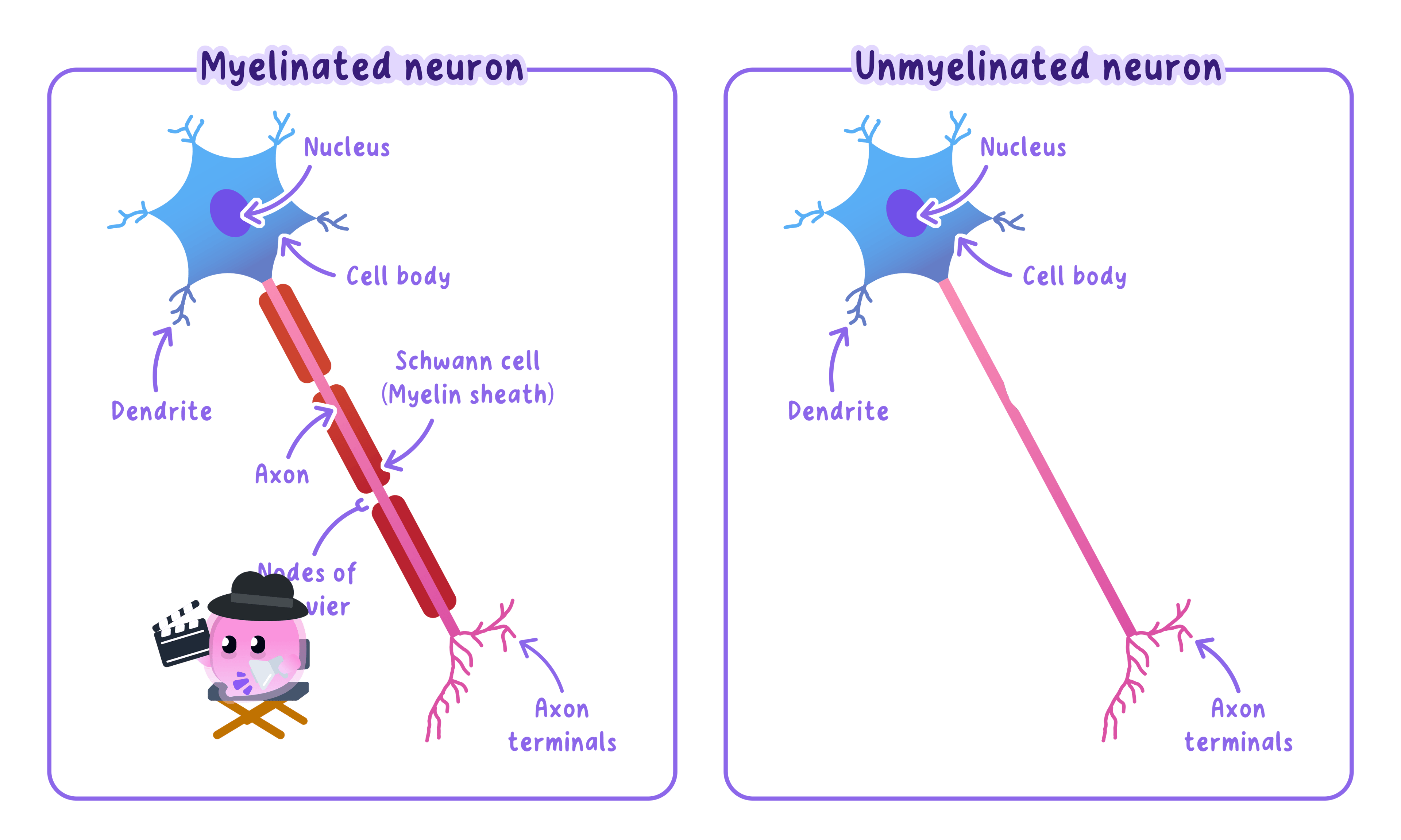
In multiple sclerosis the immune system destroys patches of myelin. Impulses then travel slowly or fail completely, which causes weakness, numbness and problems with coordination.
Effectors Carry Out the Response
Effector
A muscle or a gland that carries out the response, by contracting or by secreting a substance.
- An effector is a muscle or a gland.
- A muscle responds by contracting, which produces movement.
- A gland responds by secreting a substance such as a hormone, sweat or an enzyme.
- The impulse itself does not do anything useful until it reaches an effector, so the effector is what converts the signal into a response.
Synapses
Synapse
The junction between two neurones, where the impulse is carried across the gap by a neurotransmitter.
- Neurones do not physically touch, so there is a tiny gap between the end of one neurone and the start of the next.
- The junction, including the gap, is called a synapse.
- An electrical impulse cannot jump the gap, so the signal must be carried across in a different way.
- The signal is passed across by a chemical, which means transmission is electrical along the neurone and chemical at the synapse.
How an Impulse Crosses a Synapse
Neurotransmitter
A chemical released into a synapse that diffuses across the gap and binds to receptors on the next neurone, triggering a new electrical impulse.
- The electrical impulse arrives at the end of the first neurone.
- This causes tiny sacs called vesicles to move to the membrane and release a neurotransmitter into the gap.
- The neurotransmitter diffuses across the gap, which is fast because the gap is extremely small.
- It binds to receptor molecules on the membrane of the next neurone, and the receptors have a shape complementary to the neurotransmitter.
- Binding triggers a new electrical impulse in the second neurone, and the impulse continues on its way.
- The neurotransmitter is then broken down or reabsorbed, so the second neurone is not stimulated continuously.
Why Synapses Are Useful
- Vesicles are only present on one side of the synapse, so the impulse can travel in one direction only.
- One neurone can form synapses with many others, which allows a single input to trigger several different responses.
- Several weak inputs can be added together at a synapse, so a response happens only when the stimulus is strong enough to matter.
- The cost is a slight delay, because diffusion takes longer than conduction along an axon, which is why a pathway with fewer synapses is faster.
Do not describe the impulse as jumping across the synapse. The electrical impulse stops at the gap, and a chemical carries the signal across by diffusion.
Measuring the Speed of a Nervous Response
Investigation: The Speed of a Nervous Response
- Aim: To estimate how fast an impulse travels through the nervous system by timing a hand squeeze passed along a chain of people. Edexcel lists this as a suggested practical for Topic 2 rather than as one of the eight core practicals.
- Apparatus: A group of at least 10 volunteers, a stop clock reading to 0.01 s, a tape measure, a blindfold or a screen, a calculator.
- Method:
- Stand the volunteers in a line or a circle and have everyone hold the hand of the person on each side.
- Ask everyone except the first person to close their eyes, so nobody can see the squeeze coming and anticipate it.
- The first person starts the stop clock and immediately squeezes the hand of the next person.
- Each person squeezes the next hand as soon as they feel their own hand squeezed.
- The last person in the chain calls out, and the first person stops the clock.
- Repeat the whole run five times and calculate the mean time.
- Measure the length of the nerve pathway in each person from one hand, up the arm, across the shoulders and down to the other hand, then add these lengths together for the whole chain.
- Variables: The independent variable is the total length of the nerve pathway, set by the number of people in the chain, and the dependent variable is the time taken. Keep the same volunteers, the same hand, the eyes closed and the same instruction throughout.
- Results: The time increases as more people join the chain. Repeating the run several times usually gives a shorter mean time, because the volunteers learn to anticipate the squeeze, which is a source of error rather than a real change in nerve speed.
- Maths: Speed is calculated from the total distance and the mean time. speed=total distancemean time\text{speed} = \frac{\text{total distance}}{\text{mean time}}speed=mean timetotal distance For a chain of 10 people with a total pathway of 15 m and a mean time of 2.5 s, the calculated speed is 6 m/s6\ \text{m/s}6 m/s.
- Watch out: The calculated value is far lower than the true speed of an impulse, because most of the time is taken up by the synapses in each person's central nervous system and by the reaction time of the brain, not by conduction along the axons. Anticipation is the other major error, and it is reduced by closing eyes and by varying the interval before each run.
- Safety: Low hazard. Make sure nobody with a hand or wrist injury takes part, and squeeze gently.
- Learn the response pathway as a fixed chain and write it in order, because each correct step in stimulus, receptor, sensory neurone, CNS, motor neurone, effector, response is separately creditable.
- For a question on how an impulse crosses a synapse, the four marking points are the release of neurotransmitter, diffusion across the gap, binding to receptors, and a new impulse in the next neurone.
- State the direction of travel when you name a neurone, since a sensory neurone carries impulses to the CNS and a motor neurone carries them away from it.
- If asked why the myelin sheath is useful, give both the insulation and the increase in speed, because one alone is often only worth one mark.
- Write out the full pathway of a nervous response from stimulus to response.
- State the function of a sensory neurone, a relay neurone and a motor neurone.
- Give two functions of the myelin sheath.
- Describe how an impulse is transmitted across a synapse.
- Explain why a synapse allows an impulse to travel in one direction only.
2.3.5 Structure and function of a reflex arc
What a Reflex Action Is
Reflex action
A rapid, automatic response to a stimulus that happens without conscious thought.
- A reflex action is a rapid, automatic response to a stimulus that happens without conscious thought.
- It is involuntary, so it happens whether or not you want it to and you cannot stop it by deciding not to.
- Every person shows the same response to the same stimulus, because the pathway is not learned.
- Most reflexes are protective, and they prevent injury by acting before the brain has processed what is happening.
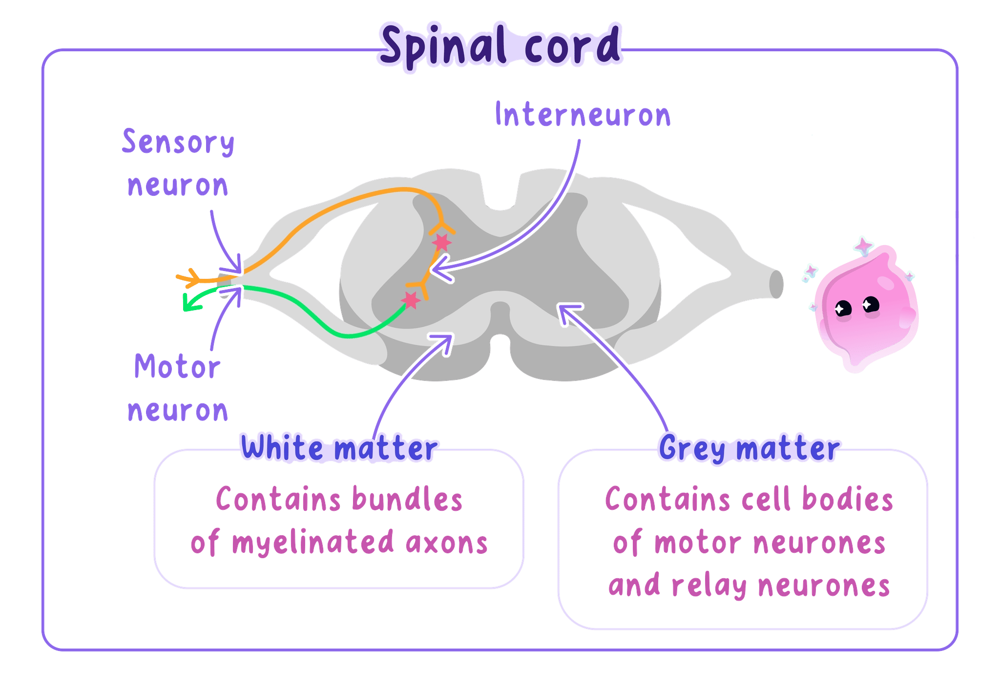
The Reflex Arc
Reflex arc
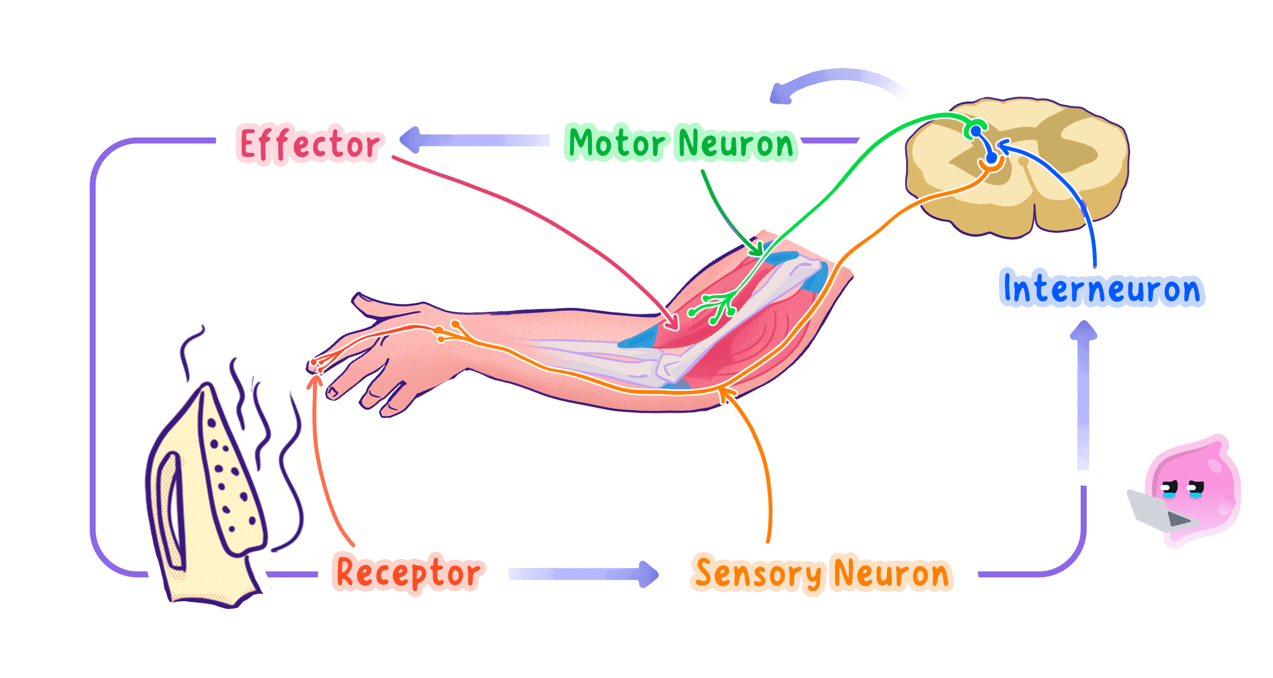
The pathway of receptor, sensory neurone, relay neurone, motor neurone and effector that brings about a reflex action.
- The reflex arc is the pathway of neurones that produces a reflex action.
- A stimulus is detected by a receptor, which converts it into an electrical impulse.
- A sensory neurone carries the impulse from the receptor to the central nervous system, usually the spinal cord.
- A relay neurone inside the CNS passes the impulse straight across to a motor neurone.
- A motor neurone carries the impulse out to the effector.
- The effector, a muscle or a gland, produces the response by contracting or by secreting.
- The impulse crosses a synapse between each pair of neurones, and a neurotransmitter carries it over each gap.
Why the Brain Is Bypassed
- The impulse travels through the spinal cord and does not pass through the conscious part of the brain first.
- Missing out the brain means fewer synapses, and each synapse adds a delay because the neurotransmitter has to diffuse across the gap.
- A shorter pathway means a shorter reaction time, so the response happens before serious damage is done.
- No decision is needed, so nothing is gained by involving conscious thought.
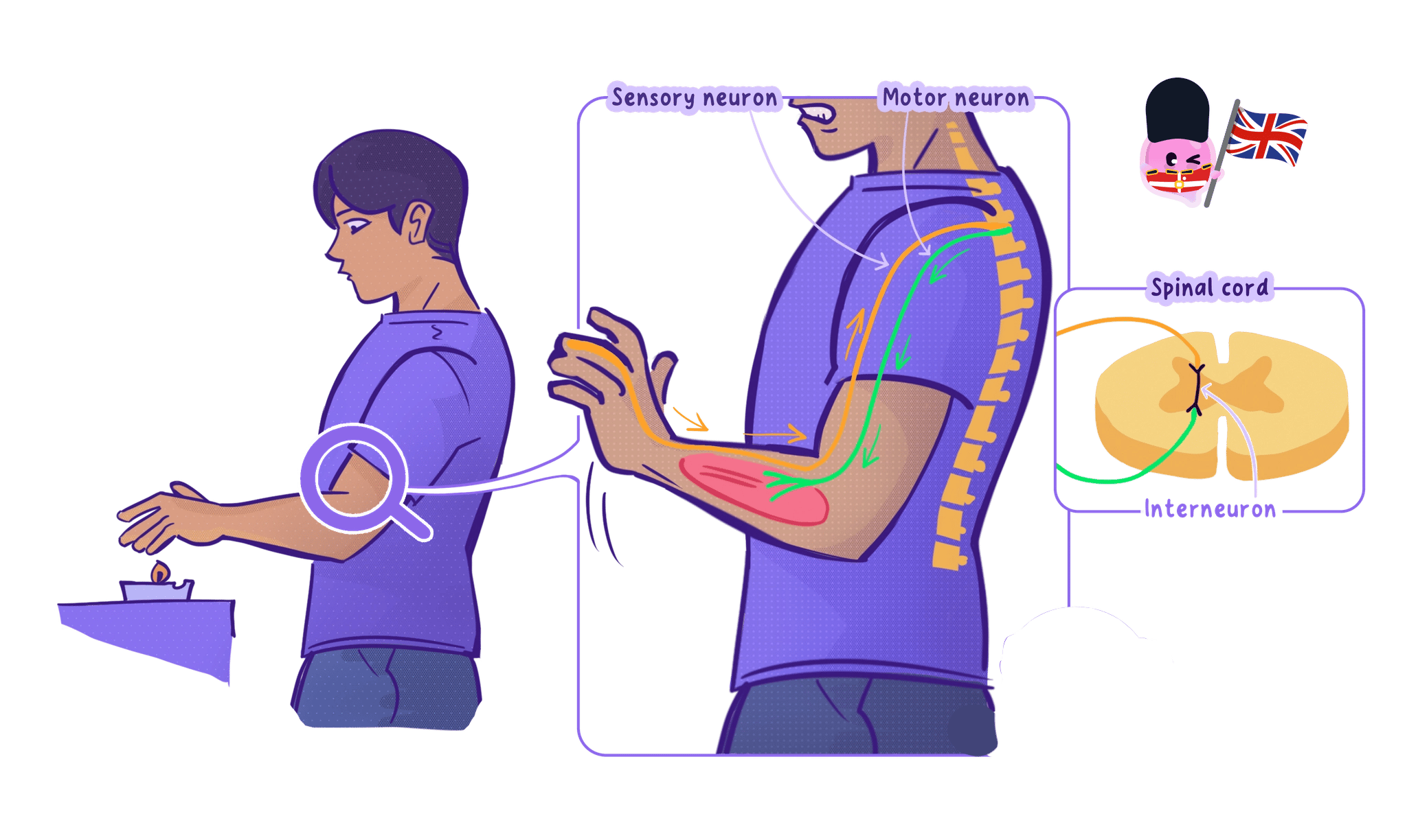
- An impulse is still sent up to the brain, which is why you feel the pain a moment after your hand has already moved.
The withdrawal is faster than the pain because the movement and the sensation travel by different routes, and the reflex route is the shorter one.
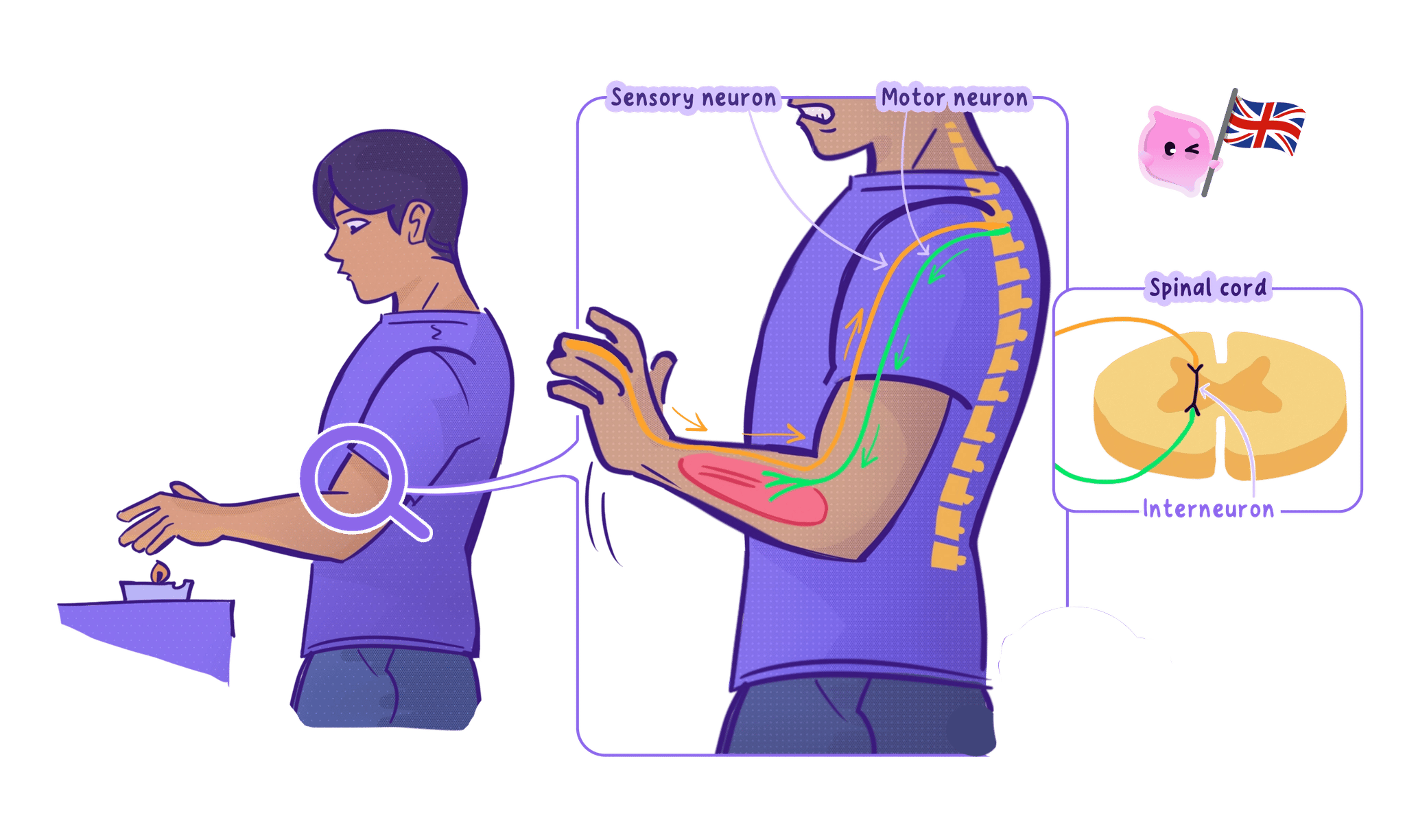
When you touch a hot object
- Stimulus. You touch a hot oven tray, and the heat is the stimulus.
- Receptor. Temperature and pain receptors in the skin of the finger detect it and generate an impulse.
- Sensory neurone. The impulse travels up the arm to the spinal cord.
- Relay neurone. In the spinal cord the impulse is passed across to a motor neurone.
- Motor neurone. The impulse travels back down the arm to the biceps muscle.
- Effector and response. The biceps contracts and the hand is pulled away from the tray.
Other Reflexes in the Body
- The pupil reflex. In bright light the iris contracts the pupil, which protects the retina from damage.
- Blinking. An object approaching the eye triggers the eyelid to close and shield the cornea.
- The knee jerk. Tapping the tendon below the knee makes the thigh muscle contract, which helps to maintain posture.
- Coughing and sneezing. Irritants in the airways trigger a sharp expulsion of air that clears them.
- Some reflexes are coordinated by the brain rather than the spinal cord, and the pupil reflex and blinking are both handled in the brain.
Do not write that a reflex does not involve the nervous system or the brain at all. A reflex uses the nervous system throughout, and the brain is bypassed only in the decision, not in the pathway.
Investigation: Measuring Reaction Time
- Aim: To measure reaction time using the ruler drop test and to find whether a factor such as caffeine, practice or a distraction changes it. Edexcel lists investigating reaction times and investigating human responses to external stimuli as suggested practicals for Topic 2.
- Apparatus: A 30 cm ruler, a chair and a table with a straight edge, a volunteer and a partner, a results table, a conversion chart or calculator.
- Method:
- The volunteer sits with their forearm flat along the table so the hand hangs over the edge, which stops them lowering the whole arm to catch the ruler.
- The partner holds the ruler vertically, with the zero mark level with the top of the volunteer's thumb and first finger and a gap of about 1 cm between them.
- The partner drops the ruler without warning and without a countdown, so the volunteer cannot anticipate it.
- The volunteer catches the ruler as fast as they can and reads the measurement level with the top of the thumb.
- Record the distance in centimetres and repeat 10 times, discarding any drop the volunteer missed.
- Calculate the mean catch distance.
- Repeat the whole set for the second condition, for example 20 minutes after a caffeinated drink, using the same volunteer.
- Variables: The independent variable is the condition being tested, such as caffeine or no caffeine, and the dependent variable is the distance the ruler falls before it is caught. Keep the same volunteer, the same hand, the same ruler, the same starting gap and the same seated position, and vary the interval before each drop.
- Results: A shorter catch distance means a shorter reaction time. Distances typically fall between 10 cm and 25 cm, and the mean usually shortens over the first few attempts as the volunteer gets used to the task.
- Maths: Calculate the mean before you compare conditions. mean distance=sum of the distancesnumber of drops\text{mean distance} = \frac{\text{sum of the distances}}{\text{number of drops}}mean distance=number of dropssum of the distances
- The distance can be converted to a time using t=2dgt = \sqrt{\frac{2d}{g}}t=g2d
- where ddd is the distance in metres and ggg is 9.8 m/s29.8\ \text{m/s}^29.8 m/s2. A catch at 0.20 m0.20\ \text{m}0.20 m gives a reaction time of about 0.20 s0.20\ \text{s}0.20 s.
- Watch out: Any countdown or eye contact lets the volunteer anticipate the drop, which measures prediction instead of reaction. Comparing two different people confounds the result with their natural differences, so use the same volunteer for both conditions.
- Safety: Low hazard. Keep the ruler away from faces and make sure the table edge is clear.
- The six mark question on this topic is marked by levels, and the top level needs the structural components linked in a complete pathway and then linked to the function of the reflex as a rapid protective response.
- Write the pathway in strict order and name every component, because a missing relay neurone or effector breaks the chain examiners are looking for.
- Bring in the myelin sheath speeding up transmission and the small number of synapses if you need extra detail for the higher levels.
- Finish with the function rather than stopping at the structure, saying that the response is rapid, automatic and protects the body from damage.
- Define a reflex action.
- List the parts of a reflex arc in order from stimulus to response.
- Explain why a reflex action is faster than a voluntary response.
- Why do you feel the pain only after you have pulled your hand away?
- Give two examples of a protective reflex other than the withdrawal reflex.
