2.1.1 Mitosis and the cell cycle
The Cell Cycle
Cell cycle
The repeating sequence of interphase, mitosis and cytokinesis by which a body cell grows, copies its DNA and divides into two genetically identical daughter cells.
- Body cells do not divide continuously. They pass through a controlled, repeating sequence of events called the cell cycle.
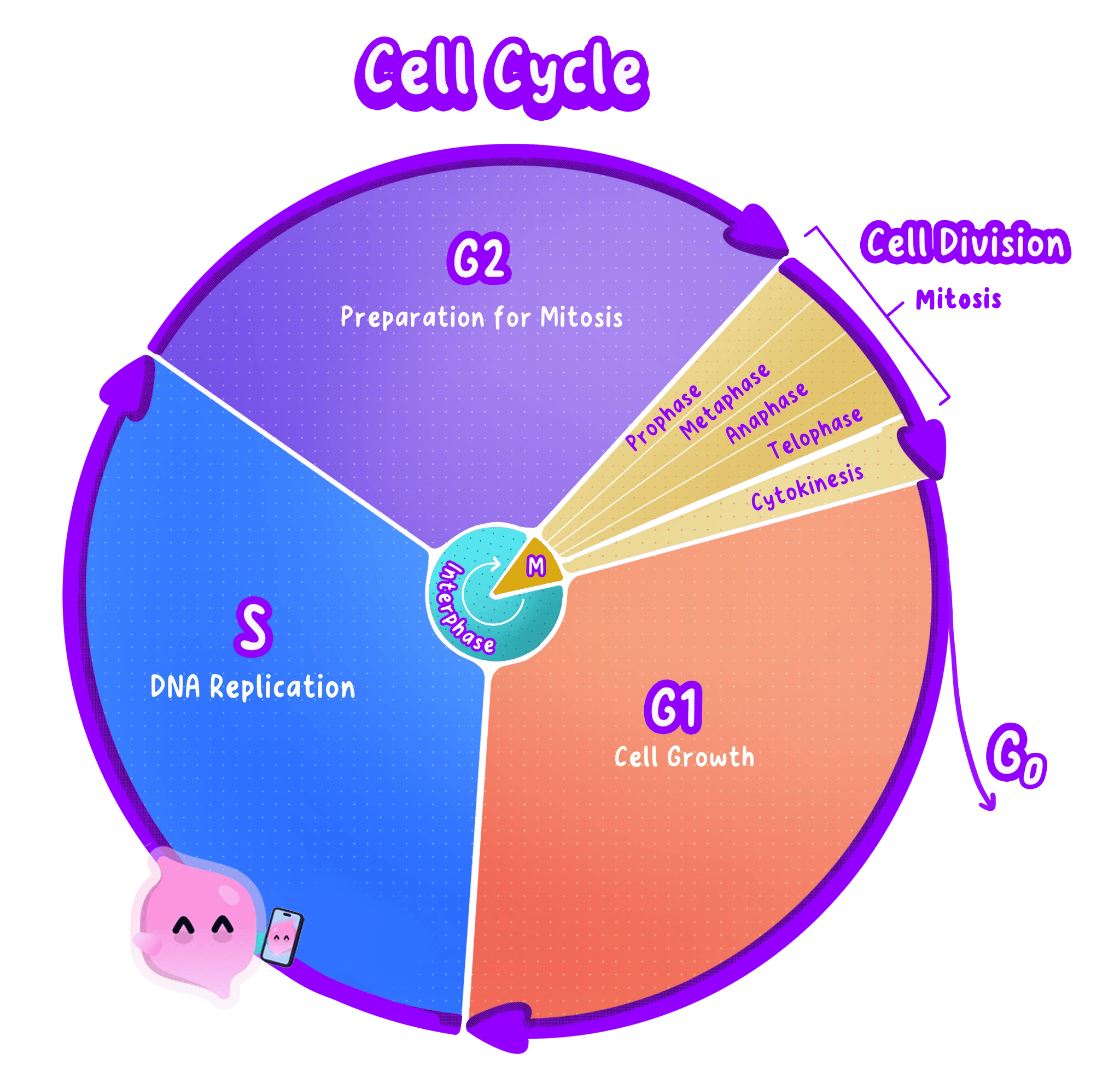
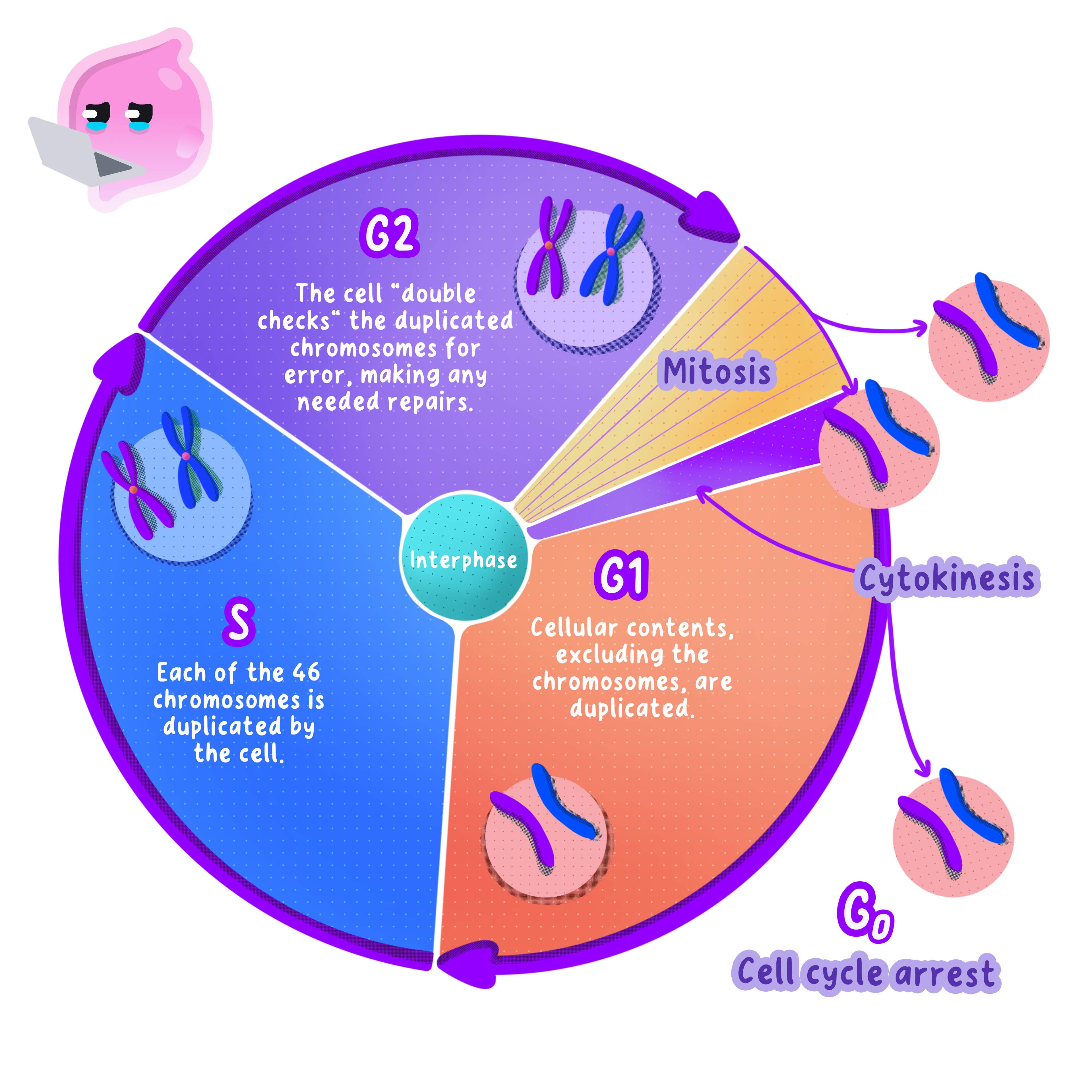
- The cell cycle has three parts: a long growth stage called interphase, then mitosis, and finally cytokinesis.
- Interphase takes up most of the cycle, often around 90 per cent of the total time in a human body cell.
- Mitosis and cytokinesis together make up the much shorter division stage, in which one parent cell becomes two.
- A cell that is not preparing to divide leaves the cycle and simply carries out its job, which is what most mature nerve and muscle cells do.
Interphase
Interphase
The stage of the cell cycle before mitosis, in which the cell grows, makes extra sub-cellular structures and replicates its DNA so that each chromosome becomes two identical sister chromatids.
- During interphase the cell grows and increases its number of sub-cellular structures, including mitochondria and ribosomes.
- More mitochondria mean more respiration, which supplies the ATP needed for the energy-demanding work of dividing.
- The DNA replicates, so that every chromosome is copied exactly.
- The cell also carries out its normal functions throughout interphase, so a liver cell in interphase is still making enzymes and storing glycogen.
Interphase is a stage of the cell cycle, not a stage of mitosis, so an answer that lists interphase as part of mitosis loses the mark.
Chromosomes Are Copied Before Division
Chromosome
A long molecule of coiled DNA found in the nucleus, carrying many genes in a fixed order.
- A human body cell contains 46 chromosomes, arranged as 23 pairs.
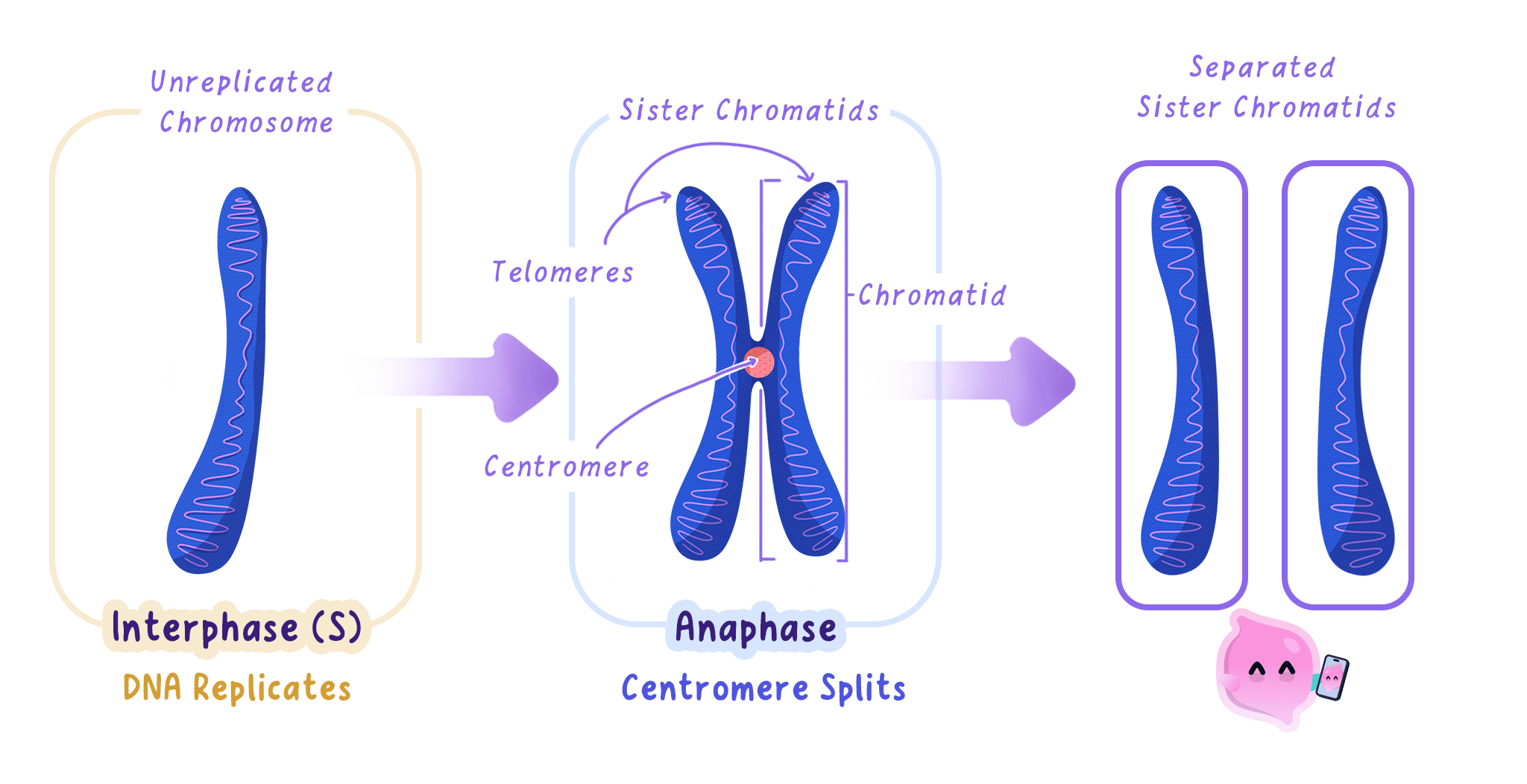
- When DNA replicates, each chromosome becomes two identical copies called sister chromatids.
- The two chromatids stay joined at a narrow region called the centromere, which is why a copied chromosome is drawn as an X shape.
- Because the copies are identical, both new cells can be given a complete and identical set of genetic information.
Do not describe replication as the chromosome splitting in half. Replication doubles the DNA, so the cell temporarily holds twice as much genetic material, not half as much.
Prophase
Sister chromatids
The two identical copies of a replicated chromosome, joined at the centromere until they are pulled apart during mitosis.
- The copied chromosomes condense, coiling up until they are short and thick enough to be seen under a light microscope.
- Each one is now visible as two sister chromatids joined at the centromere.
- The nuclear membrane breaks down, releasing the chromosomes into the cytoplasm.
- Spindle fibres begin to form from opposite ends, or poles, of the cell.
Metaphase
- The chromosomes are moved so that they line up along the equator, which is the imaginary line across the middle of the cell.
- Spindle fibres from each pole attach to the centromere of every chromosome.
- Lining the chromosomes up in a single row is what makes an equal separation possible in the next phase.
The names give the order away: chromosomes take their place in the Middle at Metaphase and move Apart at Anaphase.
Anaphase
- Each centromere divides, so the two sister chromatids are no longer held together.
- The spindle fibres shorten and pull one chromatid from each pair to opposite poles of the cell.
- Once separated, each chromatid counts as a chromosome in its own right.
- This gives each pole one complete set of chromosomes, identical to the set the parent cell started with.
Telophase
- The chromosomes reach the poles and begin to uncoil, so they can no longer be seen individually.
- A new nuclear membrane forms around each set, producing two nuclei inside one cell.
- At the end of telophase the nucleus has divided but the cell has not, so mitosis is complete and cytokinesis has yet to happen.
Mitosis is the division of the nucleus. The division of the whole cell needs cytokinesis as well.
Cytokinesis
Cytokinesis
The final stage of the cell cycle, in which the cytoplasm and cell membrane divide to form two separate daughter cells.
- The cytoplasm and the cell membrane divide to separate the two nuclei into two cells.
- In an animal cell the membrane pinches inwards at the equator until the cell splits in two.
- In a plant cell a new cell wall and membrane are built across the middle instead, because the rigid wall cannot pinch inwards.
- The result is two daughter cells, each with its own nucleus, cytoplasm and cell membrane.
Do not confuse mitosis with meiosis. Mitosis produces two genetically identical diploid cells for growth and repair, while meiosis produces four genetically different haploid gametes.
Why Mitosis Matters
- Growth. A fertilised egg becomes a multicellular organism because mitosis repeatedly increases the number of cells.
- Repair and replacement. Skin, gut lining and red blood cells are worn out or lost constantly, and mitosis replaces them with identical cells.
- Around two million red blood cells are replaced every second in an adult human, all of them produced by mitosis in the bone marrow.
- Asexual reproduction. Organisms that reproduce from a single parent use mitosis, so their offspring are genetically identical clones.
- A strawberry plant grows runners that root and form new plants, and each new plant carries exactly the same alleles as the parent.
A cut on the skin heals because cells at the edge of the wound divide by mitosis, producing identical skin cells that close the gap.
Observing Mitosis Under the Microscope
Investigation: Observing Mitosis in a Root Tip
- Aim: To prepare a stained root tip squash and identify cells in each phase of mitosis, then calculate the mitotic index. This is a standard microscopy investigation rather than one of the eight Edexcel core practicals, but Edexcel does set method and calculation questions on it.
- Apparatus: Germinating garlic or onion roots, watch glass, 1 mol/dm3 hydrochloric acid, warm water bath at 60 degrees Celsius, toluidine blue or acetic orcein stain, scalpel, mounted needle, forceps, microscope slide and coverslip, filter paper, light microscope, eye protection.
- Method:
- Cut the tip 5 mm from the end of a growing root, because only the meristem at the very tip is actively dividing and the rest of the root gives you almost nothing to look at.
- Place the tip in a watch glass of hydrochloric acid in a 60 degrees Celsius water bath for about 5 minutes, to break down the middle lamella between the cells so the tissue will spread out into a single layer.
- Rinse the tip in cold water and blot it dry.
- Transfer it to a slide and add two or three drops of stain, which binds to DNA and makes the chromosomes stand out dark against pale cytoplasm.
- Lower a coverslip at an angle with a mounted needle, so air is pushed out ahead of it rather than trapped as bubbles.
- Cover with filter paper and press down firmly and vertically with your thumb, without twisting, to squash the tissue into a layer one cell thick.
- View under low power to find the region where the cells are small and square, then switch to high power and count.
- Variables: The independent variable is the region of the root you examine and the dependent variable is the proportion of cells in mitosis. Keep the species, the age of the root, the temperature, the staining time and the total number of cells counted the same.
- Results: Most cells are in interphase, with a large round nucleus and no visible chromosomes. A smaller number show dark, condensed chromosomes: scattered in prophase, in a line across the middle in metaphase, in two separated groups in anaphase, and as two clusters with re-forming nuclei in telophase.
- Maths: The mitotic index is the proportion of cells that are in mitosis, given by mitotic index=number of cells in mitosistotal number of cells counted\text{mitotic index} = \frac{\text{number of cells in mitosis}}{\text{total number of cells counted}}mitotic index=total number of cells countednumber of cells in mitosis Multiply by 100 to express it as a percentage. Counting at least 100 cells and repeating on several fields of view reduces the effect of an unrepresentative patch of tissue.
- Watch out: Pressing down while twisting rolls the cells over each other and tears them, so the chromosomes cannot be assigned to a phase. Squashing too little leaves the tissue several cells deep, which will not come into focus at high power.
- Safety: Hydrochloric acid is an irritant, so wear eye protection and rinse any splashes immediately. Cut the root on a tile with the blade moving away from your fingers.
- When a question says describe what happens during mitosis, track the chromosomes through the four phases rather than listing the phase names, because the marks are for chromosome behaviour.
- Use the wording examiners credit: chromosomes condense, line up along the equator, are pulled apart at the centromere by spindle fibres, and gather at each pole.
- If the question asks for the stages of the cell cycle rather than mitosis, the answer is interphase, mitosis and cytokinesis, and naming prophase to telophase alone will not gain the mark.
- In a data question about the length of each phase, use the proportion of cells seen in a phase as a measure of how long that phase lasts.
- Name the three stages of the cell cycle in order.
- What two things happen to a cell during interphase?
- In which phase of mitosis do the chromosomes line up along the equator of the cell?
- What happens to the centromeres during anaphase?
- Give three roles of mitosis in a living organism.
2.1.2 Division producing identical daughter cells
What Mitosis Produces
Mitosis
The type of nuclear division that produces two daughter nuclei with chromosomes identical to those of the parent cell, used in growth, repair and asexual reproduction.
- One division by mitosis produces exactly two daughter cells from one parent cell.
- Each daughter cell receives an identical set of chromosomes to the set in the parent cell nucleus.
- Because the chromosomes carry the genes, the daughter cells also carry identical alleles, so they are genetically identical.
- The daughter cells are body cells, not gametes, and Edexcel refers to them as diploid body cells.
Diploid Body Cells
Diploid
Describes a cell that contains two copies of each chromosome, which in humans is 46 chromosomes in 23 pairs.
- A diploid cell contains two full sets of chromosomes, one set from each parent, so the chromosomes exist as matching pairs.
- In humans the diploid number is 46, made up of 23 homologous pairs.
- Mitosis keeps the diploid number constant, so a human body cell that divides gives two cells with 46 chromosomes each.
- Gametes are the exception. They are haploid with 23 chromosomes, and they are made by meiosis rather than mitosis.
Do not write that the chromosome number is halved during mitosis. Halving happens in meiosis, and an answer that says meiosis when the question is about growth or repair is rejected outright.
Why the Daughter Cells Are Identical
Daughter cell
Either of the two new cells produced when a parent cell divides.
- The DNA is replicated during interphase, before division begins, so every chromosome exists as two identical copies.
- At metaphase the copied chromosomes line up along the equator in a single row.
- At anaphase the two copies of each chromosome are separated and one copy is pulled to each pole.
- This means each pole ends up with one complete copy of every chromosome, which is why the two nuclei are identical to each other and to the original.
- Nothing is swapped between chromosomes during mitosis, so no new combinations of alleles are produced and there is no genetic variation between the daughter cells.
Mitosis is a copying process, so the only source of difference between the daughter cells is a mutation that slips through during replication.
Mitosis in Growth and Repair
- An organism grows by producing more cells, and in animals those extra cells come from mitosis.
- Identical cells are exactly what growth and repair need, because a replacement skin cell must do the same job as the cell it replaced.
- Damaged tissue is repaired the same way, so the new cells fit into the existing tissue and function normally.
- Cells that divide often, such as those lining the small intestine, are replaced every few days by mitosis.
Mitosis in Asexual Reproduction
Asexual reproduction
Reproduction involving only one parent and no fusion of gametes, producing offspring that are genetically identical to the parent.
- In asexual reproduction a single parent produces offspring by mitosis, so the offspring are clones of the parent.
- There are no gametes and no fertilisation, so no genetic material is combined from two parents.
- Spider plants produce plantlets on long stems, and each plantlet carries the same alleles as the parent plant.
- A hydra reproduces by growing a bud on its side, which detaches as a genetically identical individual.
Bacteria reproduce asexually by binary fission rather than by mitosis, because they have no nucleus. Edexcel mark schemes accept binary fission and cloning as answers about asexual reproduction, but reject meiosis.
The Advantage and the Cost of Identical Offspring
- Identical offspring are produced quickly and without the need to find a mate, so a well-adapted organism can colonise a favourable habitat rapidly.
- Every offspring inherits the parent's useful characteristics, which is why growers propagate fruit trees and potatoes from cuttings and tubers rather than seed.
- The cost is a complete lack of variation within the population.
- If the environment changes or a new disease appears, no individual has an allele that gives resistance, so the whole population is at risk.
- When a question asks how two cells produced by mitosis compare, the marks are usually for two separate points: they are genetically identical, and they are identical to the parent cell.
- If the question gives a chromosome number for the parent cell, state the same number for each daughter cell and say that this is because the DNA was replicated beforehand.
- Watch the command word. Describe wants what happens, while explain wants the replication and separation steps that make identical cells possible.
- In an advantages and disadvantages question about asexual reproduction, give at least one of each and link the disadvantage to the lack of variation.
- How many daughter cells are produced by one mitotic division?
- What does diploid mean, and what is the diploid number in humans?
- Explain why the two daughter cells produced by mitosis are genetically identical.
- Name one organism that reproduces asexually and state what its offspring are called.
- Give one disadvantage of a population made up of genetically identical individuals.
2.1.3 Cancer and uncontrolled cell division
Cancer Is Uncontrolled Cell Division
Cancer
A disease in which changes to cells cause uncontrolled cell division, producing a tumour.
- In a healthy tissue the cell cycle is tightly controlled, and cells divide only when a new cell is needed.
- Cancer begins when changes inside a cell damage the genes that control this cycle.
- The cell then divides repeatedly and uncontrollably, ignoring the signals that would normally stop it.
- The damaged control is passed on to every daughter cell, so the population of faulty cells doubles again and again.
How a Tumour Forms
Tumour
A mass of cells produced by uncontrolled cell division.
- The dividing cells build up into a mass of cells called a tumour.
- Cells inside a growing tumour need a supply of oxygen and glucose, so a tumour triggers the growth of new blood vessels towards itself.
- A tumour can press on nearby organs, blood vessels or nerves, which is often what produces the first symptoms.
- Tumours are classified as benign or malignant, and only malignant tumours are described as cancerous.
Benign Tumours
Benign tumour
A tumour that stays in one place inside a membrane and does not invade other tissues.
- A benign tumour grows in one place and is usually contained within a membrane.
- Its cells do not invade surrounding tissue and do not spread to other parts of the body.
- Benign tumours are not classed as cancer and can often be removed by surgery.
- They can still be dangerous if they grow in a confined space, which is why a benign brain tumour can press on the brain and cause serious harm.
Malignant Tumours
Malignant tumour
A cancerous tumour whose cells invade neighbouring tissue and spread in the blood to form secondary tumours elsewhere in the body.
- A malignant tumour is cancerous, and its cells invade the healthy tissue around it.
- Cells break off and are carried in the blood or the lymph to other parts of the body.
- Where they settle they divide again and form secondary tumours, and this spread is called metastasis.
- Secondary tumours are what make malignant cancers difficult to treat, because removing the original tumour no longer removes the disease.
Do not treat tumour and cancer as the same word. Every cancer is a tumour, but a benign tumour is not a cancer, and the difference that earns the mark is whether the cells spread.
What Causes the Changes in the Cells
Mutation
A random change in the sequence of bases in DNA.
- The changes are mutations in the genes that control cell division.
- Mutations happen at random during DNA replication, and most are harmless or are repaired.
- A mutation that damages a control gene may allow the cell cycle to run without a brake.
- Cancer usually requires several mutations to build up in the same cell, which is why the risk rises with age.
Carcinogens and Risk Factors
Carcinogen
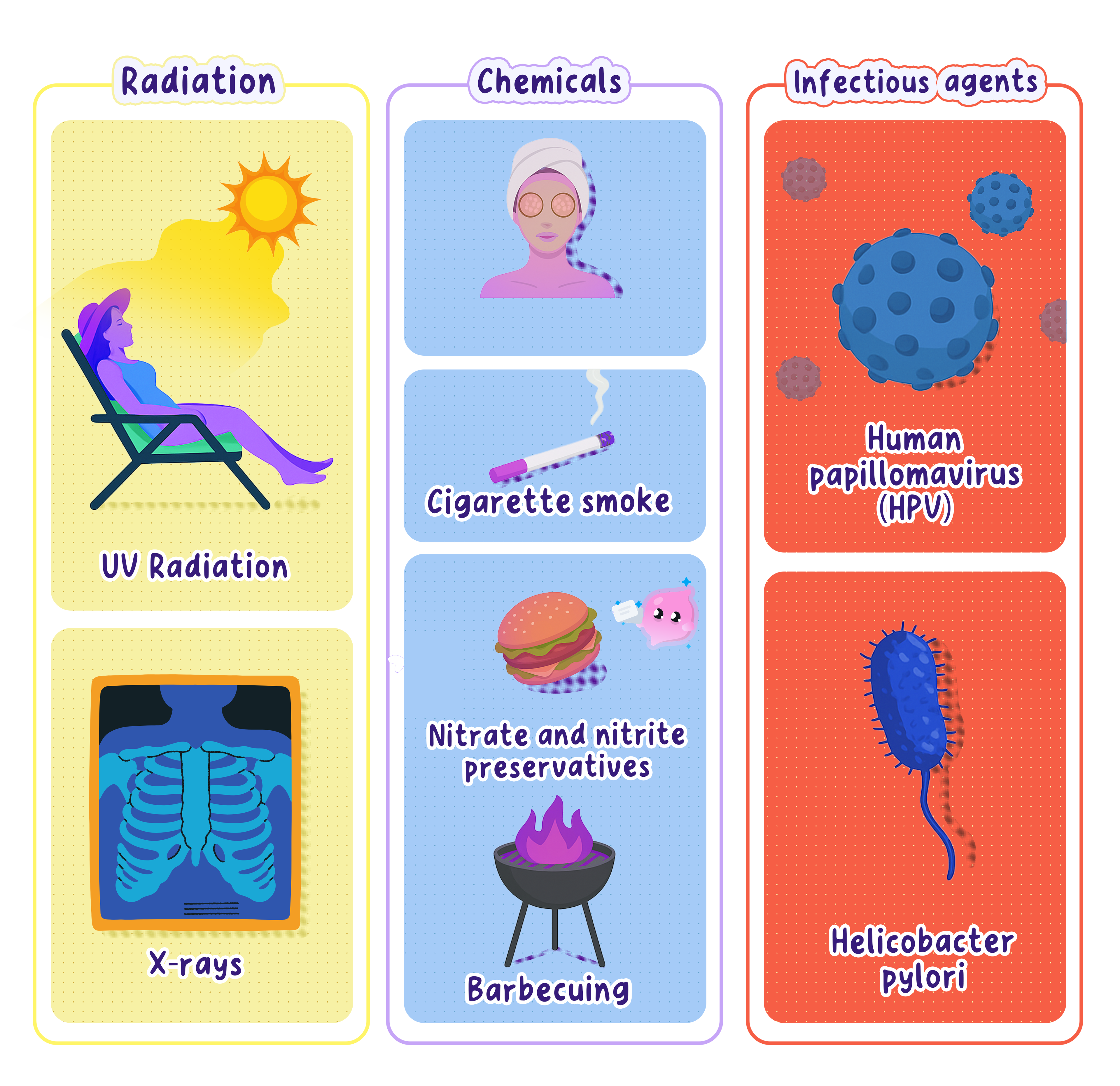
Any agent that increases the risk of cancer by causing mutations in DNA, such as the tar in tobacco smoke or ultraviolet light.
- A carcinogen is any agent that increases the rate of mutation and therefore the risk of cancer.
- Ionising radiation, including X-rays, gamma rays and the ultraviolet radiation in sunlight, damages DNA directly.
- Chemical carcinogens include the tar in tobacco smoke, asbestos fibres and some industrial solvents.
- Some viruses raise the risk, and human papillomavirus is linked to cervical cancer.
- Inherited alleles also matter, and a person who inherits a faulty BRCA1 allele has a higher risk of breast cancer.
- Lifestyle factors such as obesity and high alcohol intake raise the risk of several cancers.
A risk factor is not a cause. Exposure to a carcinogen raises the probability of cancer, so an answer must say increases the risk of rather than causes.
Interpreting Cancer Data
- Exam data on cancer is usually given as an incidence rate, meaning new cases per 100 000 people per year, so populations of different sizes can be compared.
- A correlation between a factor and cancer incidence does not on its own prove that the factor causes the cancer.
- Stronger evidence comes from a dose response, where a larger exposure is matched by a larger increase in risk.
- Percentage change is a common calculation in these questions.
- percentage change=final value−initial valueinitial value×100\text{percentage change} = \frac{\text{final value} - \text{initial value}}{\text{initial value}} \times 100percentage change=initial valuefinal value−initial value×100
- Multiple choice items define cancer as uncontrolled cell division, and the distractors swap in controlled cell division or uncontrolled organ division, so read every option to the end.
- When asked to compare benign and malignant tumours, give a paired comparison: benign stays in one place while malignant invades and spreads to form secondary tumours.
- In a data question, quote figures from the graph or table with their units and state the trend before you interpret it.
- If asked to evaluate whether a factor causes cancer, say that the data shows a correlation and name one further piece of evidence that would be needed.
- Define cancer in one sentence.
- State two differences between a benign tumour and a malignant tumour.
- What is a carcinogen? Give two examples.
- What name is given to the spread of cancer cells to another part of the body?
- Why does the risk of developing cancer increase with age?
2.1.4 Growth in animals and plants
What Growth Is
Growth
A permanent increase in the size or dry mass of an organism, brought about by cell division, cell enlargement and differentiation.
- Growth is a permanent increase in size or dry mass, and in a multicellular organism it is brought about by three processes.
- Cell division increases the number of cells, and in body cells this is mitosis.
- Cell enlargement, called elongation in plants, makes each cell bigger.
- Differentiation turns unspecialised cells into the specialised cells the organism needs.
- Animals and plants use all three processes, but they use them at different times and in different places.
Growth in Animals
Differentiation
The process by which an unspecialised cell switches on particular genes and develops the structures it needs to carry out one specific job.
- Animals grow mainly by cell division followed by differentiation.
- Most differentiation happens early, in the embryo and in the young animal, so an adult has very few cells left that can differentiate.
- Growth in animals happens all over the body rather than in a few fixed regions.
- Growth stops once the animal reaches its adult size, and it does not usually restart.
- Cell division continues in the adult, but almost entirely for repair and replacement rather than growth.
A human infant triples its birth mass in the first year, then growth slows, accelerates again during the adolescent growth spurt, and stops when the growth plates in the long bones fuse.
Growth in Plants
Cell elongation
The stage of plant growth in which a newly made cell absorbs water into its vacuole and gets longer, making the root or shoot grow.
- Plants grow by cell division, then cell elongation, then differentiation.
- Cell division happens only at the meristems, which sit at the tips of roots and shoots and in the cambium.
- Just behind the meristem the new cells elongate, taking water into the vacuole so the cell gets longer.
- Elongation is what actually pushes a root down through the soil and a shoot up towards the light, because it adds far more length than division alone.
- Behind the elongation region the cells differentiate into xylem vessels, phloem, root hair cells and the rest.
- Plants grow throughout their whole lives and keep the ability to differentiate, which is why a cutting can grow into a complete new plant.
Elongation is not the same as division. Elongation makes an existing cell longer by taking in water, and the number of cells does not change.
Comparing Animal and Plant Growth
- Where. Animal growth happens throughout the body, while plant growth is confined to the meristems.
- When. Animal growth stops at maturity, while plant growth continues for life.
- How. Animals rely on cell division and differentiation, while plants add cell elongation as the main contributor to length.
- Differentiation. Most animal cells lose the ability to differentiate, while plant cells largely keep it.
Measuring Growth
- Length or height is quick and does not harm the organism, but it measures only one dimension and misses growth in width.
- Wet mass includes everything, so it is easy to measure, but it changes with how much water the organism has taken in and is therefore unreliable.
- Dry mass is the mass after all the water has been removed, so it is the most accurate measure of the material the organism has actually made.
- The drawback of dry mass is that the organism must be killed and dried, so growth cannot be followed in the same individual over time.
Investigating the Growth of Seedlings
Investigation: Measuring the Growth of Seedlings
- Aim: To find how the dry mass of cress seedlings changes over two weeks, and to compare that with the change in height. This is a class investigation rather than one of the eight Edexcel core practicals, but Edexcel sets method, variables and percentage change questions on work of exactly this kind.
- Apparatus: Cress seeds, 15 Petri dishes lined with cotton wool, distilled water, ruler with a millimetre scale, forceps, balance reading to 0.01 g, drying oven set to 80 degrees Celsius, crucibles or foil trays, desiccator, marker pen.
- Method:
- Sow 20 cress seeds evenly on damp cotton wool in each of 15 dishes and label them day 0 to day 14.
- Keep every dish in the same place at the same temperature and light level, and add the same volume of distilled water each day.
- On day 0, and then every day, take one dish and measure the height of each seedling from the cotton wool to the tip of the tallest leaf, then calculate the mean.
- Lift the same seedlings out with forceps, rinse off the cotton wool fibres, and place them in a weighed foil tray.
- Dry the tray in the oven at 80 degrees Celsius, weigh it, return it to the oven for a further hour, and weigh it again.
- Repeat until two consecutive readings agree, because a constant mass tells you all the water has gone and the reading is a true dry mass.
- Plot mean height and mean dry mass against time on the same set of axes.
- Variables: The independent variable is time in days and the dependent variables are mean height and mean dry mass. Keep the seed variety, the number of seeds per dish, the temperature, the light intensity, the volume of water and the drying temperature the same.
- Results: Height increases rapidly at first as the cells elongate. Dry mass often falls slightly in the first two or three days, because the seedling is respiring its stored food before the leaves open, then rises steadily once photosynthesis begins.
- Maths: Growth is usually reported as a percentage change so that seedlings of different starting sizes can be compared. percentage change in mass=final mass−initial massinitial mass×100\text{percentage change in mass} = \frac{\text{final mass} - \text{initial mass}}{\text{initial mass}} \times 100percentage change in mass=initial massfinal mass−initial mass×100 A negative answer means a loss of mass.
- Watch out: A single seedling is not representative, so measure every seedling in the dish and use the mean. Returning the tray to the oven until the mass stops changing is the step most often missed, and without it the dry mass still contains water.
- Safety: The oven and the trays are hot, so use tongs and heatproof gloves. Wash your hands after handling seeds and cotton wool.
- If a question asks you to compare growth in animals and plants, answer in matched pairs so each point about the animal has the plant equivalent next to it.
- Name all three processes when asked how an organism grows, because cell division on its own rarely gets full marks.
- In a data question about a growth curve, describe the rate as well as the direction, for example rises steeply then levels off.
- Quote figures from the graph with their units when you describe a trend, since examiners credit correctly quoted data linked to the trend.
- Name the three processes that bring about growth in a plant.
- Where in a plant does cell division take place?
- Explain why dry mass is a more reliable measure of growth than wet mass.
- State two differences between growth in an animal and growth in a plant.
- A seedling has a dry mass of 0.040 g on day 3 and 0.150 g on day 10. Calculate the percentage increase in dry mass.
2.1.5 Importance of cell differentiation
What Differentiation Is
Differentiation
The process by which an unspecialised cell switches on particular genes and develops the structures it needs to carry out one specific job.
- Every body cell in an organism is produced by mitosis and therefore carries the same genes.
- Differentiation is the process that turns one of these unspecialised cells into a cell with a particular job.
- It happens because the cell switches on only some of its genes and keeps the rest switched off.
- Once a cell has differentiated it usually keeps that identity for the rest of its life.
How a Cell Becomes Specialised
- The genes that are switched on are transcribed and translated, so the cell makes a particular set of proteins.
- Those proteins determine the cell's structure, because they build the sub-cellular structures it needs.
- A cell that switches on the genes for haemoglobin fills with that protein and loses its nucleus, becoming a red blood cell.
- A neighbouring cell with the identical genome switches on a different set and becomes something else entirely.
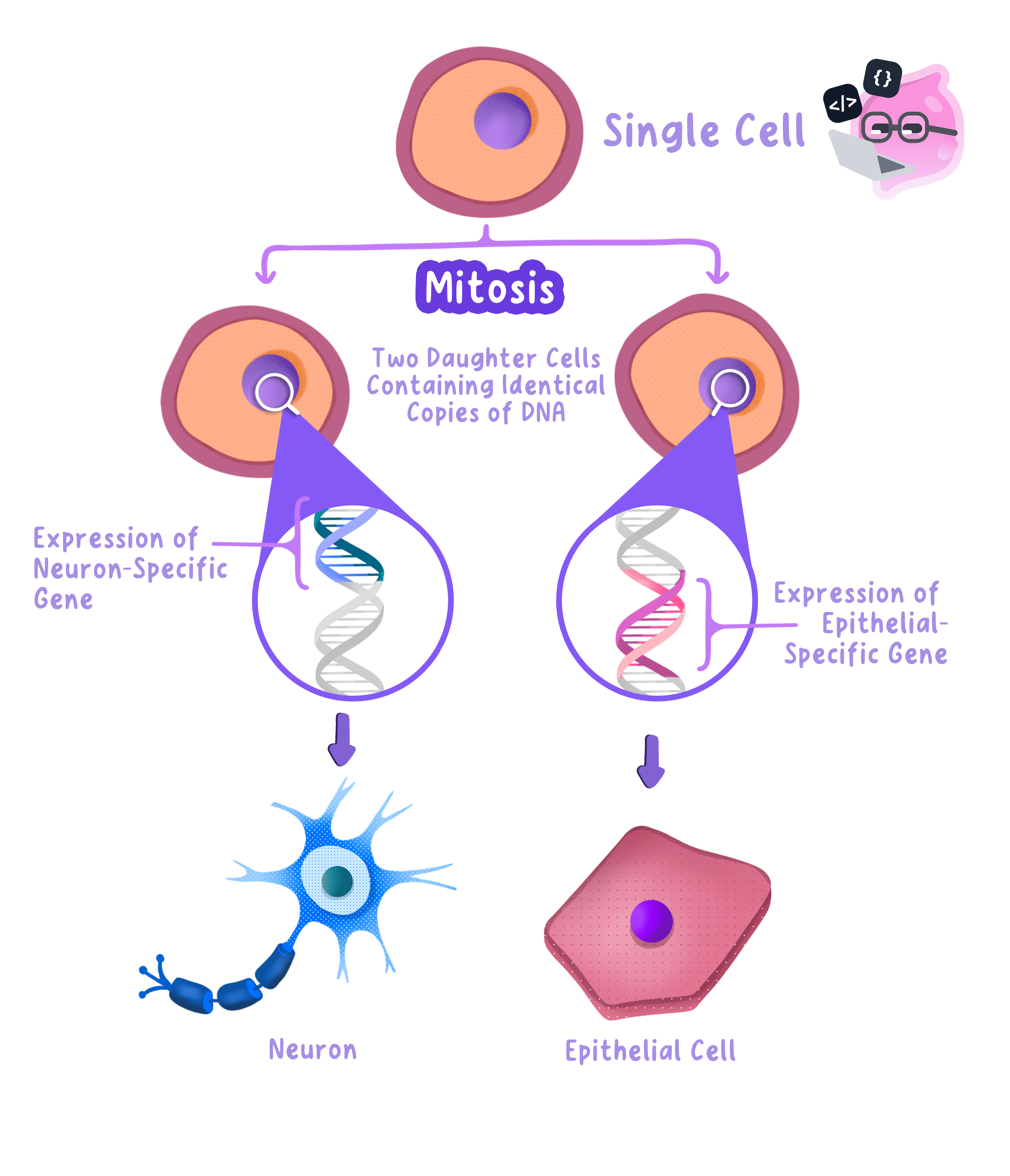
Differentiated cells do not have different genes. They have the same genes and use different ones, so an answer that says the cells lose the genes they do not need is wrong.
Why Specialisation Matters
Specialised cell
A cell whose structure is adapted so that it carries out one particular function.
- A specialised cell is far more efficient at one job than an unspecialised cell would be at everything.
- Specialisation allows a division of labour, so different cells take on different tasks and the whole organism works better.
- Specialised cells of the same type group together into a tissue, tissues form organs, and organs form organ systems.
- Without differentiation an organism could only ever be a shapeless ball of identical cells, so no tissues, organs or organ systems could exist.
- This is why differentiation is essential for the development of a fertilised egg into a complex multicellular organism.
Examples of Specialised Cells
- Sperm cell. A tail for swimming, many mitochondria to release the energy for movement, and an acrosome containing enzymes to digest through the egg membrane.
- Red blood cell. A biconcave shape for a large surface area, packed with haemoglobin, and no nucleus so there is more room for oxygen.
- Root hair cell. A long extension that increases the surface area for absorbing water and mineral ions from the soil.
- Neurone. A very long axon so an impulse can be carried a long way, and a myelin sheath that speeds transmission up.
- Palisade mesophyll cell. Tall, tightly packed and full of chloroplasts near the top of the leaf, where the light is strongest.
- Xylem vessel. Dead, hollow and open ended, with walls strengthened by lignin, forming a continuous tube for water.
For any specialised cell, the mark is for linking one named feature to the job it does, so pair each structure with its function rather than listing features on their own.
Differentiation in Animals and Plants
- In animals most differentiation happens in the embryo and in the young animal.
- Adult animals keep a small number of stem cells, in places such as bone marrow, and these are used mainly to replace worn out cells.
- In plants many cells keep the ability to differentiate throughout the plant's life.
- This is why a small cutting taken from a stem can grow roots and become a complete plant, while a piece of animal tissue cannot.
The cells that can still differentiate are stem cells in animals and meristem cells in plants. Both are covered in detail in the next section on stem cells and meristems.
When Differentiation Goes Wrong
- Cancer cells are often described as poorly differentiated, because they divide instead of taking on a specialised job.
- Under a microscope the cells of a malignant tumour look irregular and unlike the tissue they came from.
- Losing specialisation means the tissue can no longer do its job properly, which is one reason a tumour damages the organ it grows in.
- Explain the importance of differentiation is a why question, so build the chain: different genes are switched on, so different proteins are made, so the cell gains a structure suited to one job, so the organism works efficiently.
- If you are shown an unfamiliar specialised cell, describe a visible feature and suggest the function it serves, because these are application marks and there is no fact to recall.
- Do not stop at the cell. Finish the chain with tissues, organs and organ systems if the question is about the development of a whole organism.
- Define differentiation.
- Explain how two cells with identical genes can become different types of cell.
- Give two features of a sperm cell and state the function of each.
- State one difference between differentiation in animals and differentiation in plants.
- Why is differentiation essential for a multicellular organism?
2.1.6 Percentile charts to monitor growth
What a Percentile Chart Shows
Percentile chart
A growth chart showing the range of a measurement such as mass, length or head circumference at each age, so that one individual can be compared with the population.
- A percentile chart, also called a centile chart, plots a measurement against age.
- The curved lines on the chart are percentile lines, and each one was drawn from measurements of a large sample of healthy children.
- Health visitors in the United Kingdom use these charts to check that a baby is growing as expected, and the readings are recorded in the child's personal health record.
- The three measurements normally monitored are body mass, length or height, and head circumference.
- Boys and girls grow at different rates, so separate charts are used for each.
Reading a Percentile
Percentile
A value on a distribution below which a stated percentage of the population falls, so the 50th percentile is the median.
- A percentile tells you the percentage of the population that falls below a given value.
- A baby on the 50th percentile for mass is exactly average, because half of babies of that age have a smaller mass and half have a greater mass.
- A baby on the 91st percentile is heavier than 91 per cent of babies of the same age and sex, so only 9 per cent are heavier.
- A baby on the 9th percentile is heavier than only 9 per cent, so 91 per cent are heavier.
- The 50th percentile is the median, and the charts used in the United Kingdom usually print the 0.4th, 2nd, 9th, 25th, 50th, 75th, 91st, 98th and 99.6th lines.
Read the percentile as a position in a queue of 100 children of the same age. The 25th percentile means 24 children are lighter and 75 are heavier.
Using the Chart
- Find the child's age on the horizontal axis and read straight up.
- Find the measurement on the vertical axis and read straight across.
- Mark the point where the two lines cross, then read off the nearest percentile line.
- If the point lies between two printed lines, state that the child is between those two percentiles rather than guessing an exact value.
Working Through an Example
-
A boy is 9 months old and has a mass of 9.6 kg, and his point sits on the 75th percentile line.
-
This means 75 per cent of 9 month old boys have a mass below 9.6 kg, so 25 per cent are heavier.
-
At birth the same boy had a mass of 3.2 kg, so his gain in mass can be calculated.
percentage increase=9.6−3.23.2×100=200% \text{percentage increase} = \frac{9.6 - 3.2}{3.2} \times 100 = 200\% percentage increase=3.29.6−3.2×100=200% -
His mass has therefore tripled in 9 months, which is typical for a healthy infant in the first year.
Show the subtraction, the division and the multiplication as separate steps. Edexcel awards method marks for the working even when the final answer is wrong, and allows error carried forward.
Following a Percentile Line
- A healthy child normally grows along or close to the same percentile line over time.
- Staying on the 9th percentile is not a problem in itself, because it simply means the child is small and growing steadily.
- What matters is the pattern over several measurements, not a single reading.
- This is why a health visitor plots repeated measurements rather than judging a child on one visit.
When a Chart Raises Concern
- Crossing two or more percentile lines downwards may indicate a feeding problem, an illness or poor nutrition.
- Crossing two or more lines upwards may indicate excessive weight gain and a raised risk of obesity.
- A measurement below the 0.4th percentile or above the 99.6th percentile is followed up as a matter of routine.
- An unusually large head circumference, or a rapid increase in it, can be an early sign of fluid building up in the skull.
- A chart flags a child for further investigation, and it does not on its own diagnose anything.
Do not describe a child on a low percentile as unhealthy. A low percentile only means the child is small compared with the population, and the concern is a change in percentile, not the percentile itself.
Why Percentiles Are Used Rather Than Averages
- A single average value hides the natural range of sizes in a healthy population.
- Percentiles show the whole distribution, so a measurement can be judged against the spread and not just against the mean.
- They also allow a fair comparison between children of different ages and sexes, because each chart is drawn for one group.
- State the units when you read a value off a chart, since a mass in kilograms and a length in centimetres are both credited only with the unit given.
- If the question asks what a percentile means, define it in terms of the percentage of the population below that value rather than saying it is average.
- For a question about whether a child is growing normally, compare the percentile at two ages and say whether the child has stayed on, risen above or dropped below the line.
- Calculations here are percentage change or percentage of a population, so set out each step and keep the units with your answer.
- What does it mean if a child is on the 25th percentile for mass?
- Which percentile line represents the median?
- Why are separate percentile charts used for boys and girls?
- Describe one pattern on a percentile chart that would concern a health visitor.
- A baby has a mass of 3.5 kg at birth and 7.0 kg at 4 months. Calculate the percentage increase in mass.
2.2.1 Function of stem cells and meristems
What a Stem Cell Is
Stem cell
An undifferentiated cell that can keep dividing by mitosis and can differentiate into other types of cell.
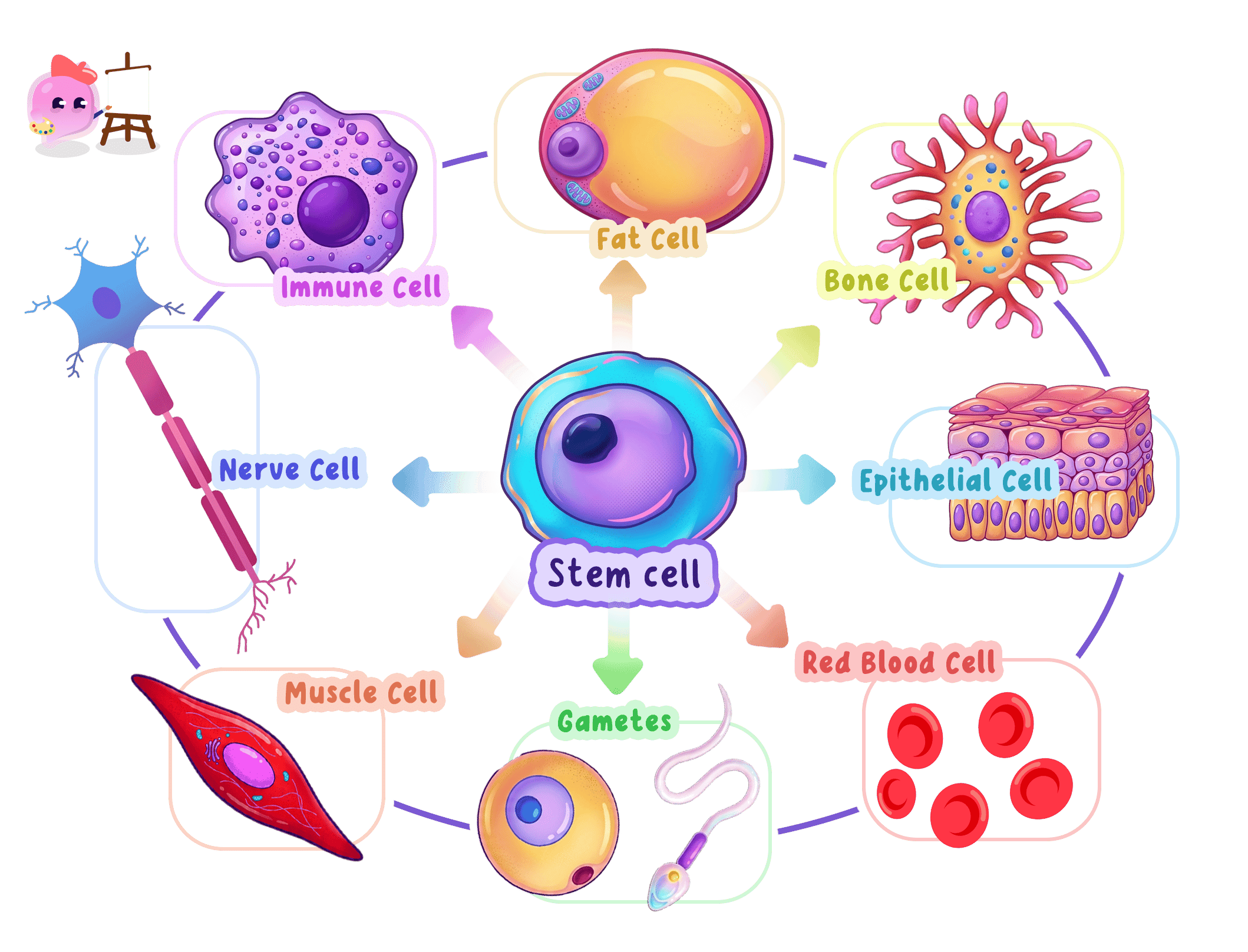
- A stem cell is an undifferentiated cell, which means it has not yet taken on a specialised job.
- It can divide by mitosis over and over again to produce more cells.
- It can also differentiate into one or more types of specialised cell.
- When a stem cell divides, one daughter cell often differentiates while the other stays a stem cell, so the supply of stem cells is not used up.
Embryonic Stem Cells
Embryonic stem cell
A stem cell taken from an early embryo, which can differentiate into any type of body cell.
- Embryonic stem cells come from a very early embryo, a few days after fertilisation, when the ball of cells is called a blastocyst.
- They are taken from the inner cell mass inside the blastocyst.
- They can differentiate into any type of body cell, which makes them the most useful stem cells for medicine.
- The embryos used are usually spare embryos left over from fertility treatment, donated with consent.
- Removing the inner cell mass destroys the embryo, which is the source of the ethical objection to using them.
Adult Stem Cells
Adult stem cell
A stem cell found in a mature tissue such as bone marrow, which can differentiate into only a limited range of cell types.
- Adult stem cells are found in mature tissues, and the best known source is the bone marrow.
- They can only differentiate into a limited range of cell types, so bone marrow stem cells produce blood cells and not nerve cells.
- Their main function in the body is to replace cells that are worn out, damaged or lost.
- Red blood cells survive only about 120 days, so bone marrow stem cells must keep producing replacements throughout life.
- They are also found in the skin, the lining of the gut and the base of hair follicles, all tissues that are constantly renewed.
- Adult stem cells can be taken from a patient without destroying an embryo, so they raise far fewer ethical objections.
Do not say that adult stem cells can turn into any cell. That is only true of embryonic stem cells, and the word to use for adult stem cells is a limited range.
Meristems in Plants
Meristem
A region of a plant, at the root and shoot tips and in the cambium, where cells stay able to divide and differentiate throughout the plant's life.
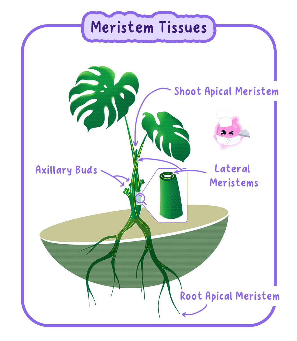
- A meristem is a region of a plant where the cells stay able to divide and differentiate.
- Apical meristems sit at the tips of roots and shoots, and they make the plant longer.
- The cambium is a meristem inside the stem and root that makes the plant wider.
- Meristem cells can differentiate into any plant cell type, including xylem, phloem and root hair cells.
- Unlike animal stem cells, meristem cells keep this ability for the whole life of the plant, which is why plants never stop growing.
Using Meristems to Clone Plants
- A small piece of meristem tissue can be grown into a whole new plant, a technique called tissue culture.
- Every plant produced is a clone of the parent, so a rare or high yielding variety can be reproduced exactly.
- Thousands of identical plants can be produced from one parent in a small space and at any time of year.
- This is how endangered plant species are conserved and how commercial growers produce uniform crops.
Using Stem Cells in Medicine
Differentiation
The process by which an unspecialised cell switches on particular genes and develops the structures it needs to carry out one specific job.
- Stem cells are given to a patient so that they differentiate into the cells the patient has lost.
- Bone marrow transplants are the longest established treatment, and they replace the stem cells that make blood cells in patients with leukaemia.
- Stem cells have been used to grow retinal cells to treat some forms of blindness caused by a damaged retina.
- Trials are looking at replacing the insulin producing cells in type 1 diabetes and the nerve cells lost in Parkinson's disease.
- Stem cells are also grown in the laboratory to test new drugs on human tissue before trials on people.
In a bone marrow transplant the patient's faulty marrow is destroyed, then donor stem cells are given into a vein, settle in the bones and divide by mitosis to produce healthy white blood cells.
Therapeutic Cloning
Therapeutic cloning
The production of an embryo with the same genes as a patient, so that stem cells taken from it will not be rejected when used to treat that patient.
- Cells transplanted from a donor carry different antigens, so the patient's immune system may attack and destroy them.
- In therapeutic cloning, the nucleus of one of the patient's own body cells is placed into an empty egg cell.
- The egg cell divides to form an early embryo whose cells carry the patient's own genes.
- Stem cells taken from this embryo have the same antigens as the patient, so they are not rejected.
- The patient also avoids a lifetime of immunosuppressant drugs, which would otherwise leave them vulnerable to infection.
- The two marks for what a stem cell does are almost always divides by mitosis and differentiates into specialised cells, so write both.
- Mark schemes reject meiosis in this topic, so never use that word about a stem cell.
- If the question names a disease, say which cell type the stem cells would differentiate into and what that cell would then do for the patient.
- When comparing embryonic and adult stem cells, contrast what each can differentiate into and where each comes from.
- Define a stem cell.
- State one difference between an embryonic stem cell and an adult stem cell.
- Where in a plant are the meristems found?
- Explain why meristem cells can be used to produce a clone of an entire plant.
- Explain how therapeutic cloning reduces the chance of transplanted cells being rejected.
2.2.2 Benefits and risks of stem cells
Why Stem Cells Are Used in Medicine
Stem cell
An undifferentiated cell that can keep dividing by mitosis and can differentiate into other types of cell.
- Many diseases are caused by a particular type of cell dying or failing to work.
- Type 1 diabetes follows the loss of the insulin producing cells in the pancreas, and Parkinson's disease follows the loss of certain nerve cells in the brain.
- Ordinary drugs treat the symptoms, because a damaged cell cannot be repaired by medication.
- Stem cells offer a different approach, because they can differentiate into the missing cell type and replace what has been lost.
The Benefits of Using Stem Cells
- Damaged tissue can be replaced rather than simply managed, so the treatment addresses the cause of the disease.
- Conditions that currently have no cure, including paralysis after a spinal injury and some forms of blindness, could be treated.
- Stem cells grown from a patient's own cells are not rejected, so there is no need for lifelong immunosuppressant drugs.
- Organs could be grown from stem cells, which would remove the shortage of donor organs and the wait for a matched donor.
- Embryonic stem cells taken from spare embryos from fertility clinics use tissue that would otherwise be destroyed.
- New drugs can be tested on human tissue grown from stem cells before they are given to people, which makes early trials safer.
Bone marrow transplants for leukaemia are an established stem cell treatment and have been used for decades, so the principle is proven and not merely theoretical.
The Risks of Using Stem Cells
Tissue rejection
The destruction of transplanted cells or tissue by the patient's immune system because their antigens are recognised as foreign.
- Tissue rejection. Stem cells from a donor carry different antigens, so the patient's immune system may destroy them.
- Uncontrolled division. Stem cells divide readily, and cells that keep dividing after transplant can form a tumour.
- Infection. Stem cells are grown in culture, and a culture contaminated with a virus or bacterium would pass that infection to the patient.
- Unpredictable results. Transplanted cells may differentiate into the wrong cell type or fail to differentiate at all.
- Long term effects are unknown, because most stem cell treatments are still at the trial stage and patients have not been followed for decades.
- Cost. The procedures are expensive and specialised, so an overstretched health service can treat only a small number of patients.
Rejection is not a risk when the stem cells came from the patient's own body. Read the question carefully, because the source of the cells decides whether rejection applies.
The Ethical Issues
- Taking stem cells from an embryo destroys that embryo.
- Some people believe that an embryo is a potential human life from the moment of fertilisation, so destroying it is destroying a life.
- Others argue that a few day old ball of cells has no nervous system and cannot feel anything, so its potential does not outweigh the suffering of a living patient.
- The embryo cannot give consent, and the donors of the egg and sperm must consent to its use in research.
- Religious and cultural beliefs lead different people to weigh these arguments differently, and there is no single agreed answer.
- Adult stem cells avoid the objection entirely, but they can only differentiate into a limited range of cell types.
How the Use of Stem Cells Is Controlled
- Stem cell research in the United Kingdom is regulated by law and licensed by a national authority.
- Embryos used in research must be spare embryos from fertility treatment, donated with the informed consent of both donors.
- An embryo used in research cannot be grown beyond 14 days, the point at which the nervous system starts to form.
- Regulation is what allows the research to continue while setting limits that most people find acceptable.
Reaching a Judgement
- A good evaluation weighs the benefit to patients against the status of the embryo and the medical risks.
- The strength of an argument depends on the source of the cells, so adult stem cells and embryonic stem cells should be judged separately.
- The evidence base also matters, because bone marrow transplants are established while brain repair is still experimental.
- A conclusion should state a position and give the reason for it, rather than listing points on both sides and stopping.
- Discuss and evaluate questions need both sides and a conclusion, so plan two or three benefits, two or three risks or ethical objections, and one sentence of judgement.
- Name the risk rather than describing it vaguely, because rejection, tumour formation and infection from culture are each separately creditable.
- Attribute an ethical view rather than stating it as fact, for example by writing that some people believe the embryo has a right to life.
- If the question specifies embryonic or adult stem cells, keep every point relevant to that type, since points about the other type earn nothing.
- Give two benefits of using stem cells to treat disease.
- Explain why a transplant of donor stem cells may be rejected.
- Why can transplanted stem cells sometimes form a tumour?
- State one ethical objection to the use of embryonic stem cells.
- Give one reason why adult stem cells are less controversial than embryonic stem cells.
