The Cell Cycle
Cell cycle
The repeating sequence of interphase, mitosis and cytokinesis by which a body cell grows, copies its DNA and divides into two genetically identical daughter cells.
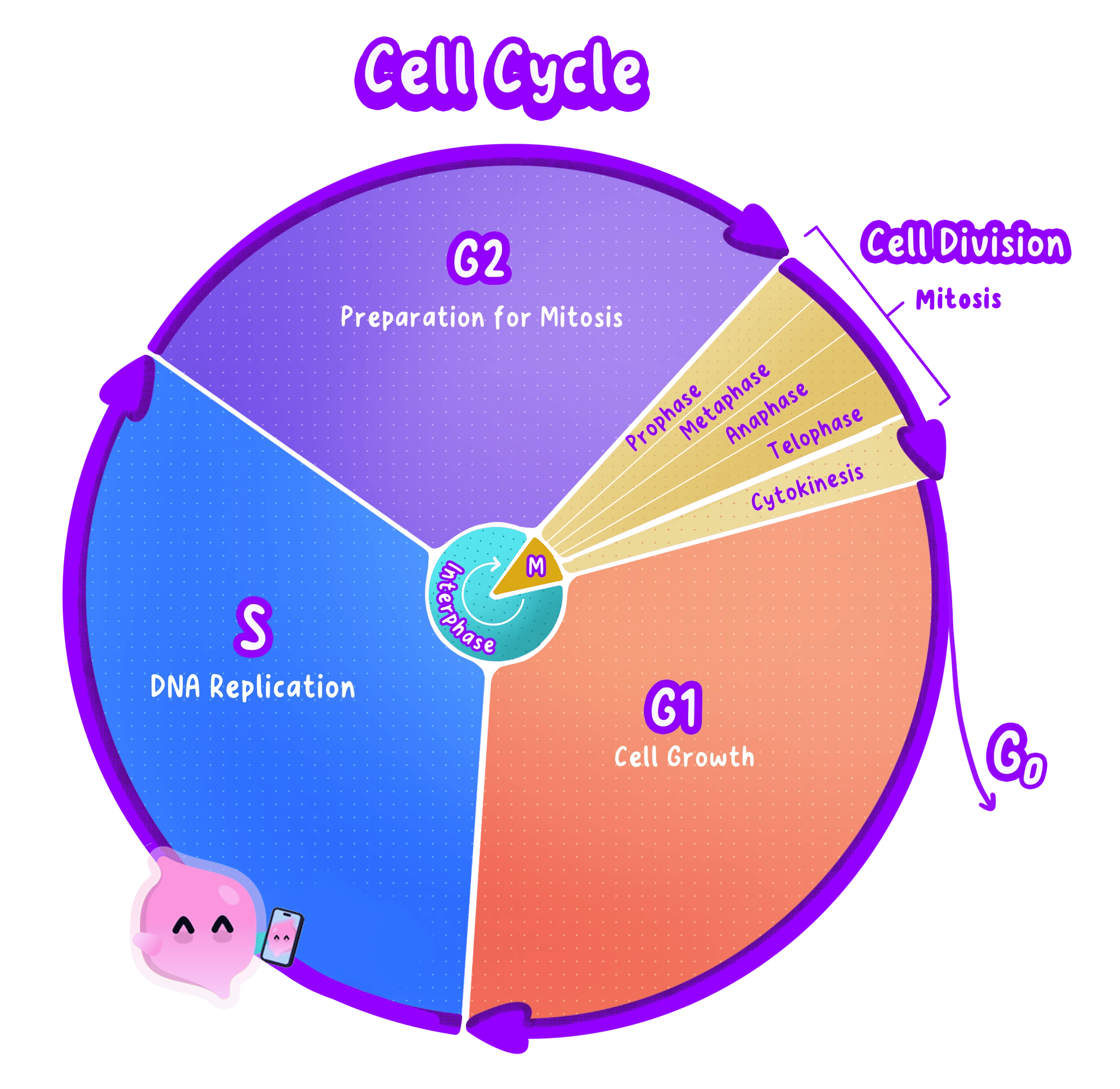
- Body cells do not divide continuously. They pass through a controlled, repeating sequence of events called the cell cycle.
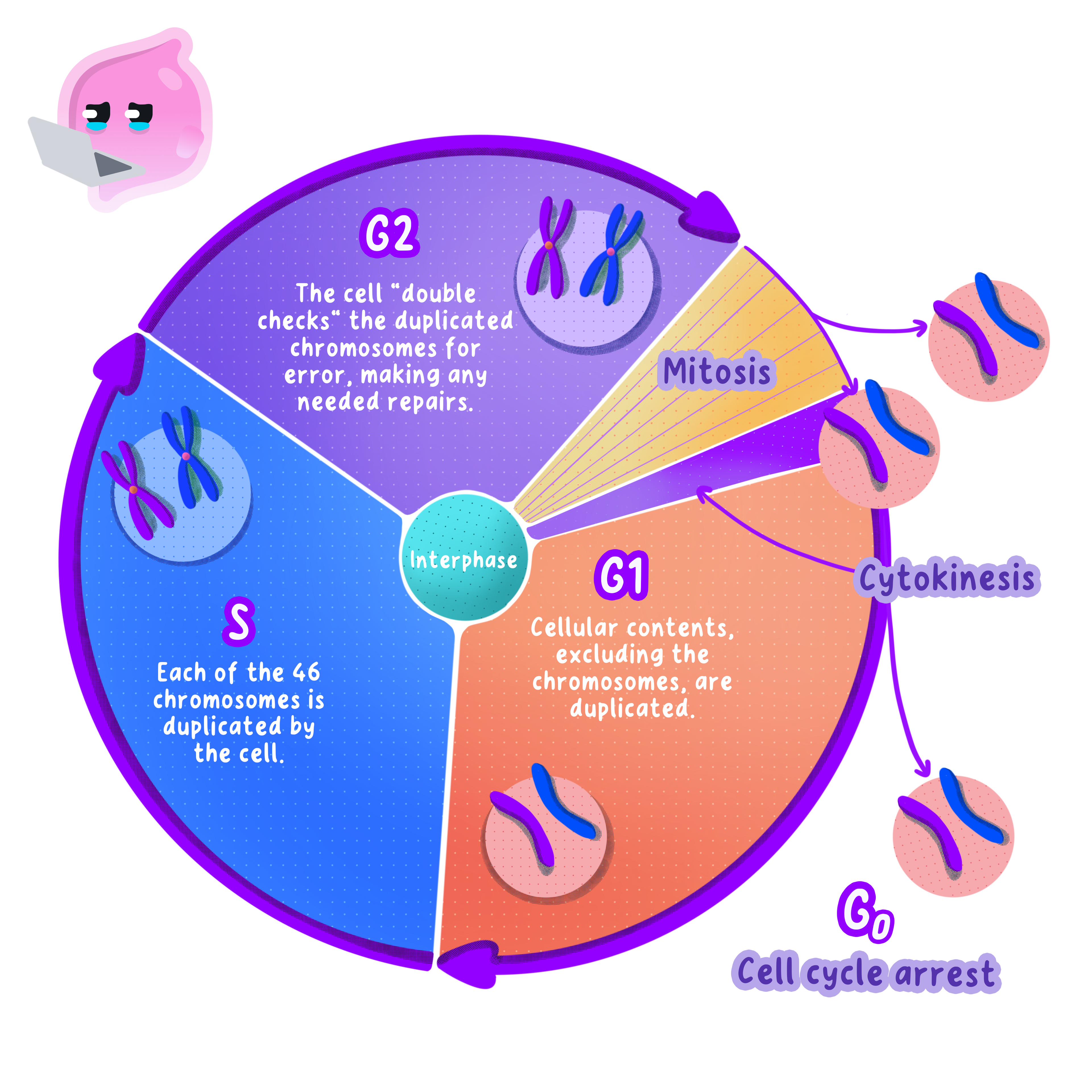
- The cell cycle has three parts: a long growth stage called interphase, then mitosis, and finally cytokinesis.
- Interphase takes up most of the cycle, often around 90 per cent of the total time in a human body cell.
- Mitosis and cytokinesis together make up the much shorter division stage, in which one parent cell becomes two.
- A cell that is not preparing to divide leaves the cycle and simply carries out its job, which is what most mature nerve and muscle cells do.
Interphase
Interphase
The stage of the cell cycle before mitosis, in which the cell grows, makes extra sub-cellular structures and replicates its DNA so that each chromosome becomes two identical sister chromatids.
- During interphase the cell grows and increases its number of sub-cellular structures, including mitochondria and ribosomes.
- More mitochondria mean more respiration, which supplies the ATP needed for the energy-demanding work of dividing.
- The DNA replicates, so that every chromosome is copied exactly.
- The cell also carries out its normal functions throughout interphase, so a liver cell in interphase is still making enzymes and storing glycogen.
Interphase is a stage of the cell cycle, not a stage of mitosis, so an answer that lists interphase as part of mitosis loses the mark.
Chromosomes Are Copied Before Division
Chromosome
A long molecule of coiled DNA found in the nucleus, carrying many genes in a fixed order.
- A human body cell contains 46 chromosomes, arranged as 23 pairs.
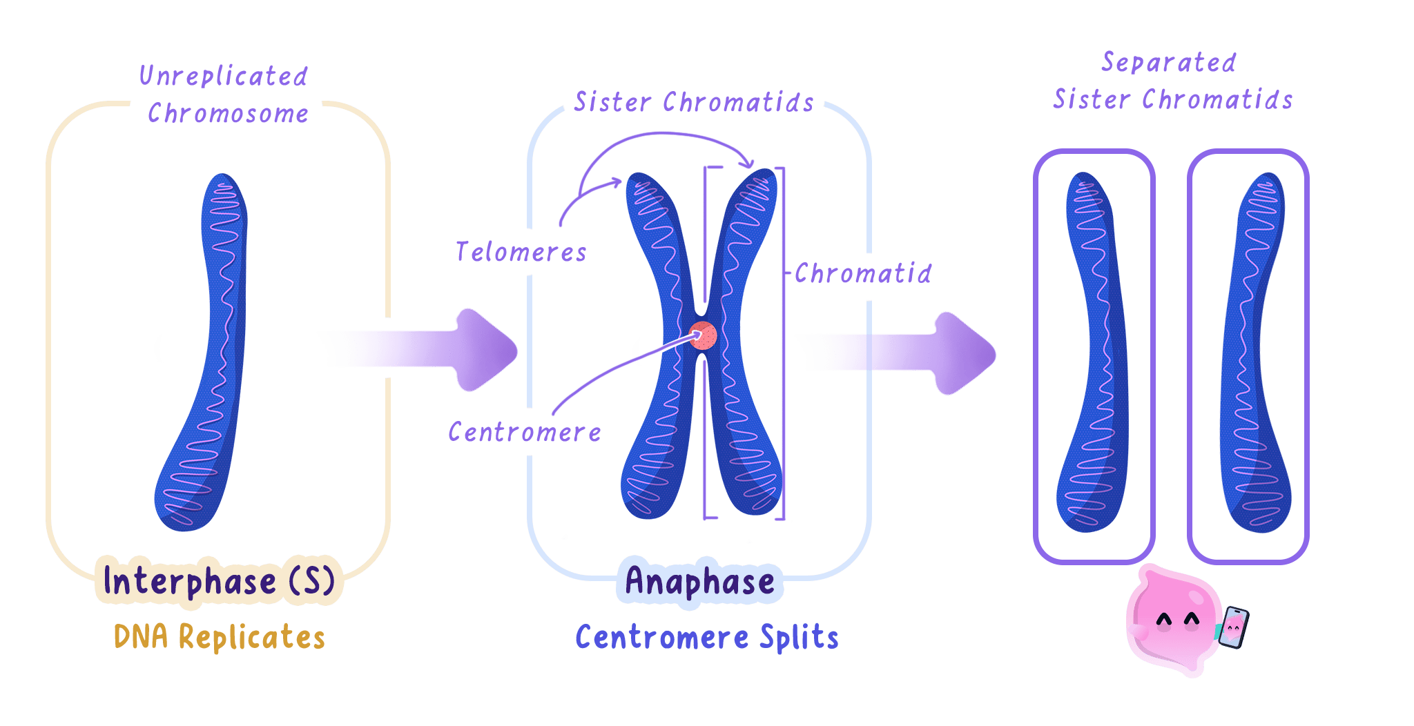
- When DNA replicates, each chromosome becomes two identical copies called sister chromatids.
- The two chromatids stay joined at a narrow region called the centromere, which is why a copied chromosome is drawn as an X shape.
- Because the copies are identical, both new cells can be given a complete and identical set of genetic information.
Do not describe replication as the chromosome splitting in half. Replication doubles the DNA, so the cell temporarily holds twice as much genetic material, not half as much.
Prophase
Sister chromatids
The two identical copies of a replicated chromosome, joined at the centromere until they are pulled apart during mitosis.
- The copied chromosomes condense, coiling up until they are short and thick enough to be seen under a light microscope.
- Each one is now visible as two sister chromatids joined at the centromere.
- The nuclear membrane breaks down, releasing the chromosomes into the cytoplasm.
- Spindle fibres begin to form from opposite ends, or poles, of the cell.
Metaphase
- The chromosomes are moved so that they line up along the equator, which is the imaginary line across the middle of the cell.
- Spindle fibres from each pole attach to the centromere of every chromosome.
- Lining the chromosomes up in a single row is what makes an equal separation possible in the next phase.
The names give the order away: chromosomes take their place in the Middle at Metaphase and move Apart at Anaphase.
Anaphase
- Each centromere divides, so the two sister chromatids are no longer held together.
- The spindle fibres shorten and pull one chromatid from each pair to opposite poles of the cell.
- Once separated, each chromatid counts as a chromosome in its own right.
- This gives each pole one complete set of chromosomes, identical to the set the parent cell started with.
Telophase
- The chromosomes reach the poles and begin to uncoil, so they can no longer be seen individually.
- A new nuclear membrane forms around each set, producing two nuclei inside one cell.
- At the end of telophase the nucleus has divided but the cell has not, so mitosis is complete and cytokinesis has yet to happen.
Mitosis is the division of the nucleus. The division of the whole cell needs cytokinesis as well.
Cytokinesis
Cytokinesis
The final stage of the cell cycle, in which the cytoplasm and cell membrane divide to form two separate daughter cells.
- The cytoplasm and the cell membrane divide to separate the two nuclei into two cells.
- In an animal cell the membrane pinches inwards at the equator until the cell splits in two.
- In a plant cell a new cell wall and membrane are built across the middle instead, because the rigid wall cannot pinch inwards.
- The result is two daughter cells, each with its own nucleus, cytoplasm and cell membrane.
Do not confuse mitosis with meiosis. Mitosis produces two genetically identical diploid cells for growth and repair, while meiosis produces four genetically different haploid gametes.
Why Mitosis Matters
- Growth. A fertilised egg becomes a multicellular organism because mitosis repeatedly increases the number of cells.
- Repair and replacement. Skin, gut lining and red blood cells are worn out or lost constantly, and mitosis replaces them with identical cells.
- Around two million red blood cells are replaced every second in an adult human, all of them produced by mitosis in the bone marrow.
- Asexual reproduction. Organisms that reproduce from a single parent use mitosis, so their offspring are genetically identical clones.
- A strawberry plant grows runners that root and form new plants, and each new plant carries exactly the same alleles as the parent.
A cut on the skin heals because cells at the edge of the wound divide by mitosis, producing identical skin cells that close the gap.
Observing Mitosis Under the Microscope
Investigation: Observing Mitosis in a Root Tip
- Aim: To prepare a stained root tip squash and identify cells in each phase of mitosis, then calculate the mitotic index. This is a standard microscopy investigation rather than one of the eight Edexcel core practicals, but Edexcel does set method and calculation questions on it.
- Apparatus: Germinating garlic or onion roots, watch glass, 1 mol/dm3 hydrochloric acid, warm water bath at 60 degrees Celsius, toluidine blue or acetic orcein stain, scalpel, mounted needle, forceps, microscope slide and coverslip, filter paper, light microscope, eye protection.
- Method:
- Cut the tip 5 mm from the end of a growing root, because only the meristem at the very tip is actively dividing and the rest of the root gives you almost nothing to look at.
- Place the tip in a watch glass of hydrochloric acid in a 60 degrees Celsius water bath for about 5 minutes, to break down the middle lamella between the cells so the tissue will spread out into a single layer.
- Rinse the tip in cold water and blot it dry.
- Transfer it to a slide and add two or three drops of stain, which binds to DNA and makes the chromosomes stand out dark against pale cytoplasm.
- Lower a coverslip at an angle with a mounted needle, so air is pushed out ahead of it rather than trapped as bubbles.
- Cover with filter paper and press down firmly and vertically with your thumb, without twisting, to squash the tissue into a layer one cell thick.
- View under low power to find the region where the cells are small and square, then switch to high power and count.
- Variables: The independent variable is the region of the root you examine and the dependent variable is the proportion of cells in mitosis. Keep the species, the age of the root, the temperature, the staining time and the total number of cells counted the same.
- Results: Most cells are in interphase, with a large round nucleus and no visible chromosomes. A smaller number show dark, condensed chromosomes: scattered in prophase, in a line across the middle in metaphase, in two separated groups in anaphase, and as two clusters with re-forming nuclei in telophase.
- Maths: The mitotic index is the proportion of cells that are in mitosis, given by mitotic index=number of cells in mitosistotal number of cells counted\text{mitotic index} = \frac{\text{number of cells in mitosis}}{\text{total number of cells counted}}mitotic index=total number of cells countednumber of cells in mitosis Multiply by 100 to express it as a percentage. Counting at least 100 cells and repeating on several fields of view reduces the effect of an unrepresentative patch of tissue.
- Watch out: Pressing down while twisting rolls the cells over each other and tears them, so the chromosomes cannot be assigned to a phase. Squashing too little leaves the tissue several cells deep, which will not come into focus at high power.
- Safety: Hydrochloric acid is an irritant, so wear eye protection and rinse any splashes immediately. Cut the root on a tile with the blade moving away from your fingers.
- When a question says describe what happens during mitosis, track the chromosomes through the four phases rather than listing the phase names, because the marks are for chromosome behaviour.
- Use the wording examiners credit: chromosomes condense, line up along the equator, are pulled apart at the centromere by spindle fibres, and gather at each pole.
- If the question asks for the stages of the cell cycle rather than mitosis, the answer is interphase, mitosis and cytokinesis, and naming prophase to telophase alone will not gain the mark.
- In a data question about the length of each phase, use the proportion of cells seen in a phase as a measure of how long that phase lasts.
- Name the three stages of the cell cycle in order.
- What two things happen to a cell during interphase?
- In which phase of mitosis do the chromosomes line up along the equator of the cell?
- What happens to the centromeres during anaphase?
- Give three roles of mitosis in a living organism.