1.3.1 Mechanism of enzyme action
What an enzyme is
Enzyme
A protein that acts as a biological catalyst, speeding up a specific reaction without being used up or changed by it.
- Every enzyme is a protein, made on the ribosomes from a chain of amino acids.
- An enzyme works as a biological catalyst, speeding a reaction up without being used up or changed by it.
- Because it is not used up, a single enzyme molecule catalyses the same reaction thousands of times over.
- Without enzymes the reactions inside a cell would run far too slowly at body temperature to keep an organism alive.
- Some enzymes act inside cells, such as those that catalyse respiration, and others are released to act outside cells, such as the digestive enzymes in the gut.
- Enzyme names usually end in -ase and are built from the substrate, so lactase acts on lactose.
A catalyst lowers the energy a reaction needs to get started, which is why an enzyme lets a reaction run quickly at 37 ∘C37\,^{\circ}\text{C}37∘C instead of needing high heat.
The active site
Active site
The part of an enzyme with a specific shape into which the substrate fits.
- The chain of amino acids in an enzyme folds into a precise three-dimensional shape, held in place by bonds between different parts of the chain.
- That folding leaves a small groove on the surface of the molecule, and this groove is the active site.
- The shape of the active site is complementary to the shape of one particular substrate.
- Complementary means the two shapes match each other, in the same way a hand matches a glove, rather than being identical.
- Everything an enzyme does depends on that shape being maintained, which is why heat and pH matter so much.
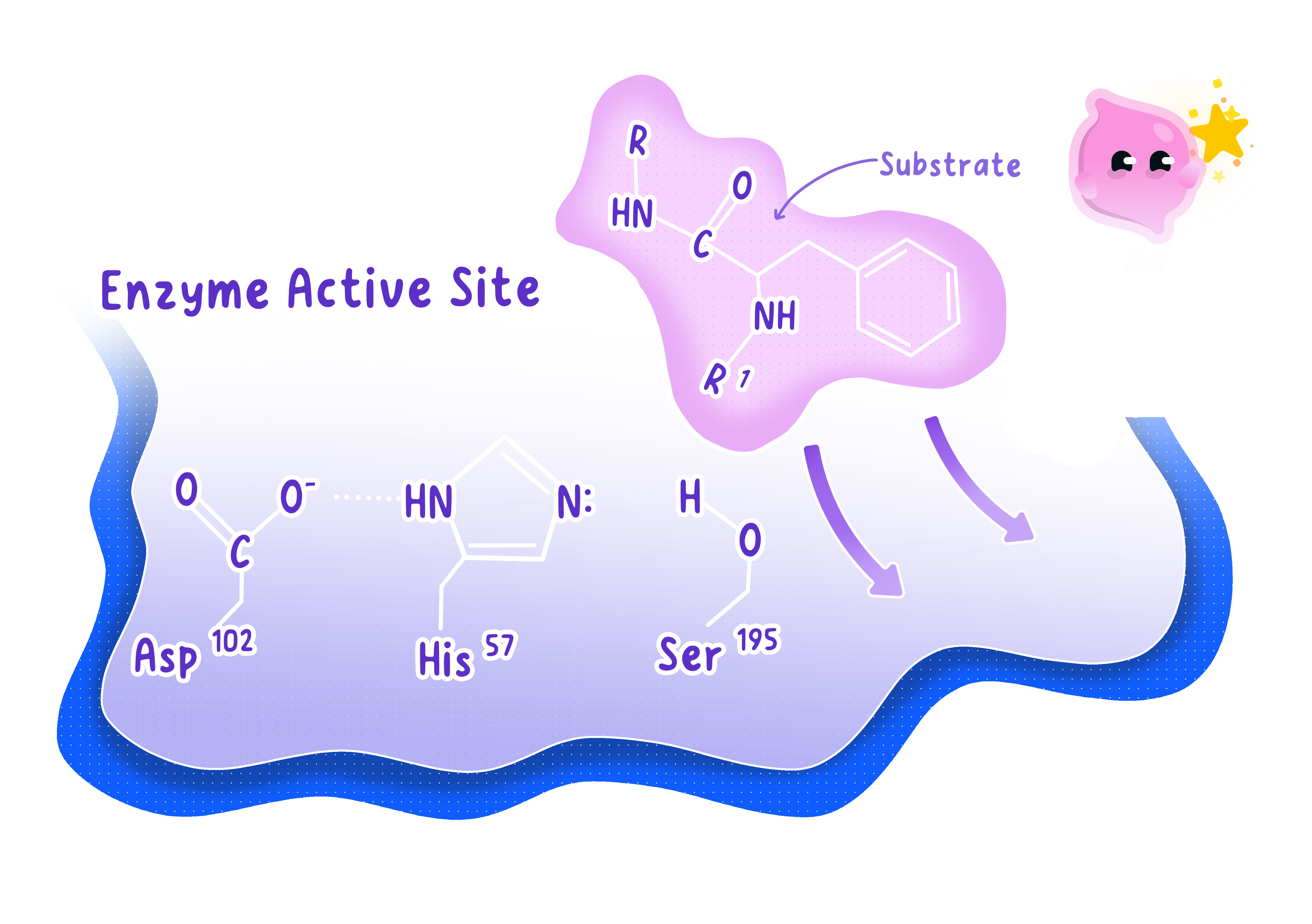
The shape of the active site is the whole explanation for how an enzyme works, why it works on only one substrate, and why it stops working when conditions change.
The lock and key model
Substrate
The molecule on which an enzyme acts.
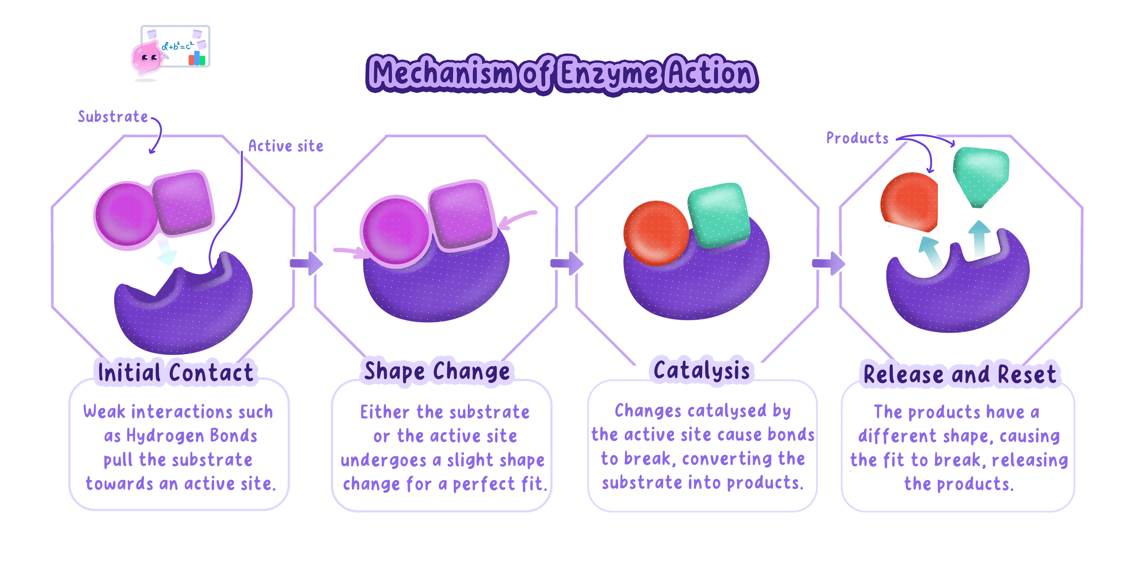
- The substrate moves randomly through the solution until it collides with the enzyme.
- If the collision happens at the active site and in the right orientation, the substrate fits into it and binds.
- The structure formed at that moment is the enzyme-substrate complex.
- Holding the substrate in the active site makes the reaction happen much more readily, so the substrate is either split apart or joined to another molecule.
- The products no longer fit the active site, so they leave it.
- The active site is then empty and unchanged, ready for the next substrate molecule.
- This description is called the lock and key model, because only one key has the shape that opens a given lock.
Enzyme-substrate complex
The structure formed when a substrate binds to the active site of an enzyme.
A six-step description of collide, fit, form the complex, react, release the products and free the active site will cover almost any mark scheme on enzyme action.
Why enzymes are specific
- Specificity means that one enzyme catalyses one reaction and no others.
- The reason is the shape of the active site, which is complementary to just one substrate.
- A molecule with a different shape cannot bind, so no enzyme-substrate complex forms and the reaction is not catalysed.
- Amylase, for example, breaks down starch but has no effect at all on protein, even in the same test tube.
- Specificity is what allows a cell to control which reactions happen, because it makes only the enzymes it needs.
Enzymes are molecules and not living things, so write that an enzyme is specific to its substrate rather than saying it chooses, wants or recognises one.
Enzymes that build large molecules
- In a synthesis reaction an enzyme holds two small molecules together so that they join, releasing water.
- Glucose molecules are joined into the carbohydrates starch in plants, glycogen in animals, and cellulose for plant cell walls. many glucose→starch, glycogen or cellulose\text{many glucose} \rightarrow \text{starch, glycogen or cellulose}many glucose→starch, glycogen or cellulose
- Amino acids are joined in a particular order to build proteins, which include enzymes themselves. many amino acids→protein\text{many amino acids} \rightarrow \text{protein}many amino acids→protein
- One glycerol molecule is joined to three fatty acids to make a lipid. glycerol+3 fatty acids→lipid\text{glycerol} + 3\ \text{fatty acids} \rightarrow \text{lipid}glycerol+3 fatty acids→lipid
Synthesis reactions are how a growing organism turns absorbed nutrients into its own tissue, so this is the chemistry behind growth.
Enzymes that break molecules down
- In a breakdown reaction an enzyme splits a large molecule into its smaller subunits by adding water.
- Carbohydrases break carbohydrates into sugars, and amylase breaking starch into maltose is the example Edexcel uses. starch→ amylase sugars\text{starch} \xrightarrow{\ \text{amylase}\ } \text{sugars}starch amylase sugars
- Proteases break proteins into amino acids. protein→ protease amino acids\text{protein} \xrightarrow{\ \text{protease}\ } \text{amino acids}protein protease amino acids
- Lipases break lipids into fatty acids and glycerol. lipid→ lipase fatty acids+glycerol\text{lipid} \xrightarrow{\ \text{lipase}\ } \text{fatty acids} + \text{glycerol}lipid lipase fatty acids+glycerol
- Breakdown matters in digestion because large food molecules are insoluble and too big to pass through the gut wall.
- The small soluble products can be absorbed into the blood and then reassembled into whatever the body needs.
- Learn the three enzyme groups with both their substrate and their products, since questions swap freely between naming the enzyme and naming the product.
- The mark most often dropped on digestion questions is the reason, so add that the products are small and soluble enough to be absorbed.
- If a question invents an enzyme name, use the -ase rule to work out its substrate rather than assuming it is off the specification.
- An enzyme is not used up in the reaction, so never write that it is broken down or consumed.
- The active site is complementary to the substrate, not the same shape as it.
- Enzymes speed up a reaction that would happen anyway, rather than causing a reaction that could never occur.
- Substrate and product are different things, so keep the two words apart when describing the complex.
- Define an enzyme in one sentence.
- Describe the lock and key model from the collision to the release of the products.
- Explain why amylase cannot break down protein.
- Name the products formed when a protease and a lipase act on their substrates.
- Which three molecules are joined together to make a lipid?
1.3.2 Denaturation of enzymes
What denaturation is
Denaturation
A permanent change in the shape of an enzyme's active site, caused by high temperature or extreme pH, so that the substrate no longer fits.
- An enzyme holds its folded shape because of bonds formed between different parts of its amino acid chain.
- In denaturation those bonds are broken, so the chain unfolds and takes up a different shape.
- The part of the molecule that matters is the active site, and it changes shape along with the rest.
- The substrate is no longer complementary to the altered active site, so it cannot bind.
- No enzyme-substrate complex can form, so the reaction is no longer catalysed and the rate falls to zero.
A full denaturation answer runs bonds break, active site changes shape, substrate no longer fits, no complex forms, so the reaction stops.
Denaturation by heat
- Raising the temperature gives the enzyme molecule more kinetic energy, so it vibrates more.
- Beyond the optimum temperature the vibration is violent enough to break the bonds holding the folded shape together.
- The active site distorts, and every molecule that distorts is permanently out of action, so the rate drops away steeply.
- Most human enzymes have an optimum of about 37 ∘C37\,^{\circ}\text{C}37∘C and are badly denatured by around 50 ∘C50\,^{\circ}\text{C}50∘C.
- Boiling denatures almost every enzyme, which is why boiled enzyme solution is used as a negative control in experiments.
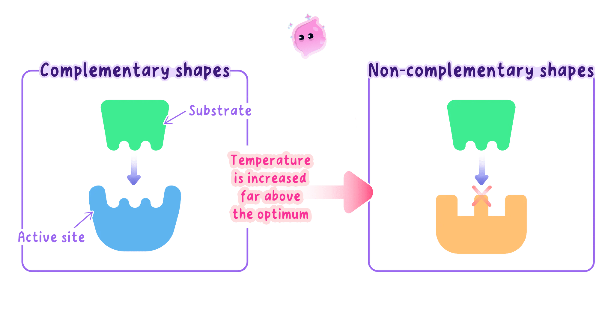
On a temperature graph the rise before the optimum is gradual while the fall after it is sudden, because the rise comes from faster collisions and the fall comes from molecules being destroyed.
Denaturation by pH
- Every enzyme also has an optimum pH at which its active site holds exactly the right shape.
- Away from that pH there are too many hydrogen ions or too many hydroxide ions in the solution.
- These ions interfere with the bonds between the charged parts of the amino acid chain, so the folded shape is pulled out of position.
- The active site changes shape once more, and the same chain of consequences follows as with heat.
- Optimum pH depends on where the enzyme works, so the protease in the stomach works best at about pH 222 while amylase in the mouth works best near pH 777.
- A small shift either side of the optimum slows the enzyme down, and a large shift denatures it completely.
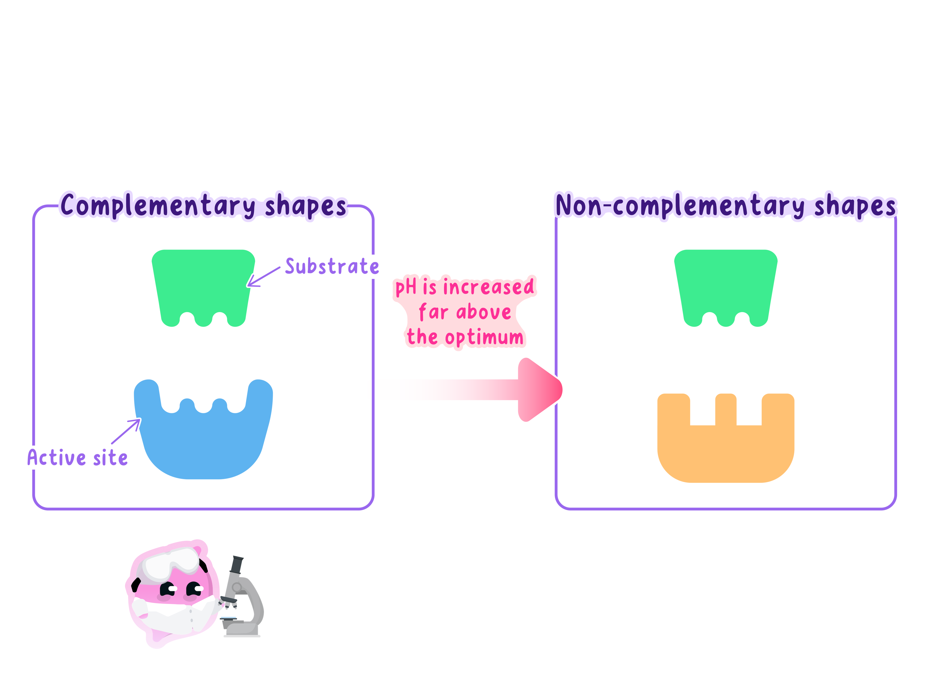
An enzyme with a low optimum pH is not denatured by acid at all, so never write that acid always denatures enzymes when stomach protease needs acid to work.
Why denaturation is permanent
- Once the bonds have broken, the chain does not refold into its original arrangement when conditions return to normal.
- Cooling a denatured enzyme, or returning it to its optimum pH, therefore does not bring the activity back.
- This is different from an enzyme kept in a fridge, which is only slowed down and works normally again once it warms up.
- At low temperature the molecules simply have less kinetic energy, so collisions are less frequent and no bonds are broken.
- Cooking an egg shows the same change in a protein you can see, because the clear albumen turns solid and white and never turns back.
- Write that the enzyme is denatured, never that it is killed or dead, because an enzyme is a protein rather than a living organism.
- The mark most often dropped is the middle step, so always state that the active site changes shape rather than jumping from heat straight to no reaction.
- If a question asks whether an effect is reversible, say that cooling reverses a slow-down but does not reverse denaturation.
Spotting denaturation in data
- On a rate graph, denaturation appears as the rate falling steeply towards zero beyond the optimum.
- A rate that stays at zero after conditions are restored is the clearest evidence that denaturation, rather than a slow-down, has happened.
- In an enzyme investigation, a tube of boiled enzyme should give no reaction at all, and that result confirms the reaction seen elsewhere was caused by the enzyme.
- A reading that is much lower than its neighbours at a mild pH or temperature is more likely to be an anomaly than denaturation, so check the pattern before concluding.
- Describe what happens to an enzyme molecule when it is denatured.
- Explain in full why a denatured enzyme cannot catalyse its reaction.
- Name the two conditions that cause denaturation.
- Why does cooling an enzyme that has been boiled fail to restore its activity?
- Give one reason why it is wrong to say a denatured enzyme has been killed.
1.3.3 Factors affecting enzyme activity
Temperature and enzyme activity
Optimum
The temperature or pH at which an enzyme catalyses its reaction fastest.
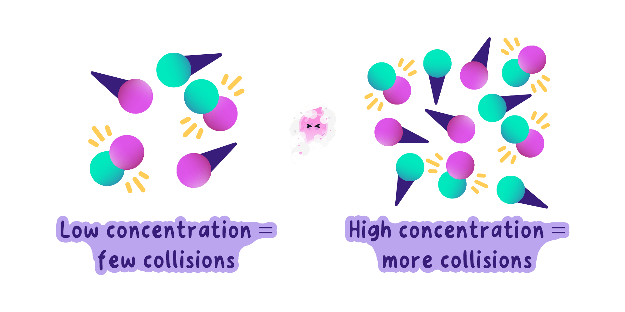
- Warming a mixture gives both the enzyme and the substrate more kinetic energy, so the molecules move faster.
- Faster movement means more successful collisions between substrate molecules and active sites every second.
- More enzyme-substrate complexes form per second, so the rate of reaction rises.
- The rate keeps climbing until the optimum temperature, which is about 37 ∘C37\,^{\circ}\text{C}37∘C for most human enzymes.
- Above the optimum the enzyme is denatured, the active site changes shape, and the rate falls steeply to zero.
- Enzymes from other organisms have different optima, so bacteria living in hot springs have enzymes that work well above 70 ∘C70\,^{\circ}\text{C}70∘C.
The two halves of a temperature curve have completely different causes, since the rise is about collision frequency and the fall is about the active site being destroyed.
pH and enzyme activity
- Plotting rate against pH gives a peak, because each enzyme has an optimum pH at which its active site is exactly the right shape.
- Moving a little either side of the optimum distorts the active site slightly, so fewer substrate molecules fit and the rate falls.
- Moving far from the optimum breaks the bonds holding the shape altogether, and the enzyme is denatured.
- The optimum matches where the enzyme normally works, so the protease in the acidic stomach peaks near pH 222.
- Salivary amylase peaks close to neutral, and the enzymes in the small intestine work best in slightly alkaline conditions near pH 888.
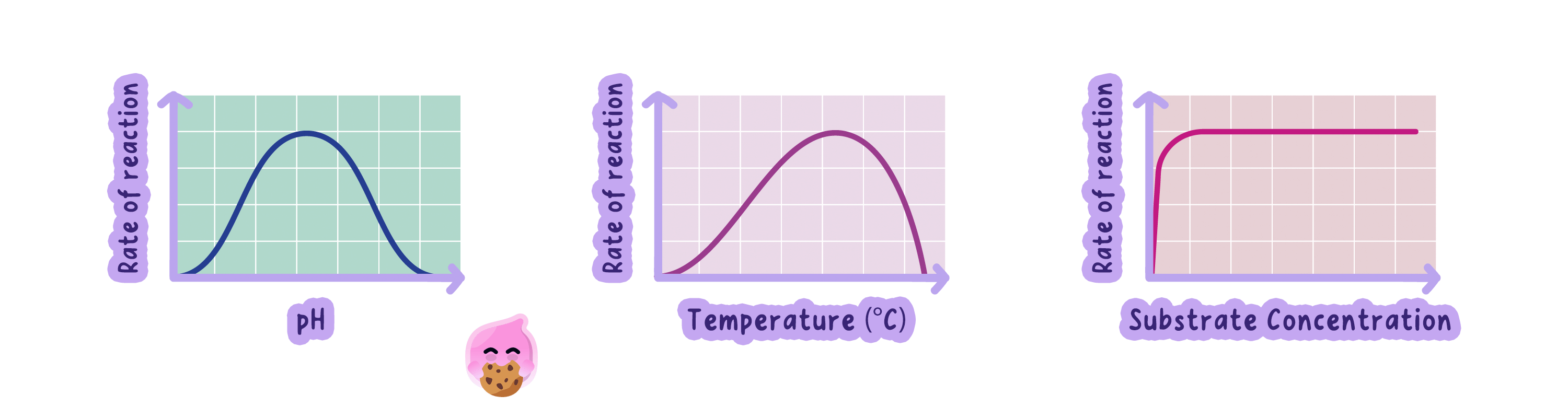
Read the optimum straight off the peak of the curve, and quote it with its unit, so a pH graph peaking at 6.56.56.5 gives an optimum pH of 6.56.56.5.
Substrate concentration and enzyme activity
- At low substrate concentration the rate rises in direct proportion to the amount of substrate added.
- More substrate molecules in the same volume means more collisions with active sites each second, so more complexes form.
- Beyond a certain point the line levels off into a plateau and adding more substrate makes no difference.
- Every active site is already occupied, so the enzyme is described as saturated and the extra substrate has to wait its turn.
- The number of enzyme molecules is what limits the rate once the plateau is reached.
- Adding more enzyme therefore raises the height of the plateau, while adding more substrate does not.
A substrate concentration graph levels off but never falls, so a curve that drops back down is showing denaturation and cannot be a substrate concentration graph.
Calculating a rate
Rate of reaction
The amount of substrate used up, or product formed, in a given time.
- When you can measure how much product forms, divide that amount by the time it took.
- rate=change in quantitytime taken\text{rate} = \dfrac{\text{change in quantity}}{\text{time taken}}rate=time takenchange in quantity
- When you can only time how long a reaction takes to reach an end point, use the reciprocal of the time instead.
- rate=1000time in seconds\text{rate} = \dfrac{1000}{\text{time in seconds}}rate=time in seconds1000
- Multiplying by 100010001000 simply avoids awkward decimals, and the values are treated as arbitrary units.
- A shorter time always means a faster rate, which is the check to run before writing an answer down.
Take a mean of the repeats before calculating a rate, and ignore any anomalous result when working that mean out.
- Starch takes 40 s40\,\text{s}40s to disappear at pH 666, so use the reciprocal form.
- rate=100040=25 arbitrary units\text{rate} = \dfrac{1000}{40} = 25\ \text{arbitrary units}rate=401000=25 arbitrary units
- At pH 444 the same reaction takes 125 s125\,\text{s}125s, giving a rate of 888, so the enzyme works around three times faster at pH 666.
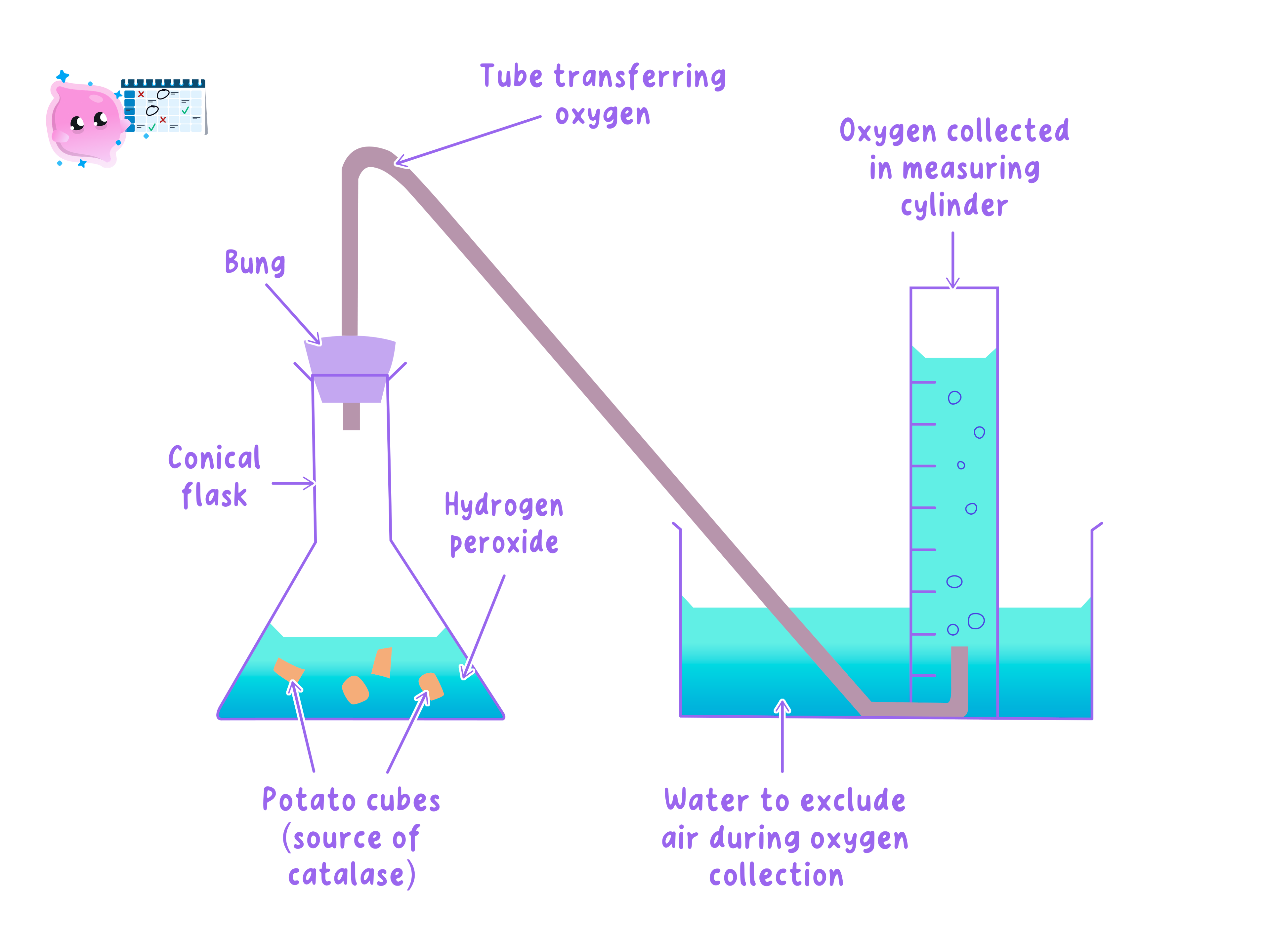
- Catalase releases 24 cm324\,\text{cm}^324cm3 of oxygen in 60 s60\,\text{s}60s, and here you know the quantity, so divide it by the time.
- rate=2460=0.4 cm3/s\text{rate} = \dfrac{24}{60} = 0.4\,\text{cm}^3/\text{s}rate=6024=0.4cm3/s
- Give the unit with the answer whenever the quantity has one, so a gas volume over time is written in cm3/s\text{cm}^3/\text{s}cm3/s.
- Write the equation, then the substitution, then the answer, because the method mark survives an arithmetic slip only if the equation is on the page.
- When a question asks you to explain a rise in rate, name the collisions, because saying only that the particles move faster earns half the credit.
- For a plateau, say that all the active sites are occupied and name enzyme concentration as the factor now limiting the rate.
pH and enzyme activity
- Aim: to find how pH affects how fast amylase breaks starch down, by timing how long the starch takes to disappear.
- Apparatus: spotting tile, 0.01 mol/dm30.01\,\text{mol/dm}^30.01mol/dm3 iodine solution, 1% amylase solution, 1% starch solution, buffer solutions across a pH range, one 5 cm35\,\text{cm}^35cm3 syringe for each solution, stirring rod, test tubes, a water bath at about 30 ∘C30\,^{\circ}\text{C}30∘C, stop clock and eye protection.
- Method:
- Put a drop of iodine solution into every well of the spotting tile.
- Using its own syringe, put 2 cm32\,\text{cm}^32cm3 of amylase into a test tube and add 1 cm31\,\text{cm}^31cm3 of the pH 4 buffer.
- Stand the tube in the 30 ∘C30\,^{\circ}\text{C}30∘C water bath for a few minutes, so it reaches temperature and the pH is already set before the reaction begins.
- Add 2 cm32\,\text{cm}^32cm3 of starch solution, start the clock at that moment, and stir.
- After 10 s10\,\text{s}10s transfer one drop of the mixture into the first well of iodine.
- Repeat every 10 s10\,\text{s}10s into the next well along.
- While starch is present the iodine goes blue-black, and the moment a drop leaves the iodine orange-brown all the starch has been digested, so record that time.
- Repeat at pH 5, 6, 7 and 8, keeping every volume, the temperature and the stirring the same, and do three runs at each pH to take a mean.
- Variables: you change the pH and you measure the time for the starch to disappear, while keeping the temperature, the volume and concentration of amylase and starch, the stirring, the drop size and the sampling interval the same.
- Results: the time is shortest near amylase's optimum of about pH 6 to 7, so the rate is highest there and falls away on both sides, and at an extreme pH some wells stay blue-black for the whole run because the enzyme has been denatured.
- Maths: rate =1000÷= 1000 \div=1000÷ time in seconds, then plot rate against pH and read the optimum off the peak of the curve.
- Watch out: use a separate syringe for every solution because one drop of amylase in the starch bottle spoils the whole class set, start the clock as the starch goes in rather than before, and remember that sampling every 10 s10\,\text{s}10s means the true end point could be up to 10 s10\,\text{s}10s out.
- Safety: wear eye protection because iodine stains and irritates, take care with the hot water bath, and wash your hands afterwards.
- Temperature below the optimum slows an enzyme down but does not denature it, so the effect is reversible.
- A plateau on a substrate concentration graph is caused by saturated active sites, not by denaturation.
- A shorter time means a faster rate, so a graph of time against pH is the mirror image of a graph of rate against pH.
- There is no single optimum shared by all enzymes, so quote the optimum of the enzyme in the question rather than assuming 37 ∘C37\,^{\circ}\text{C}37∘C and pH 777.
- Explain, in terms of collisions, why raising the temperature below the optimum speeds an enzyme up.
- Why does the rate level off at high substrate concentration?
- A reaction finishes in 50 s50\,\text{s}50s, so what is the rate in arbitrary units?
- In the pH core practical, what colour change tells you all the starch has been digested?
- Name three variables that must be controlled in that practical.
1.3.4 Measuring energy in food by calorimetry
Why food stores energy
- Carbohydrates, lipids and proteins all hold energy in the chemical bonds between their atoms.
- Your cells release that energy in respiration, in a controlled series of small enzyme-catalysed steps.
- Burning the food releases the same energy in one uncontrolled step, as heat and light.
- That is what makes measurement possible, because heat transferred to water can be worked out from a temperature rise.
- Energy is measured in joules, and food labels usually quote kilojoules per 100 g100\,\text{g}100g.
- Lipids store roughly twice as much energy per gram as carbohydrates or proteins, so crisps and nuts give much larger temperature rises than bread.
Calorimetry measures the energy in a food indirectly, by measuring what the burning food does to the temperature of a known mass of water.
Measuring energy by calorimetry
Calorimetry
A method of measuring the energy stored in a food by burning it and measuring the temperature rise it causes in a known mass of water.
- The food is burned underneath a measured volume of water, and the rise in water temperature is recorded.
- The mass of the food must be measured as well, so the result can be given per gram and different foods compared fairly.
Measuring energy in food
- Aim: to compare the energy stored in different dry foods by burning them under a known mass of water.
- Apparatus: boiling tube, clamp and stand, 25 cm325\,\text{cm}^325cm3 measuring cylinder, thermometer reading to 0.5 ∘C0.5\,^{\circ}\text{C}0.5∘C, balance reading to 2 decimal places, mounted needle, Bunsen burner, heatproof mat, dry food samples such as pasta, bread and a crisp, and eye protection.
- Method:
- Measure 20 cm320\,\text{cm}^320cm3 of water into a boiling tube and clamp it at an angle above the mat.
- Record the starting temperature of the water.
- Weigh the food sample and write its mass down to 2 decimal places.
- Push the sample onto a mounted needle.
- Set it alight in a Bunsen flame and move it immediately under the boiling tube, holding it as close as you safely can.
- If the flame goes out, relight it and return it at once, and keep going until the food will not burn any more.
- Stir the water gently with the thermometer and record the highest temperature it reaches.
- Variables: you change the type of food and you measure the temperature rise, while keeping the volume of water, the starting temperature, the distance from the flame to the tube and the type of thermometer the same.
- Results: a crisp burns readily with a bright flame and produces a large temperature rise, while a piece of pasta of the same mass is harder to keep alight and warms the water far less.
- Maths: find the energy transferred with Q=m×c×ΔTQ = m \times c \times \Delta TQ=m×c×ΔT, then divide by the mass of food burned to get the energy per gram.
- Watch out: most of the heat escapes into the air rather than reaching the water, and food that stops burning has not released all its energy, so every school result is an underestimate.
- Safety: wear eye protection, tie long hair back, keep the burning food away from your skin and the bench, let hot apparatus cool before moving it, and check for nut allergies in the room before any nut is burned.
The calorimetry calculation
- The energy transferred to the water is found from the mass of water, its specific heat capacity and the temperature rise.
- Q=m×c×ΔTQ = m \times c \times \Delta TQ=m×c×ΔT
- In that equation QQQ is the energy in joules, mmm is the mass of water in grams, ccc is 4.2 J/g/∘C4.2\,\text{J/g}/^{\circ}\text{C}4.2J/g/∘C and ΔT\Delta TΔT is the temperature rise.
- Water has a density of 1 g/cm31\,\text{g/cm}^31g/cm3, so 20 cm320\,\text{cm}^320cm3 of water has a mass of 20 g20\,\text{g}20g.
- Dividing by the mass of food burned then gives a figure that can be compared between foods.
- energy per gram=Qmass of food burned\text{energy per gram} = \dfrac{Q}{\text{mass of food burned}}energy per gram=mass of food burnedQ
The mass in the first equation is the mass of the water and the mass in the second is the mass of the food, and swapping them is the single most common error in this calculation.
- A 0.50 g0.50\,\text{g}0.50g crisp is burned under 20 cm320\,\text{cm}^320cm3 of water, and the temperature rises from 21 ∘C21\,^{\circ}\text{C}21∘C to 39 ∘C39\,^{\circ}\text{C}39∘C.
- The temperature rise is ΔT=39−21=18 ∘C\Delta T = 39 - 21 = 18\,^{\circ}\text{C}ΔT=39−21=18∘C, and the mass of water is 20 g20\,\text{g}20g.
- Q=20×4.2×18=1512 JQ = 20 \times 4.2 \times 18 = 1512\,\text{J}Q=20×4.2×18=1512J
- Now divide by the mass of the crisp to get the energy per gram.
- energy per gram=15120.50=3024 J/g\text{energy per gram} = \dfrac{1512}{0.50} = 3024\,\text{J/g}energy per gram=0.501512=3024J/g
- That is about 3.0 kJ/g3.0\,\text{kJ/g}3.0kJ/g, which is well below the figure on a crisp packet and shows how much energy the experiment loses.
Divide by 100010001000 to turn joules into kilojoules, since packet values are quoted in kilojoules.
Why the answer is too low
- Most of the heat from the flame goes into the surrounding air rather than into the water.
- Heat is also absorbed by the glass of the boiling tube and lost again from its surface.
- The food rarely burns completely, so some of its chemical energy is never released at all.
- Every one of these losses makes the calculated value smaller, which is why the result is always an underestimate.
- Improvements include insulating the tube, using a draught screen, holding the food closer, and stirring the water so the temperature is even.
- A commercial bomb calorimeter burns the food in pure oxygen inside a sealed insulated chamber, which is why manufacturers get much higher values.
- When asked why the value is lower than the packet, name a specific route for the heat loss rather than writing that heat was lost.
- Every improvement you suggest should be paired with the error it fixes, so insulation goes with heat lost through the glass.
- Give energy per gram rather than total energy whenever two foods are compared, because the samples are rarely the same mass.
Reducing sugar
A sugar such as glucose or maltose that produces a brick-red precipitate when heated with Benedict's solution.
Testing food for nutrients
- Aim: to test a range of foods for starch, reducing sugars, protein and lipids, and record the colour change each test produces.
- Apparatus: test tubes, rack and bungs, spotting tile, a water bath at 707070 to 80 ∘C80\,^{\circ}\text{C}80∘C, pestle and mortar, distilled water, spatula, measuring cylinder, dropping pipettes, iodine solution, Benedict's solution, biuret solution, ethanol, food samples such as powdered potato, milk powder and glucose powder, and eye protection.
- Method:
- Preparing the food: grind the solid food with distilled water in a pestle and mortar to make a suspension, and filter it if a clear solution is needed.
- Starch test: add 2 or 3 drops of iodine solution to 2 cm32\,\text{cm}^32cm3 of the food solution, and a change from orange-brown to blue-black shows starch is present.
- Reducing sugar test: add 2 cm32\,\text{cm}^32cm3 of Benedict's solution to 2 cm32\,\text{cm}^32cm3 of the food solution and heat in the water bath for about five minutes, and the blue goes green, then yellow, then orange, then brick-red as the concentration rises.
- Protein test: add 2 cm32\,\text{cm}^32cm3 of biuret solution to 2 cm32\,\text{cm}^32cm3 of the food solution and shake gently, and a change from blue to purple or lilac shows protein is present.
- Lipid test: add 2 cm32\,\text{cm}^32cm3 of ethanol to 2 cm32\,\text{cm}^32cm3 of the food and shake to dissolve any lipid, leave it to settle, then pour the ethanol layer into a tube of distilled water, where a cloudy white emulsion shows lipid is present.
- Controls: run each test on distilled water instead of food as a negative control, and it should give no colour change, which is what lets you say the change came from the food.
- Watch out: use a clean pipette and spatula for every reagent and every food because cross-contamination gives convincing false positives, always heat the Benedict's test or it stays blue and you record a false negative, and remember that a sugary food can still fail the test because sucrose is not a reducing sugar.
- Safety: wear eye protection throughout, keep ethanol well away from naked flames because it is highly flammable and use an electric water bath, treat Benedict's and biuret solutions as irritants, and never taste any sample.
- The Benedict's test detects reducing sugars only, so a negative result does not prove a food is sugar-free.
- Iodine solution is orange-brown to begin with, so describing the change means naming both the starting and the final colour.
- The emulsion test needs the ethanol layer to be added to water, since the cloudiness appears in the water and not in the ethanol.
- Calorimetry gives the energy in the food and not the energy your body obtains from it, because some food passes through undigested.
- Write the equation used to calculate the energy transferred to the water.
- A 0.4 g0.4\,\text{g}0.4g sample raises 25 g25\,\text{g}25g of water by 10 ∘C10\,^{\circ}\text{C}10∘C, so what is the energy per gram?
- Give two reasons why the calculated energy is lower than the value on the packet.
- Name the reagent and the positive colour for each of the four food tests.
- Why must the Benedict's test be heated?
