1.1.1 Sub-cellular structures and functions
Eukaryotic and prokaryotic cells
Eukaryotic cell
A cell whose genetic material is enclosed inside a nucleus, as in animal, plant and fungal cells.
- Every living organism is built from cells, and every cell belongs to one of two groups.
- In a eukaryotic cell the genetic material is held inside a nucleus, and animal, plant and fungal cells are all eukaryotic.
- In a prokaryotic cell there is no nucleus, so the genetic material lies free in the cytoplasm as one circular chromosome.
- Bacteria are the prokaryotes named in the Edexcel specification, and they are the only ones you are examined on.
- Eukaryotic cells are also far larger, usually 101010 to 100 μm100\,\mu\text{m}100μm across, while a bacterial cell is typically 111 to 5 μm5\,\mu\text{m}5μm across.
- Each structure inside a cell is a sub-cellular structure, also called an organelle.
The shape and contents of an organelle explain the function it carries out, and Edexcel writes its questions to test that link rather than the list of names.
Structures in an animal cell
Organelle
A sub-cellular structure that carries out a particular function inside a cell.
- The nucleus is a large organelle enclosed by its own membrane that holds the cell's chromosomal DNA.
- That DNA carries the instructions for making proteins, which is why the nucleus controls the activities of the whole cell.
- The cell membrane is a thin, partially permeable layer that forms the outer boundary and controls which substances enter and leave.
- Mitochondria are rod-shaped organelles in the cytoplasm and they are the site of aerobic respiration, which releases the energy the cell needs.
- Cells with a high energy demand, such as muscle cells and sperm cells, contain far more mitochondria than cells that use little energy.
- Ribosomes are the smallest organelles, and protein synthesis takes place on them.
- The cytoplasm is the jelly-like fluid that fills the cell and is where most of its chemical reactions happen.
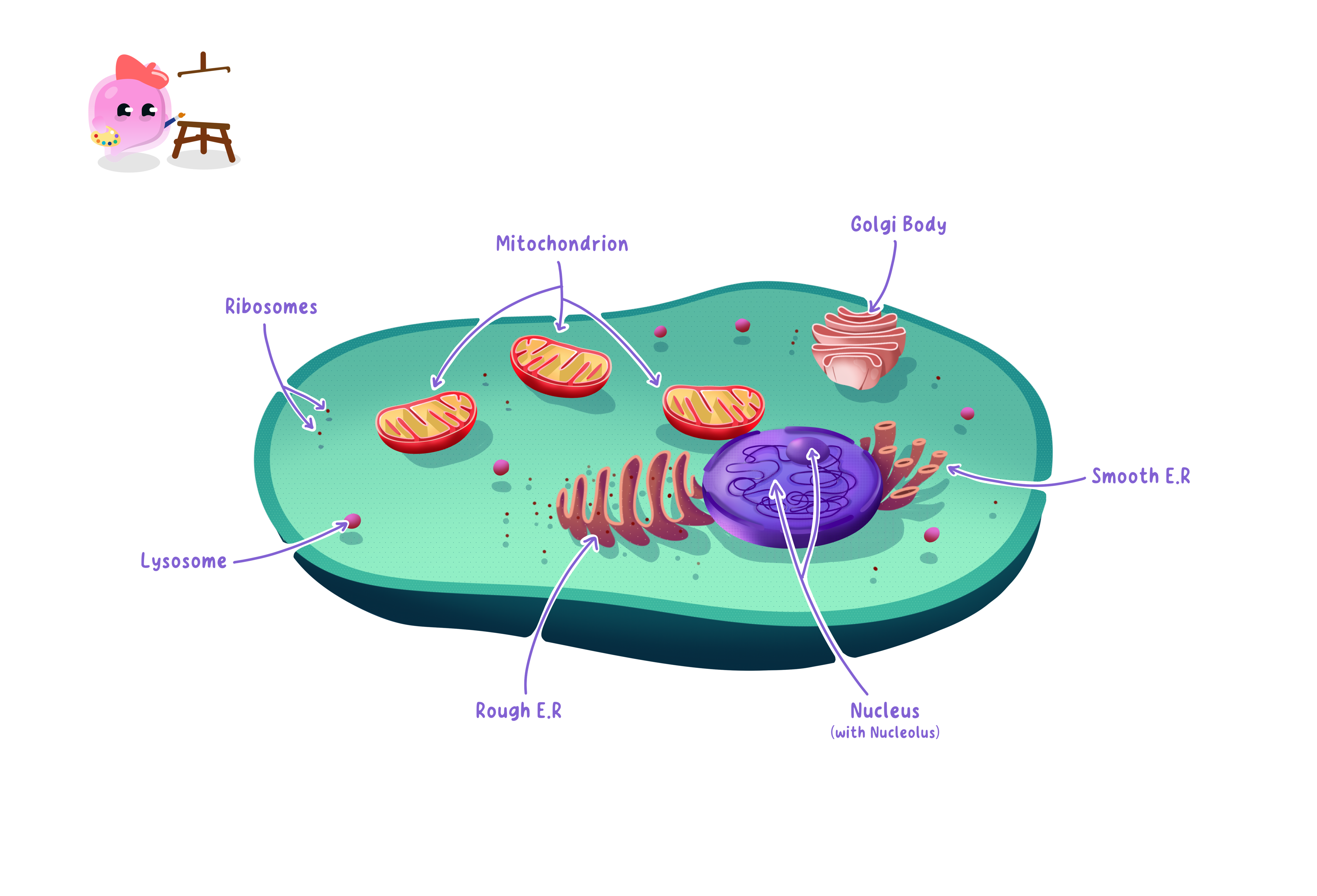
An animal cell has no cell wall, no chloroplasts and no permanent vacuole, so a diagram drawn with a thick rigid outer layer is not an animal cell.
Extra structures in a plant cell
- A plant cell contains every structure an animal cell has, and then three more.
- The cell wall is a rigid layer of cellulose outside the cell membrane.
- It supports the cell and stops it bursting when water moves in, which is why plant tissue stays firm.
- Chloroplasts contain the green pigment chlorophyll, which absorbs light so that photosynthesis can take place.
- The permanent vacuole is a large sac of cell sap whose pressure pushes outwards on the cell wall and keeps the cell firm.
- Not every plant cell has chloroplasts, because root cells are underground and receive no light.
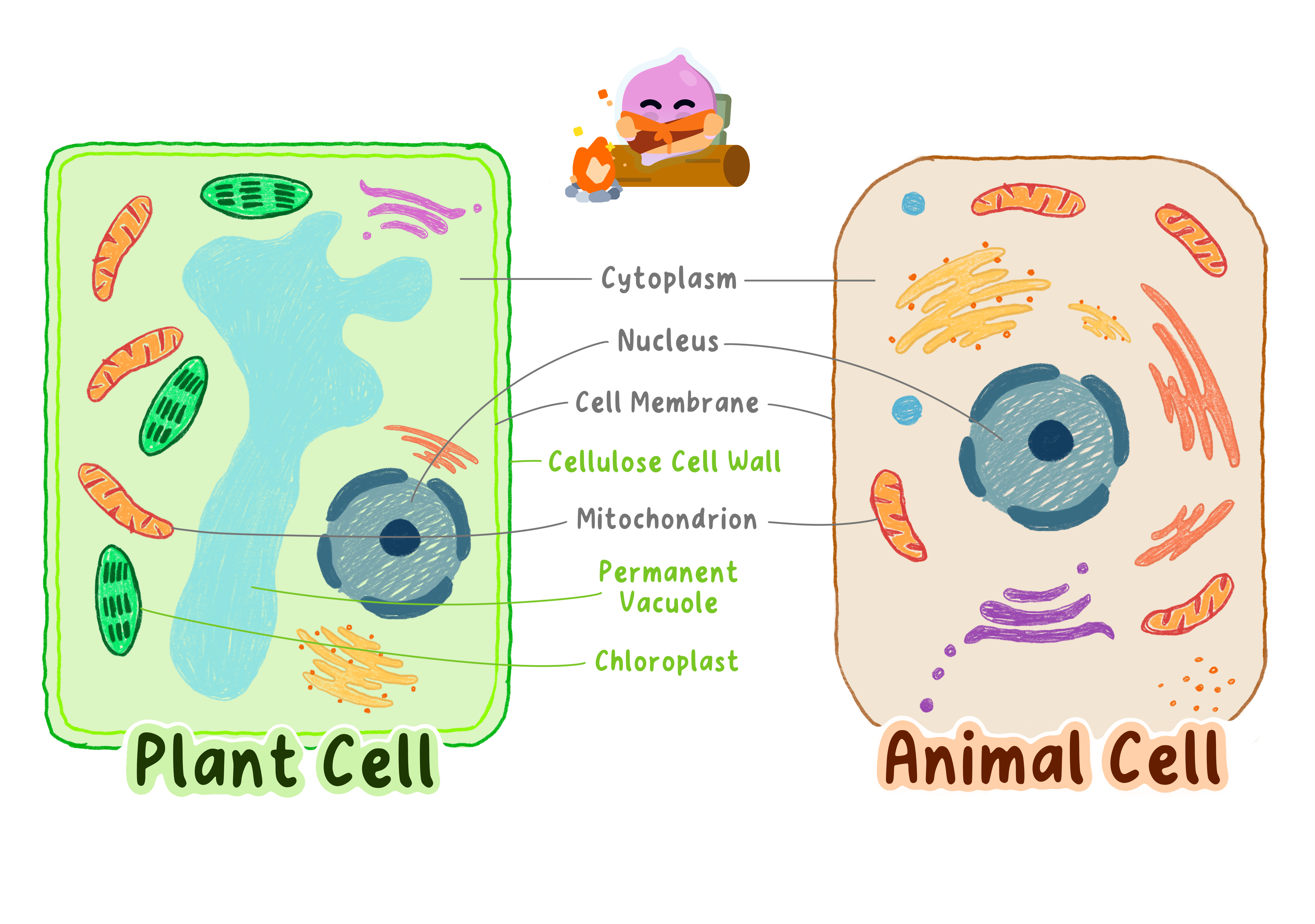
The cell wall and the cell membrane are different structures: the wall is rigid and made of cellulose, and the membrane is the thin partially permeable layer just inside it.
Structures in a bacterial cell
Prokaryotic cell
A cell that has no nucleus, its genetic material instead lying free in the cytoplasm as a single circular chromosome, as in bacteria.
- A bacterial cell holds its chromosomal DNA as one long circular strand floating freely in the cytoplasm.
- It also carries plasmid DNA, small separate circles of DNA holding extra genes such as those for antibiotic resistance.
- A cell membrane surrounds the cytoplasm and controls what passes in and out, exactly as it does in a eukaryotic cell.
- Ribosomes are present, so a bacterium makes its own proteins, although they are smaller than eukaryotic ribosomes.
- Many bacteria also have one or more flagella, long whip-like tails that rotate to drive the cell through liquid.
- There is no nucleus and there are no mitochondria, so a bacterium respires using enzymes held in its cytoplasm and membrane.
Bacteria have a cell wall as well, but it is not made of cellulose, so a cell wall on its own never proves that a cell is a plant cell.
Comparing the three cell types
- Only eukaryotic cells have a nucleus, which is the clearest single difference between them and prokaryotic cells.
- Cell membrane, cytoplasm and ribosomes appear in animal, plant and bacterial cells alike.
- Mitochondria are found in eukaryotic cells only.
- A cell wall is present in plant and bacterial cells but absent from animal cells.
- Chloroplasts and a permanent vacuole are found only in plant cells.
- Check the command word: state wants the name of the organelle, while explain wants the name, the job and the reason the cell needs it.
- The mark most often dropped is the reason, so follow mitochondria with so more energy is released by aerobic respiration for the cell to use.
- Given an unfamiliar cell, read off what it contains and work backwards to its function instead of trying to recall the cell by name.
- Cells do not make energy, they transfer it, so write that respiration releases energy rather than creates it.
- The nucleus stores the instructions for proteins, but the proteins themselves are assembled on the ribosomes.
- Bacterial DNA is described as free in the cytoplasm, never as being inside a nucleus.
- A leaf cell contains chloroplasts and mitochondria together, because plants respire as well as photosynthesise.
- What is the difference between a eukaryotic and a prokaryotic cell?
- State the function of the nucleus, the mitochondria and the ribosomes.
- Name the three structures a plant cell has that an animal cell does not.
- What is plasmid DNA and how does it differ from chromosomal DNA?
- Explain why a muscle cell contains many mitochondria.
1.1.2 Specialised cells and their adaptations
How cells become specialised
Specialised cell
A cell whose structure is adapted so that it carries out one particular function.
- Every cell in a multicellular organism carries the same genes, because they all came from one fertilised egg.
- A cell becomes specialised by differentiation, switching on only the genes it needs and switching the rest off.
- Different genes being used produces different proteins, and different proteins give the cell a different shape and different contents.
- An adaptation is a feature that helps a cell carry out its particular job well.
- Because structure follows function, you can usually work out what an unfamiliar cell does from its shape and contents alone.
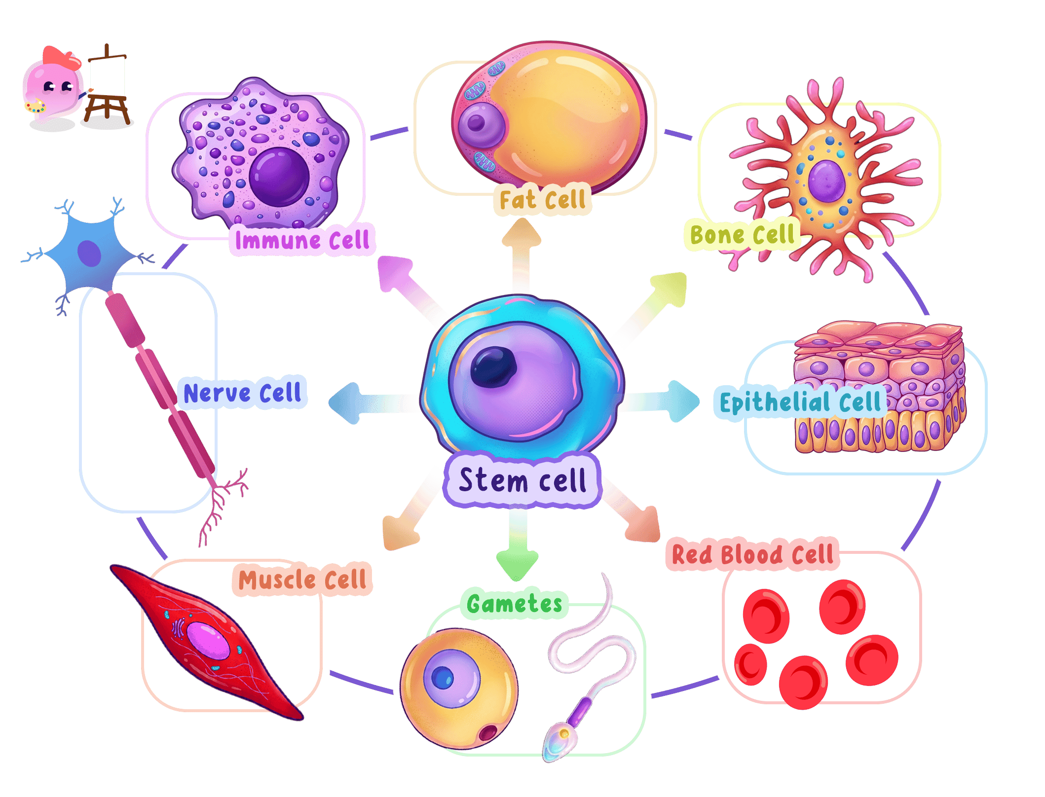
Edexcel names three specialised cells in this part of the specification, sperm cells, egg cells and ciliated epithelial cells, and expects each adaptation to be linked to the function it serves.
Sperm cells
Acrosome
The vesicle at the tip of a sperm cell that contains enzymes to digest the outer layers of an egg cell.
- A sperm cell is the male gamete, and its job is to reach an egg cell and fuse with it.
- The acrosome at the tip stores digestive enzymes.
- The enzymes break down the outer layers of the egg cell so the sperm can push through and reach the egg membrane.
- The haploid nucleus holds one set of chromosomes, which is 232323 in a human rather than the usual 464646.
- Fusing a haploid sperm nucleus with a haploid egg nucleus restores the full number of chromosomes in the fertilised egg.
- Many mitochondria are packed into the middle section of the sperm.
- They release energy by aerobic respiration, and that energy drives the tail.
- The tail beats from side to side so the sperm can swim towards the egg.
A sperm cell carries almost no cytoplasm, which keeps it light and makes swimming easier.
Egg cells
- An egg cell is the female gamete, and its job is to be fertilised and then supply the early embryo with what it needs.
- The cytoplasm is large and packed with nutrients.
- These nutrients feed the embryo while it divides and travels down the oviduct, before it implants and gains a blood supply.
- The haploid nucleus holds one set of chromosomes, matching the single set brought by the sperm.
- As soon as one sperm has entered, the cell membrane changes so that it becomes impossible for any other sperm to get in.
- This keeps the chromosome number correct, because a second sperm would give the zygote too many chromosomes and it would not develop.
The membrane change happens straight after fertilisation, not before it, so describing the egg as blocking sperm from the start loses the mark.
Ciliated epithelial cells
Ciliated epithelial cell
A lining cell covered in tiny hair-like cilia that beat together to sweep mucus and trapped particles along a tube such as an airway.
- Ciliated epithelial cells line the trachea and bronchi, and also the oviducts.
- The surface facing into the tube is covered with cilia, tiny hair-like projections that beat together in a wave.
- In the airways, goblet cells release mucus that traps dust, pollen and bacteria in the air you breathe in.
- The cilia then sweep that mucus up towards the throat, where it is swallowed, so the trapped microorganisms never reach the lungs.
- In the oviduct the same beating action moves an egg cell along towards the uterus.
- The cells contain many mitochondria, because beating cilia continuously needs a steady supply of energy from respiration.
Cigarette smoke paralyses and destroys cilia, so mucus and the microorganisms in it collect in the airways instead of being cleared, which is why smokers cough and catch chest infections more easily.
- Other specialised cells you should know include neurones, root hair cells, xylem, phloem and red blood cells.
- Neurones are covered in detail with the nervous system in Topic 2.
- Root hair cells, xylem and phloem are covered in detail with plant transport in Topic 6.
- Red blood cells are covered in detail with the circulatory system in Topic 8.
- What is differentiation?
- Give the function of the acrosome and say why a sperm cell needs it.
- Why do both gametes have a haploid nucleus?
- What happens to the egg cell membrane after fertilisation, and why?
- Explain how ciliated epithelial cells help keep the lungs free of bacteria.
1.1.3 Microscope technology and cell structures
Magnification and resolution
Resolution
The smallest distance between two points that can still be seen as two separate points.
- A microscope has to do two separate jobs, and they are easily confused.
- Magnification is how many times bigger the image is than the real object.
- Resolution is the smallest gap between two points that still appears as two points rather than one blur.
- Once the magnification goes past what the resolution can support, the image gets bigger but no clearer.
- Resolution is therefore what decides which structures can be seen at all, and it is the reason new microscope technology mattered.
Higher resolution means finer detail, and every advance in what scientists knew about organelles followed an advance in resolution.
The light microscope
Light microscope
A microscope that uses a beam of light focused by glass lenses to produce a magnified image of a specimen.
- A light microscope passes a beam of light through a thin specimen and focuses it with glass lenses.
- Its useful magnification stops at roughly ×1500\times 1500×1500, and its resolution is about 200 nm200\,\text{nm}200nm.
- That limit exists because two points closer together than about half the wavelength of light cannot be separated, however good the lenses are.
- A light microscope shows the nucleus, cytoplasm, cell membrane, cell wall, chloroplasts and the vacuole.
- It cannot show ribosomes, plasmids, the inside of a mitochondrion or any detail of the membrane itself.
- It is still used every day because it is cheap, portable, simple to use, and it can show living cells in colour.
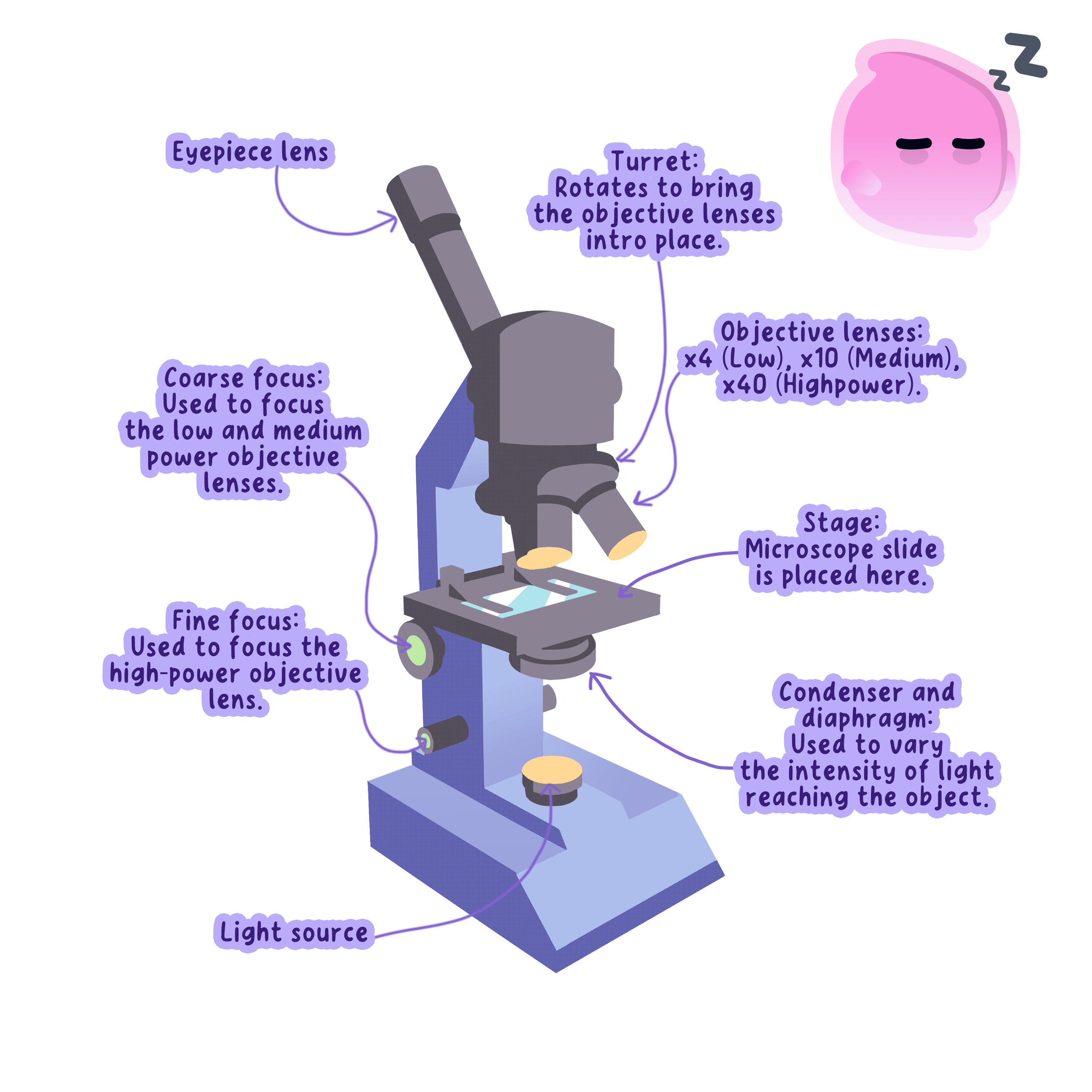
Total magnification is the eyepiece lens multiplied by the objective lens, so a ×10\times 10×10 eyepiece with a ×40\times 40×40 objective gives ×400\times 400×400.
The electron microscope
Electron microscope
A microscope that uses a beam of electrons rather than light, giving far higher magnification and resolution than a light microscope.
- Electron microscopes were developed in the 1930s and use a beam of electrons in place of light.
- An electron beam has a far shorter wavelength than light, so the resolution improves to about 0.2 nm0.2\,\text{nm}0.2nm.
- That is roughly a thousand times better than a light microscope, and magnifications above ×1 000 000\times 1\,000\,000×1000000 become useful.
- Specimens must be dead, chemically fixed and placed in a vacuum, because air would scatter the electron beam.
- The instruments are large, expensive and need trained operators, and the images they produce are black and white until colour is added artificially.
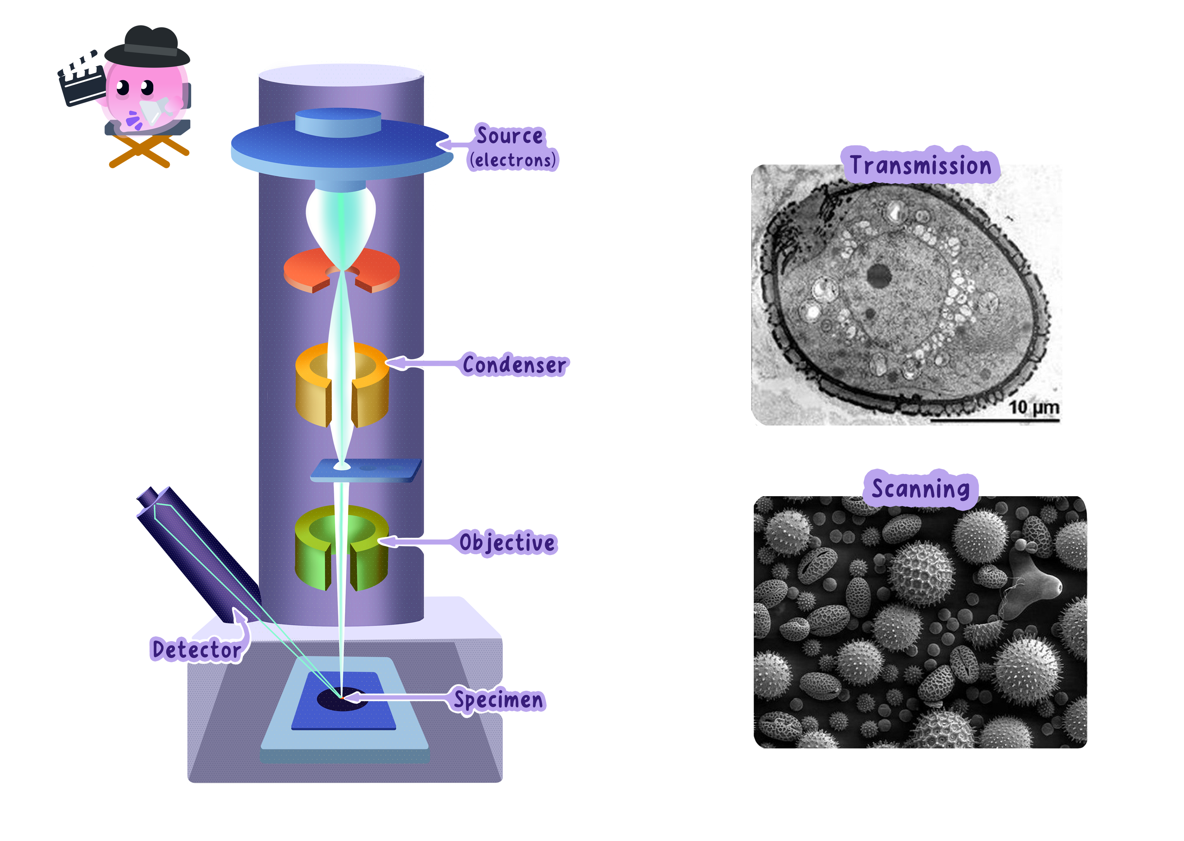
Saying only that an electron microscope has a higher magnification is not enough for the marks, because the reason more can be seen is the higher resolution.
What better microscopes revealed
- Under a light microscope a mitochondrion was no more than a dot, and its job was unknown.
- Electron microscopy showed a double membrane with the inner one deeply folded, and that folded surface was then linked to the reactions of aerobic respiration.
- Ribosomes were invisible before electron microscopy, and only once they could be seen was protein synthesis traced to them.
- Chloroplasts turned out to contain stacks of internal membranes, which explained how they hold enough chlorophyll to absorb light efficiently.
- The cell membrane was resolved as a distinct double layer, which supported the model of a partially permeable barrier.
- Plasmids and viruses became visible for the first time, and both later became central to medicine and genetic engineering.
- The pattern is the one Edexcel wants you to describe: better technology gave better evidence, and better evidence changed the accepted explanation.
- A question asking how microscope technology changed our understanding wants a named organelle, what was newly seen inside it, and the function that was then worked out.
- Use both words correctly in the same answer, because credit is usually split between a magnification point and a resolution point.
- If asked to evaluate the two microscopes, give a strength of the light microscope as well, since cost, portability and living specimens are creditable answers.
Looking at Cells
- Aim: to prepare slides of onion epidermis and cheek cells, observe them under a light microscope, draw them, and calculate magnification and actual size.
- Apparatus: light microscope and lamp, glass slides, coverslips, mounted needle, forceps, pipette, iodine solution, methylene blue, an onion, a sterile wooden spatula, paper towels, a beaker of 1% Virkon, gloves and eye protection.
- Method, plant slide:
- Peel one thin layer of epidermis from the inside of an onion scale with forceps.
- Lay it flat in a drop of water on the slide with no folds, because folded tissue is too thick for light to pass through and will never come into focus.
- Add one drop of iodine solution to stain it, which raises the contrast so the nucleus and cell wall stand out.
- Rest one edge of the coverslip on the slide and lower it slowly at about 45∘45^\circ45∘ with a mounted needle, so air is pushed out ahead of it instead of being trapped as bubbles.
- Blot the excess stain with a paper towel.
- Method, cheek cells:
- Rub a sterile wooden spatula gently along the inside of your cheek.
- Smear it thinly onto a clean slide.
- Add a drop of methylene blue and lower a coverslip in the same way.
- Drop the used spatula straight into 1% Virkon.
- Method, focusing:
- Clip the slide onto the stage.
- Start on the lowest-power objective.
- Watching from the side, turn the coarse focus to bring the objective close to the slide.
- Now look down the eyepiece and turn the coarse focus the other way, moving the lens away until the cells come into view.
- Switch to a higher-power objective and sharpen using the fine focus only.
- Results: onion cells appear as neat rectangular boxes with a clear cell wall and a stained nucleus, while cheek cells are irregular blobs with a nucleus, cytoplasm and membrane and no wall.
- Maths: total magnification = eyepiece ×\times× objective, and actual size = image size ÷\div÷ magnification. To estimate real cell size, lay a transparent ruler across the low-power field of view, measure the field diameter in mm, convert to μm\mu\text{m}μm by multiplying by 100010001000, then judge what fraction of that diameter one cell takes up.
- Watch out: trapped air bubbles have thick dark outlines and are regularly mistaken for cells, the specimen has to be a single cell layer thick, and the coarse focus must never be used at high power because the objective can crack the slide.
- Safety: wear eye protection because iodine and methylene blue stain skin and clothes, never re-use a spatula, put anything that touched a mouth into 1% Virkon, and handle coverslips carefully because they snap easily.
Scientific drawings
- Use a sharp pencil and draw clean single lines, with no shading and no sketchy repeated strokes.
- Make the drawing large enough to fill the space you are given.
- Draw label lines straight, without arrowheads, and do not let them cross one another.
- Give the drawing a title and state the magnification you used.
- Draw only what is actually on your slide, not the textbook version of the cell.
- Give the difference between magnification and resolution.
- State the approximate resolution of a light microscope and of an electron microscope.
- Name two structures visible with an electron microscope but not with a light microscope.
- Explain why the coverslip is lowered at an angle when preparing a slide.
- Describe one way electron microscopy changed what scientists knew about an organelle.
1.2.1 Number, size and scale with estimations
The scale of cells and organelles
- Cells and organelles are far too small to measure comfortably in millimetres, so biologists work in micrometres and nanometres.
- A typical animal cell is around 20 μm20\,\mu\text{m}20μm across, and a plant cell is usually larger at 505050 to 100 μm100\,\mu\text{m}100μm.
- A bacterial cell is about 2 μm2\,\mu\text{m}2μm long, roughly ten times smaller than an animal cell.
- Inside a cell, a nucleus is around 6 μm6\,\mu\text{m}6μm across and a mitochondrion about 2 μm2\,\mu\text{m}2μm long.
- A ribosome is only about 20 nm20\,\text{nm}20nm across, a thousand times smaller again, which is why a light microscope cannot resolve one.
- Being able to put these in order matters more than memorising exact values, because Edexcel asks you to compare scales rather than recite figures.
Each factor of ten is one order of magnitude, so a 20 μm20\,\mu\text{m}20μm animal cell is about one order of magnitude larger than a 2 μm2\,\mu\text{m}2μm bacterium and three orders of magnitude larger than a 20 nm20\,\text{nm}20nm ribosome.
The magnification equation
Magnification
The number of times larger an image is than the actual object it shows.
- Magnification links the size of an image to the real size of the object it shows.
- magnification=image sizeactual size\text{magnification} = \dfrac{\text{image size}}{\text{actual size}}magnification=actual sizeimage size
- Rearranging it gives the form you need whenever a question supplies the magnification and asks for the real size.
- actual size=image sizemagnification\text{actual size} = \dfrac{\text{image size}}{\text{magnification}}actual size=magnificationimage size
- Magnification has no units, because it is one length divided by another, and it is written with a multiplication sign such as ×400\times 400×400.
- Both lengths have to be in the same unit before you divide.
- On a light microscope the total magnification is the eyepiece lens multiplied by the objective lens in use.
Almost every lost mark on these questions comes from dividing a length in millimetres by a length in micrometres, so convert the units first and write the conversion down.
- A drawing of a cheek cell measures 60 mm60\,\text{mm}60mm across and is labelled ×1000\times 1000×1000.
- Convert the image size into micrometres, since the answer will be a cell size: 60 mm=60×1000=60 000 μm60\,\text{mm} = 60 \times 1000 = 60\,000\,\mu\text{m}60mm=60×1000=60000μm.
- Now divide the image size by the magnification.
- actual size=60 0001000=60 μm\text{actual size} = \dfrac{60\,000}{1000} = 60\,\mu\text{m}actual size=100060000=60μm
- Check the answer against what you know, because 60 μm60\,\mu\text{m}60μm is a sensible size for a cheek cell.
An answer in the millions or in tiny fractions is usually a sign that a unit conversion has been missed.
- An electron micrograph shows a mitochondrion measuring 40 mm40\,\text{mm}40mm long, and its actual length is 2 μm2\,\mu\text{m}2μm.
- Convert the image size into the same unit as the actual size: 40 mm=40 000 μm40\,\text{mm} = 40\,000\,\mu\text{m}40mm=40000μm.
- Divide the image size by the actual size.
- magnification=40 0002=20 000\text{magnification} = \dfrac{40\,000}{2} = 20\,000magnification=240000=20000
- Write the answer as ×20 000\times 20\,000×20000 and give it no units.
- Set the working out in three lines, equation, substitution, answer, because a conversion error still earns method marks if the examiner can see the equation.
- Measure the image yourself with a ruler when the question tells you to, and give the measurement in millimetres before converting.
- Read the unit demanded in the answer line, since a question that asks for micrometres will not credit an answer left in millimetres.
When to use an estimate
Estimate
An approximate value worked out from rounded figures, used when an exact measurement is not needed or not possible.
- Use an estimate when an exact measurement is not possible, such as when cells overlap or run off the edge of the field of view.
- Use one when the quantity naturally varies, because cells in the same tissue are not all the same size and a single exact value would be misleading.
- Use one to check a calculated answer, since an estimate quickly shows whether a result is the right order of magnitude.
- To estimate a cell size, lay a transparent ruler across the field of view and measure its diameter, then count how many cells span it.
- If the field of view is 4.5 mm4.5\,\text{mm}4.5mm across and 151515 onion cells fit end to end along it, convert to 4500 μm4500\,\mu\text{m}4500μm and divide.
- estimated cell length=450015=300 μm\text{estimated cell length} = \dfrac{4500}{15} = 300\,\mu\text{m}estimated cell length=154500=300μm
- Say clearly that the value is an estimate and give one reason, for example that the cells are not all identical in length.
- Write the magnification equation and both of its rearranged forms.
- Why does magnification have no units?
- An image is 30 mm30\,\text{mm}30mm wide at ×500\times 500×500, so what is the actual width in micrometres?
- Put a bacterium, a ribosome and a plant cell in order of size, smallest first.
- Give two situations in which an estimate is more appropriate than an exact measurement.
1.2.2 Quantitative units in relation to cells
The four prefixes you need
Micrometre
A unit of length equal to one millionth of a metre, written µm, used for measuring cells and organelles.
- A prefix in front of a unit is a shorthand for multiplying that unit by a power of ten.
- Milli means 10−310^{-3}10−3, so 1 mm1\,\text{mm}1mm is one thousandth of a metre and is the unit you use for an image measured with a ruler.
- Micro means 10−610^{-6}10−6, so 1 μm1\,\mu\text{m}1μm is one millionth of a metre and is the working unit for whole cells and large organelles.
- Nano means 10−910^{-9}10−9, so 1 nm1\,\text{nm}1nm is one thousand millionth of a metre and is used for ribosomes, membranes and viruses.
- Pico means 10−1210^{-12}10−12, and at that scale you are measuring individual atoms rather than biological structures.
- Each prefix in that list is 100010001000 times smaller than the one before it.
The symbol for micro is the Greek letter mu, written μ\muμ, so a micrometre is μm\mu\text{m}μm and is sometimes called a micron.
Converting between units
- Moving down one prefix step means multiplying by 100010001000, and moving up one step means dividing by 100010001000.
- 1 m=1000 mm1 mm=1000 μm1 μm=1000 nm1 nm=1000 pm1\,\text{m} = 1000\,\text{mm} \qquad 1\,\text{mm} = 1000\,\mu\text{m} \qquad 1\,\mu\text{m} = 1000\,\text{nm} \qquad 1\,\text{nm} = 1000\,\text{pm}1m=1000mm1mm=1000μm1μm=1000nm1nm=1000pm
- Going from a larger unit to a smaller one always makes the number bigger, which is a quick way to check the conversion went the right way.
- Going from millimetres straight to nanometres skips a step, so you multiply by 1000×1000=1061000 \times 1000 = 10^{6}1000×1000=106.
Write the conversion out as a separate line of working, because an examiner can award a method mark for a correct conversion even when the final answer is wrong.
- A plant cell is measured as 0.045 mm0.045\,\text{mm}0.045mm long, and the question asks for the length in micrometres.
- Millimetres to micrometres is one step down, so multiply by 100010001000.
- 0.045 mm×1000=45 μm0.045\,\text{mm} \times 1000 = 45\,\mu\text{m}0.045mm×1000=45μm
- To reach nanometres, multiply by 100010001000 again to give 45 000 nm45\,000\,\text{nm}45000nm.
- The answer of 45 μm45\,\mu\text{m}45μm is a believable plant cell length, which confirms the conversion ran in the right direction.
Dividing when you should multiply gives an answer one million times out, so always ask whether the number should have grown or shrunk before writing it down.
Standard form on higher tier
Standard form
A way of writing a number as a value between 1 and 10 multiplied by a power of ten.
- Calculations using standard form are printed in bold in the Edexcel specification, which means they are assessed on higher tier only.
- A number in standard form is written as a value between 111 and 101010 multiplied by a power of ten.
- 45 μm=4.5×10⁻⁵ m20 nm=2×10⁻⁸ m45\,\mu\text{m} = 4.5 \times 10^{-5}\,\text{m} \qquad 20\,\text{nm} = 2 \times 10^{-8}\,\text{m}45μm=4.5×10⁻⁵m20nm=2×10⁻⁸m
- A negative power means the number is smaller than one, and the size of the power tells you how many places the decimal point moves.
- Standard form is what makes cell sizes comparable, because 2×10−82 \times 10^{-8}2×10⁻⁸ is instantly recognisable as smaller than 4.5×10−54.5 \times 10^{-5}4.5×10⁻⁵.
Calculating in standard form
- To multiply, multiply the front numbers and add the powers.
- To divide, divide the front numbers and subtract the powers.
- An electron micrograph of a mitochondrion measures 4.5×10⁻² m4.5 \times 10^{-2}\,\text{m}4.5×10⁻²m, and the real mitochondrion is 3×10⁻⁶ m3 \times 10^{-6}\,\text{m}3×10⁻⁶m long.
- magnification=4.5×10−23×10−6=1.5×10⁴\text{magnification} = \dfrac{4.5 \times 10^{-2}}{3 \times 10^{-6}} = 1.5 \times 10^{4}magnification=3×10−64.5×10−2=1.5×10⁴
- Divide 4.54.54.5 by 333 to get 1.51.51.5, then subtract the powers so that −2−(−6)=4-2 - (-6) = 4−2−(−6)=4.
- The magnification is therefore ×15 000\times 15\,000×15000, and both forms of the answer are acceptable.
- Use the standard form button on your calculator rather than typing out strings of zeros, since a miscounted zero is the commonest error here.
- Convert everything into metres before you start when a question mixes millimetres, micrometres and nanometres.
- Check that a front number sits between 111 and 101010, because 15×10315 \times 10^{3}15×103 is not properly in standard form.
Choosing a sensible unit
- Pick the unit that keeps the number easy to read, so a cell is quoted in micrometres rather than as 0.00002 m0.00002\,\text{m}0.00002m.
- Measure images in millimetres, quote cells and large organelles in micrometres, and quote ribosomes, membranes and viruses in nanometres.
- Keep the same unit throughout a comparison, because two sizes in different units cannot be compared directly.
- Always write the unit next to the number, since an unlabelled answer is treated as incomplete.
- State the power of ten that each of milli, micro, nano and pico stands for.
- How many nanometres are there in one micrometre?
- Convert 0.008 mm0.008\,\text{mm}0.008mm into micrometres and then into nanometres.
- Write 250 nm250\,\text{nm}250nm in metres in standard form.
- Which of these two quantities is larger, 3×10⁻⁶ m3 \times 10^{-6}\,\text{m}3×10⁻⁶m or 8×10⁻⁸ m8 \times 10^{-8}\,\text{m}8×10⁻⁸m?
