8.2.1 Structure and function of the blood
Blood Has Four Main Components
- Plasma is the liquid that suspends the blood cells and carries dissolved materials and heat.
- Red blood cells, also called erythrocytes, transport oxygen.
- White blood cells defend the body against pathogens.
- Platelets are cell fragments involved in blood clotting.
Red Blood Cells Carry Oxygen
Red blood cell
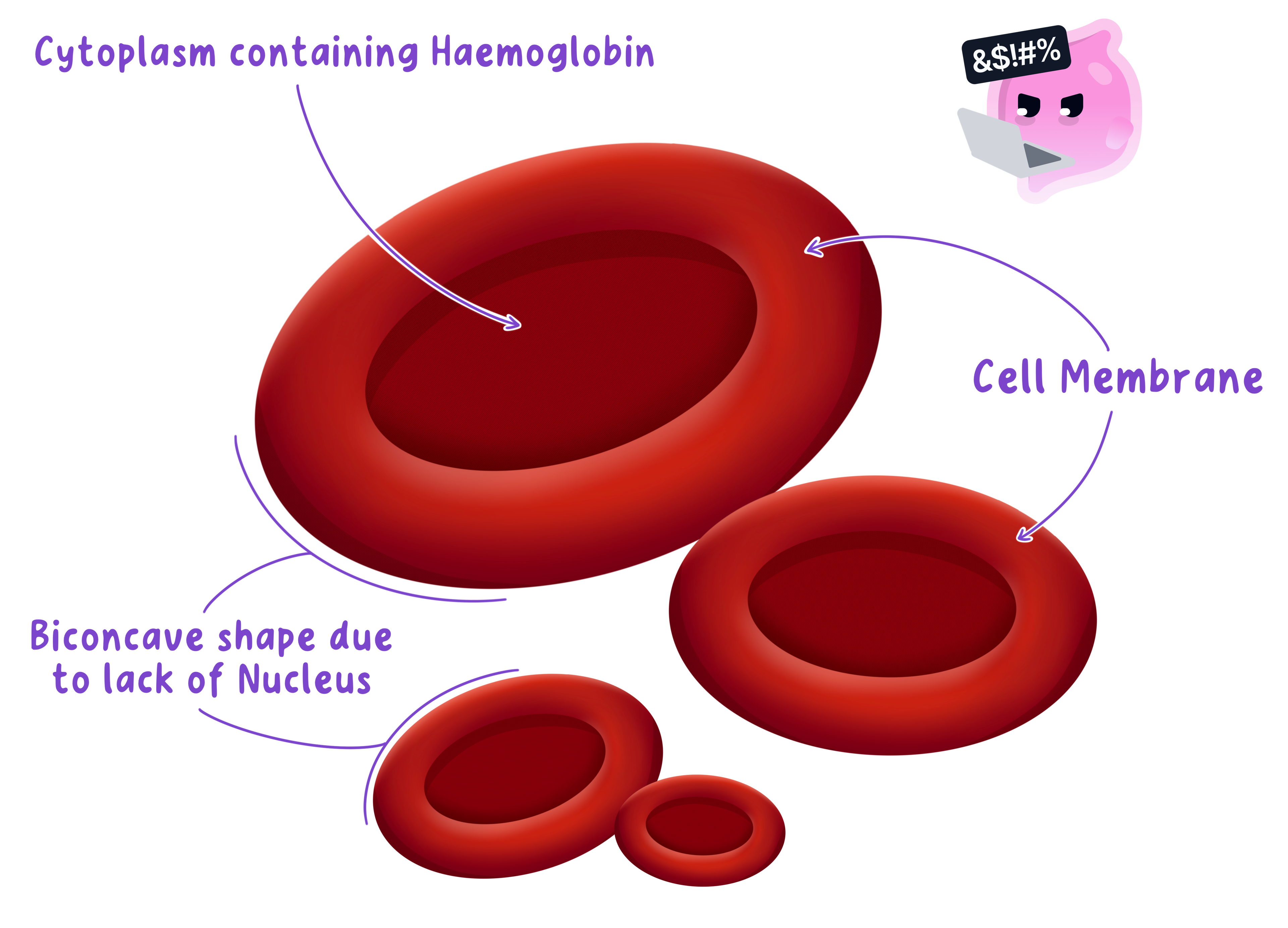
A biconcave blood cell with no nucleus that contains haemoglobin and transports oxygen around the body.
Haemoglobin
A red protein in red blood cells that binds reversibly to oxygen for transport in the blood.
- A red blood cell is a biconcave disc, so its curved surfaces give a large surface area relative to its volume for rapid oxygen diffusion.
- The thin centre gives oxygen a short diffusion distance into and out of the cell.
- A mature red blood cell has no nucleus, leaving more internal space for haemoglobin.
- Haemoglobin binds oxygen reversibly in the lungs to form oxyhaemoglobin and releases oxygen where cells are respiring.
- The cell is small and flexible, allowing it to bend through capillaries that are narrower than its resting diameter.
Phagocytes Engulf Pathogens
White blood cell
A nucleated blood cell that defends the body against pathogens through the actions of phagocytes and lymphocytes.
Phagocyte
A white blood cell that engulfs and digests pathogens by phagocytosis.
- White blood cells have a nucleus and can change shape as they move through tissues.
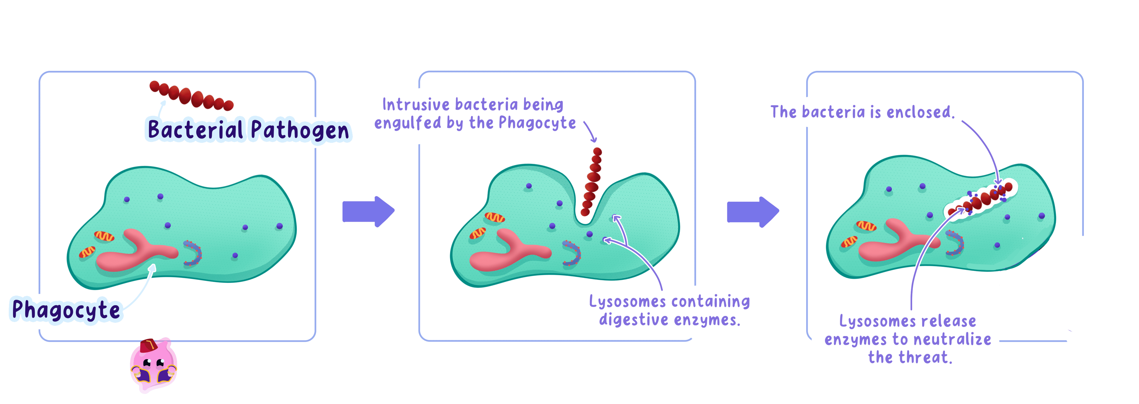
- A phagocyte detects a pathogen, surrounds it with its cell membrane and encloses it inside the cell.
- Digestive enzymes then break down the engulfed pathogen in a process called phagocytosis.
- The defence is non-specific because a phagocyte can engulf different types of pathogen.
Lymphocytes Make Specific Antibodies
Lymphocyte
A white blood cell that produces antibodies specific to antigens on pathogens.
Antibody
A protein produced by lymphocytes that binds to a specific antigen because their shapes are complementary.
- A lymphocyte recognises an antigen, a molecule on the surface of a pathogen.
- The lymphocyte produces an antibody with a binding site complementary to that antigen.
- The antibody binds only to its matching antigen, which helps the immune system identify and destroy the pathogen.
- This response is specific because a different antigen requires a different antibody.
- When comparing white blood cells, state that phagocytes engulf and digest pathogens, whereas lymphocytes produce antibodies.
- Do not write that every white blood cell makes antibodies, because this is the role of lymphocytes.
Platelets Form Blood Clots
Platelet
A cell fragment that helps form a blood clot at a wound, reducing blood loss and pathogen entry.
- When a blood vessel is damaged, platelets gather at the wound and trigger a series of clotting reactions.
- A mesh of insoluble fibres forms and traps blood cells, producing a clot.
- The clot reduces blood loss and blocks pathogens from entering through the break in the skin.
- The clot later dries into a scab while the tissue underneath repairs.
Plasma Carries Dissolved Materials
Plasma
The liquid part of blood that transports blood cells, dissolved substances and heat around the body.
- Plasma carries carbon dioxide from respiring tissues towards the lungs.
- It transports soluble products of digestion, including glucose and amino acids, from the small intestine to cells.
- It carries urea from the liver to the kidneys for excretion.
- It transports hormones from endocrine glands to their target organs.
- It distributes heat from active organs to other parts of the body.
- Blood cells and platelets are suspended in plasma and carried through the circulation.
Structure Answers Need a Function Link
- For each adaptation, name the structure first and then state exactly how it helps the component perform its function.
- A complete red blood cell link is: no nucleus, more space for haemoglobin, more oxygen can be transported.
- A complete platelet link is: cell fragments collect at damage, a clot forms, blood loss and pathogen entry are reduced.
- What are the four main components of blood?
- How does the biconcave shape of a red blood cell improve oxygen transport?
- How does a phagocyte destroy a pathogen?
- Why are antibodies described as specific?
- How does a blood clot protect the body?
8.2.2 Structure of blood vessels
Arteries Carry Blood at High Pressure
Artery
A blood vessel that carries blood away from the heart.
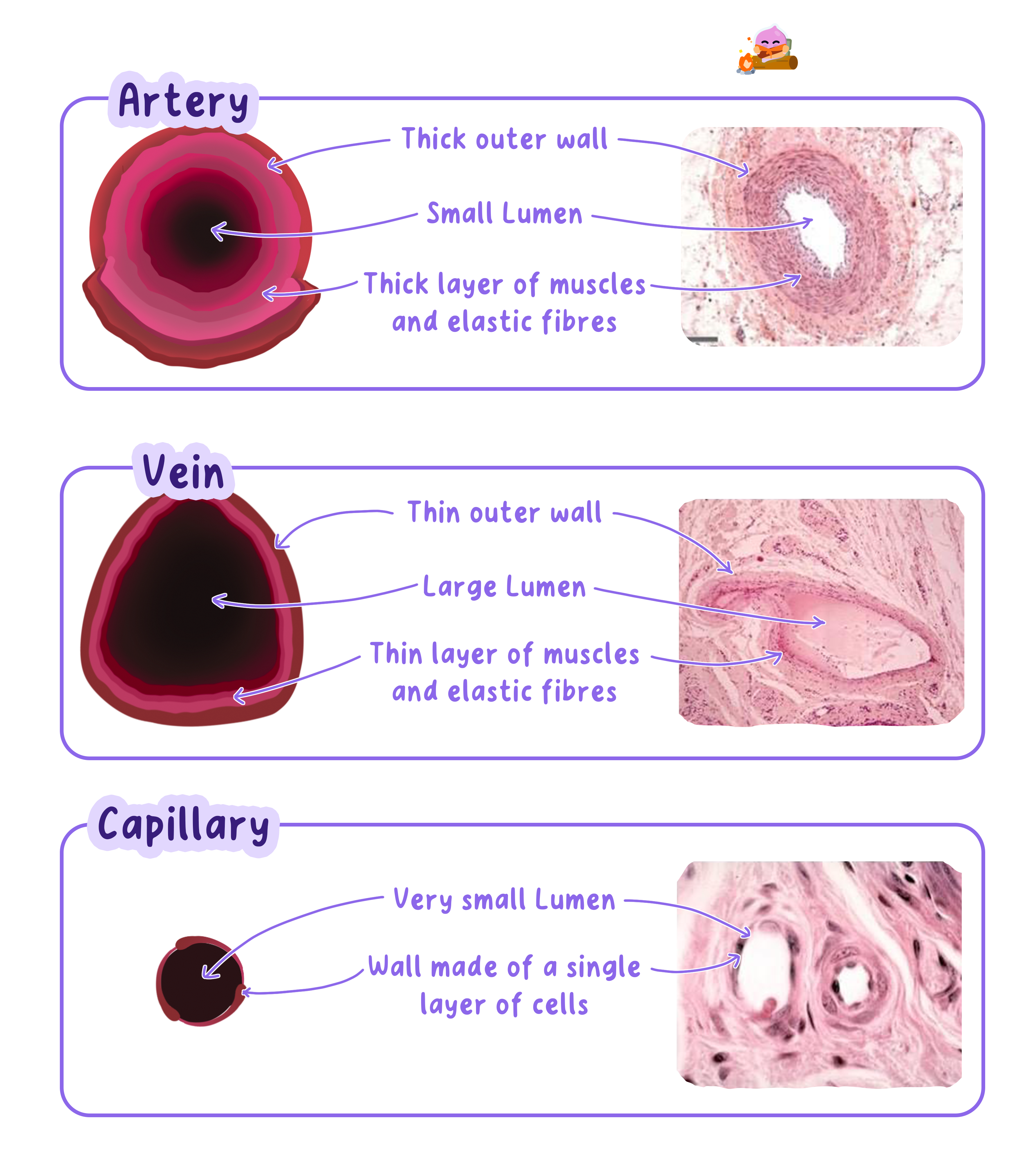
- Arteries carry blood away from the heart in pulses produced by ventricular contractions.
- Their walls contain thick layers of smooth muscle that withstand high pressure and can alter the vessel diameter.
- Their walls contain many elastic fibres that stretch when pressure rises and recoil between heartbeats, helping maintain blood flow.
- A relatively small lumen helps the vessel maintain high pressure.
- A tough outer layer prevents the wall from bursting.
Veins Return Blood at Low Pressure
Vein
A blood vessel that carries blood towards the heart.
- Veins carry blood towards the heart after pressure has fallen through the capillary network.
- Their walls are thinner and contain less muscle and elastic tissue because they do not experience arterial pressure.
- A large lumen reduces resistance to flow at low pressure.
- Valves open towards the heart and close if blood begins to move backwards.
- Contractions of surrounding skeletal muscles squeeze veins, and the valves make this pressure move blood towards the heart.
Capillaries Allow Rapid Exchange
Capillary
A microscopic blood vessel with a wall one cell thick where substances are exchanged between blood and tissues.
- Capillaries form extensive networks close to body cells, giving a large total surface area for exchange.
- Their walls consist of a single layer of flattened cells, so substances cross a very short diffusion distance.
- Their lumen is narrow, which brings red blood cells close to the wall and slows their passage through the network.
- Oxygen and glucose diffuse from the blood into tissues, while carbon dioxide diffuses from tissues into the blood.
- Small gaps between capillary wall cells allow plasma and dissolved substances to move into tissue fluid, but blood cells remain inside.
- An artery is defined by carrying blood away from the heart, not by carrying oxygenated blood.
- A vein is defined by carrying blood towards the heart, not by carrying deoxygenated blood.
Pulmonary Vessels Are Exceptions
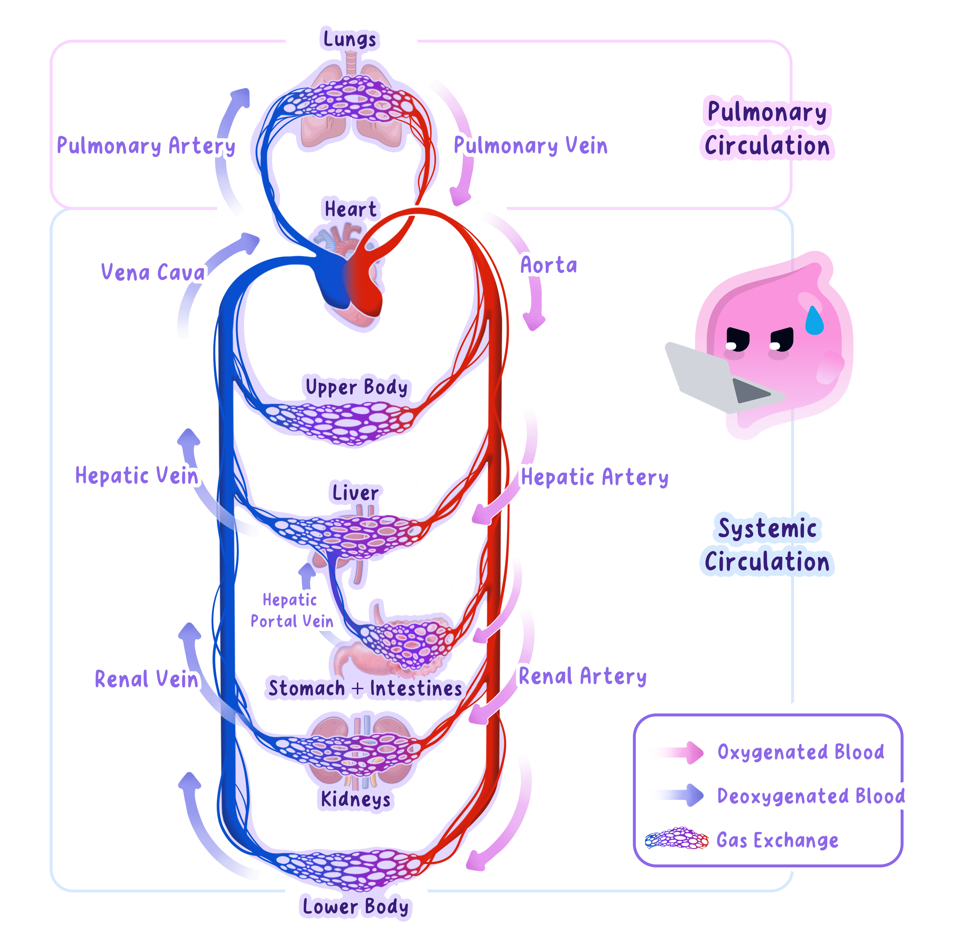
- The pulmonary artery carries deoxygenated blood from the right ventricle to the lungs.
- The pulmonary vein carries oxygenated blood from the lungs to the left atrium.
- Most systemic arteries carry oxygenated blood and most systemic veins carry deoxygenated blood, but direction remains the reliable definition.
Compare Vessels by Function
- For an artery, link high pressure to a thick muscular and elastic wall, then explain that recoil maintains blood flow.
- For a vein, link low pressure to a large lumen and explain that valves prevent backflow.
- For a capillary, link exchange to a one-cell-thick wall, narrow lumen and large network.
- A structure-and-function answer needs a causal link, such as one-cell-thick wall, shorter diffusion distance, faster diffusion.
- Naming a feature without explaining its consequence often leaves the explanation incomplete.
Pressure and Exchange Explain Structure
- Arteries need strength and elasticity because they receive blood directly from the ventricles.
- Veins need help preventing reverse flow because their blood pressure is low.
- Capillaries sacrifice thick walls for rapid exchange because pressure has already fallen before blood enters them.
- In which direction does an artery carry blood?
- Why do arteries contain elastic fibres?
- How do valves help blood return through veins?
- How does a capillary wall speed diffusion?
- Why is the pulmonary artery an exception to the usual oxygen pattern?
8.2.3 Structure of the heart and circulatory system
The Heart Is a Double Pump
Double circulatory system
A circulatory system in which blood passes through the heart twice during one complete circuit of the body.
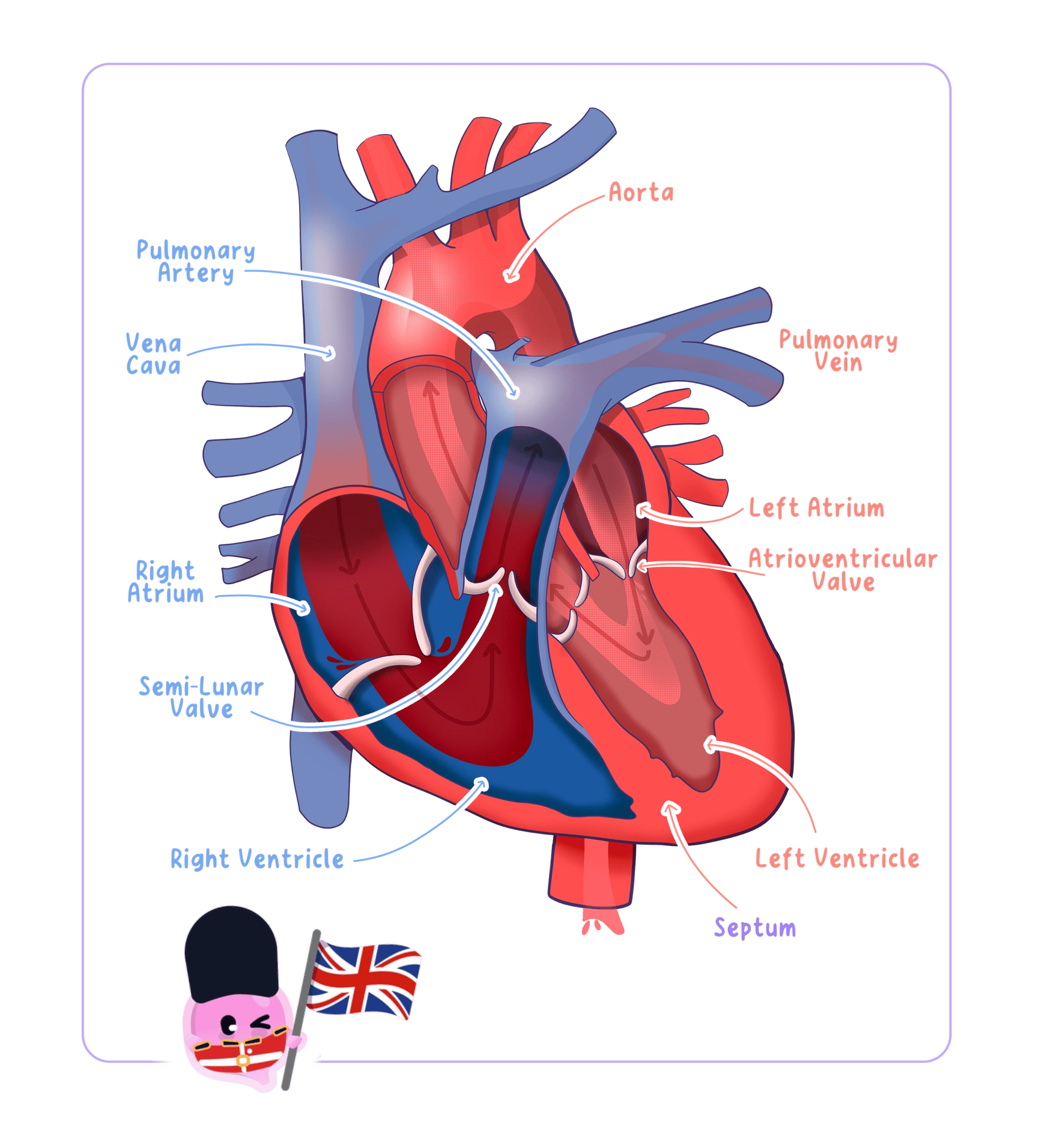
- The right side pumps deoxygenated blood to the lungs in the pulmonary circulation.
- The left side pumps oxygenated blood to the rest of the body in the systemic circulation.
- Blood returns to the heart between the two circuits, so it passes through the heart twice in one complete journey.
- Separate pumps allow lower pressure through delicate lung capillaries and higher pressure through the longer systemic circuit.
Chambers Control Blood Flow
Atrium
An upper chamber of the heart that receives blood returning to the heart.
Ventricle
A lower chamber of the heart that contracts to pump blood out of the heart.
- The right atrium receives deoxygenated blood from the body through the vena cava.
- The right atrium contracts and moves blood through an atrioventricular valve into the right ventricle.
- The right ventricle contracts and pumps blood through the pulmonary artery to the lungs.
- The left atrium receives oxygenated blood from the lungs through the pulmonary vein.
- The left atrium contracts and moves blood through an atrioventricular valve into the left ventricle.
- The left ventricle contracts and pumps blood through the aorta to the body.
Valves Prevent Backflow
Heart valve
A flap of tissue that opens in one direction to prevent the backflow of blood through the heart.
- Atrioventricular valves lie between each atrium and ventricle.
- They open when pressure is greater in an atrium than in its ventricle, allowing blood to move forwards.
- They close when ventricular pressure rises, preventing blood from returning to the atria.
- Valves at the exits of the ventricles prevent blood in the pulmonary artery and aorta from flowing back into the heart.
- Valves open and close because of pressure differences, rather than contracting by themselves.
The Septum Prevents Mixing
Septum
The muscular wall that separates the right and left sides of the heart and prevents oxygenated and deoxygenated blood from mixing.
- The septum keeps oxygenated blood on the left side separate from deoxygenated blood on the right side.
- This separation allows blood sent to the body to retain a high oxygen concentration.
- A steep oxygen concentration gradient can therefore be maintained between systemic capillaries and respiring cells.
Pressure Determines Wall Thickness
- Atria have thin walls because they push blood only a short distance into the ventricles.
- The right ventricle has a thicker wall than an atrium because it pumps blood out of the heart to the lungs.
- The left ventricle has the thickest muscular wall because it must produce high pressure to drive blood around the whole body.
- The lower pressure produced by the right ventricle protects the thin exchange surfaces in the lungs.
- When describing a route, include the starting chamber, the named vessel and the destination.
- When explaining wall thickness, state the distance or pressure requirement that the chamber must meet.
- Do not say that the heart adds oxygen to blood, because gas exchange occurs in the lungs.
Contraction Produces One-Way Flow
- Atrial contraction raises pressure and pushes blood into the ventricles.
- Ventricular contraction raises pressure, closes the atrioventricular valves and opens the exit valves.
- Relaxation lowers pressure, allowing the chambers to refill before the next heartbeat.
- What are the two circuits in a double circulatory system?
- Which vessel carries blood from the right ventricle to the lungs?
- How do heart valves prevent backflow?
- Why is the left ventricular wall thicker than the right?
- What is the function of the septum?
8.2.4 Cardiac output
Heart Rate Means Beats per Minute
Heart rate
The number of times the heart beats each minute.
- Heart rate is measured in beats min−1\mathrm{beats\,min^{-1}}beatsmin−1, often written as bpm\mathrm{bpm}bpm.
- Each pressure wave felt in an artery is a pulse produced by one heartbeat.
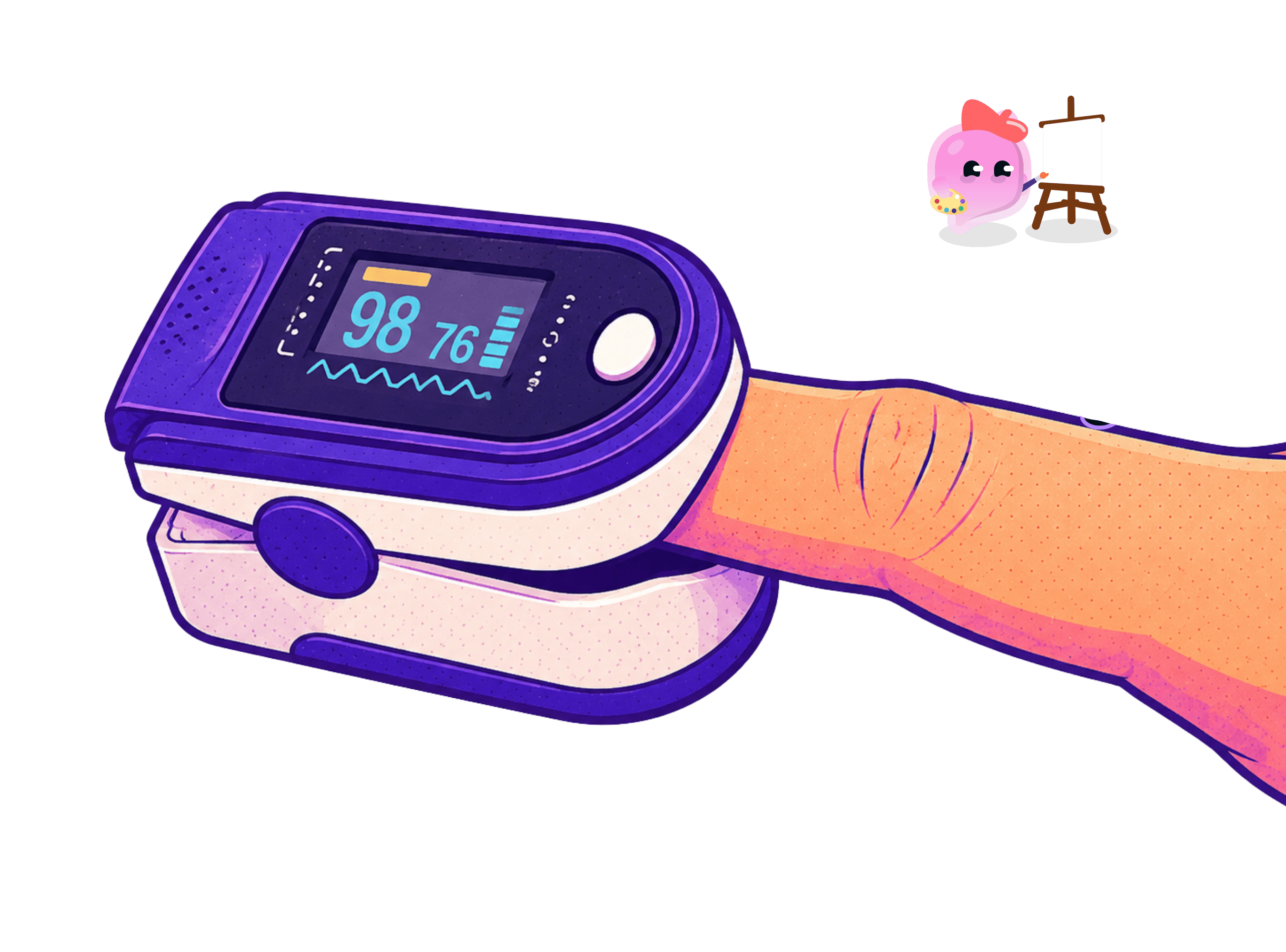
- A pulse oximeter can display heart rate electronically, while a pulse can also be counted at the wrist or neck.
Measuring Heart Rate
- Aim: Measure resting heart rate, then determine how a fixed period of exercise changes heart rate and recovery.
- Apparatus: Stop clock, chair, clear floor space or a low exercise step, and a pulse oximeter if available.
- Method:
- Sit quietly for 5 min5\,\mathrm{min}5min so your heart rate settles.
- Place your index and middle fingers lightly over the radial artery on the thumb side of the wrist.
- Count beats for 60 s60\,\mathrm{s}60s, or for 30 s30\,\mathrm{s}30s and multiply by 222.
- Repeat three times and calculate a mean resting heart rate.
- Complete the same stepping exercise for 2 min2\,\mathrm{min}2min at a fixed pace.
- Measure heart rate immediately, then at fixed 1 min1\,\mathrm{min}1min intervals until it approaches the resting value.
- Variables: Change whether the measurement is taken before or after exercise, measure heart rate, and keep the exercise duration, pace, counting interval, posture, person and room conditions constant.
- Results: Heart rate rises after exercise and then falls during recovery because active muscles need faster delivery of oxygen and glucose and faster removal of carbon dioxide and lactic acid.
- Maths: For a 30 s30\,\mathrm{s}30s count, heart rate=beats counted×2\text{heart rate}=\text{beats counted}\times2heart rate=beats counted×2; for repeats, mean heart rate=sum of heart ratesnumber of readings\text{mean heart rate}=\frac{\text{sum of heart rates}}{\text{number of readings}}mean heart rate=number of readingssum of heart rates.
- Watch out: Do not use your thumb because its own pulse can be counted, measure immediately after exercise because recovery begins at once, and repeat readings to reduce random error.
- Safety: Use moderate exercise in a clear space, stop if you feel pain or dizziness, and never press both carotid arteries at the same time.
Stroke Volume Is Blood per Beat
Stroke volume
The volume of blood pumped from a ventricle in one heartbeat.
- Stroke volume is usually measured in cm3 beat−1\mathrm{cm^3\,beat^{-1}}cm3beat−1.
- A stronger ventricular contraction ejects a larger stroke volume.
- During exercise, increased venous return and stronger contractions can raise stroke volume.
Cardiac Output Is Blood per Minute
Cardiac output
The volume of blood pumped by a ventricle each minute.
- Cardiac output combines how often the heart beats with how much blood leaves a ventricle on each beat.
- The equation is cardiac output=stroke volume×heart rate\text{cardiac output}=\text{stroke volume}\times\text{heart rate}cardiac output=stroke volume×heart rate.
- In symbols, Q=SV×HRQ=SV\times HRQ=SV×HR.
- Useful rearrangements are SV=QHRSV=\frac{Q}{HR}SV=HRQ and HR=QSVHR=\frac{Q}{SV}HR=SVQ.
- If stroke volume is in cm3 beat−1\mathrm{cm^3\,beat^{-1}}cm3beat−1 and heart rate is in beats min−1\mathrm{beats\,min^{-1}}beatsmin−1, cardiac output is in cm3 min−1\mathrm{cm^3\,min^{-1}}cm3min−1.
Exercise Raises Cardiac Output
- Contracting muscles use more oxygen and glucose for aerobic respiration.
- A higher heart rate and stroke volume increase cardiac output, delivering these reactants faster.
- The faster blood flow also removes more carbon dioxide and lactic acid from muscle tissue.
- The increased rate of aerobic respiration releases more energy for muscle contraction.
- Write the equation before substituting values, then give the final unit.
- Convert dm3\mathrm{dm^3}dm3 to cm3\mathrm{cm^3}cm3 before combining values expressed in different volume units.
- A correct numerical answer needs a sensible scale, so a resting adult value is usually several dm3 min−1\mathrm{dm^3\,min^{-1}}dm3min−1, not several cm3 min−1\mathrm{cm^3\,min^{-1}}cm3min−1.
Check Units Before Calculating
- The word per represents division, so cm3 beat−1\mathrm{cm^3\,beat^{-1}}cm3beat−1 means cubic centimetres for each beat.
- Multiplying cm3 beat−1\mathrm{cm^3\,beat^{-1}}cm3beat−1 by beats min−1\mathrm{beats\,min^{-1}}beatsmin−1 cancels beats and leaves cm3 min−1\mathrm{cm^3\,min^{-1}}cm3min−1.
- Use 1 dm3=1000 cm31\,\mathrm{dm^3}=1000\,\mathrm{cm^3}1dm3=1000cm3 when converting cardiac output.
- What is heart rate?
- What is stroke volume?
- Write the equation for cardiac output.
- What unit results from multiplying cm3 beat−1\mathrm{cm^3\,beat^{-1}}cm3beat−1 by beats min−1\mathrm{beats\,min^{-1}}beatsmin−1?
- Why does cardiac output rise during exercise?
8.3.1 Aerobic and anaerobic respiration
Respiration Releases Energy Continuously
Cellular respiration
The continuous exothermic reactions in living cells that break down glucose and release energy for metabolic processes.
Exothermic reaction
A chemical reaction that transfers energy to the surroundings.
- Living cells respire continuously because they need a constant transfer of energy.
- Respiration is exothermic because chemical energy from glucose is transferred to the surroundings and to processes in the cell.
- Cells use the released energy for muscle contraction, active transport and building larger molecules from smaller ones.
- Energy released by respiration also helps mammals and birds maintain a stable body temperature.
- Respiration is a series of enzyme-controlled reactions, not a single reaction and not the same process as breathing.
Aerobic Respiration Uses Oxygen
Aerobic respiration
Cellular respiration that uses oxygen to break down glucose completely, producing carbon dioxide and water and releasing a large amount of energy.
Mitochondrion
An organelle in which most aerobic respiration takes place.
- Aerobic respiration uses oxygen and completely breaks down glucose.
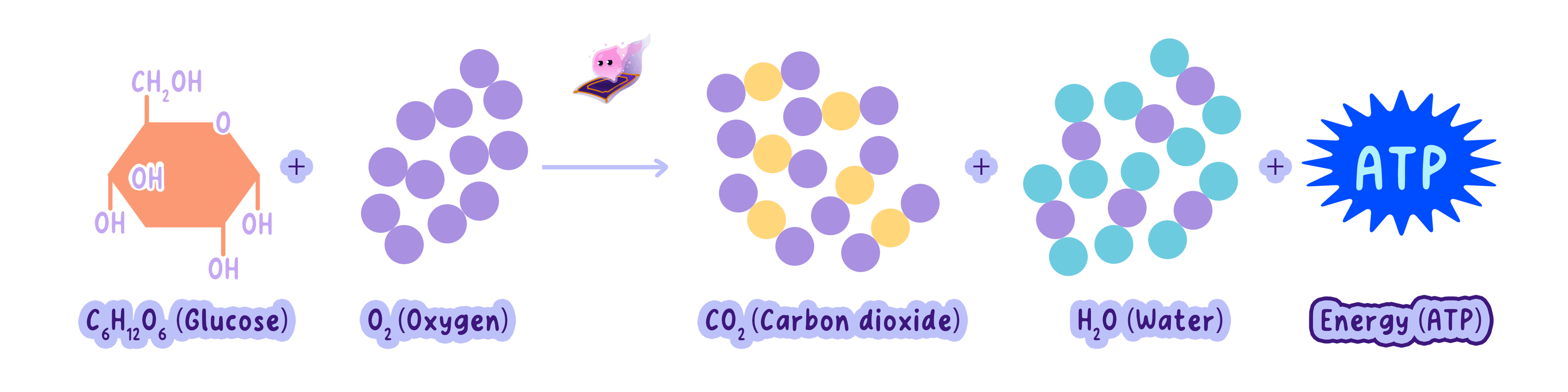
- The word equation is glucose+oxygen→carbon dioxide+water\text{glucose}+\text{oxygen}\rightarrow\text{carbon dioxide}+\text{water}glucose+oxygen→carbon dioxide+water.
- The balanced symbol equation is C6H12O6+6O2→6CO2+6H2O\mathrm{C_6H_{12}O_6+6O_2\rightarrow6CO_2+6H_2O}C6H12O6+6O2→6CO2+6H2O.
- Most aerobic respiration occurs in mitochondria, so cells with high energy demands often contain many mitochondria.
- Complete breakdown of glucose releases much more energy than anaerobic respiration.
Anaerobic Respiration Uses No Oxygen
Anaerobic respiration
Cellular respiration that occurs without oxygen and breaks down glucose incompletely, releasing less energy than aerobic respiration.
Lactic acid
The product formed when animal muscle cells respire anaerobically.
- Anaerobic respiration occurs when oxygen supply cannot meet the demand for aerobic respiration.
- Glucose is broken down incompletely, so less energy is released from each molecule of glucose.
- In animal muscle cells, the word equation is glucose→lactic acid\text{glucose}\rightarrow\text{lactic acid}glucose→lactic acid.
- Lactic acid can accumulate during vigorous exercise and is associated with muscle fatigue.
- In yeast and plant cells, the word equation is glucose→ethanol+carbon dioxide\text{glucose}\rightarrow\text{ethanol}+\text{carbon dioxide}glucose→ethanol+carbon dioxide.
- Anaerobic respiration occurs in the cytoplasm rather than the mitochondria.
The Two Pathways Have Clear Differences
- Oxygen: aerobic respiration requires oxygen, while anaerobic respiration does not.
- Breakdown: aerobic respiration breaks glucose down completely, while anaerobic respiration breaks it down incompletely.
- Energy: aerobic respiration releases much more energy per molecule of glucose.
- Animal products: aerobic respiration produces carbon dioxide and water, while anaerobic respiration produces lactic acid.
- Yeast and plant products: anaerobic respiration produces ethanol and carbon dioxide.
- Location: most aerobic respiration occurs in mitochondria, while anaerobic respiration occurs in the cytoplasm.
- Both aerobic and anaerobic respiration use glucose, so do not claim that glucose is unique to either pathway.
- For two differences, give two independent contrasts rather than reversing the same oxygen statement twice.
- Use the correct products for the organism named, because animal muscles make lactic acid while yeast and plant cells make ethanol and carbon dioxide.
- Write that respiration releases energy, because energy is not a chemical product stored on the right side of the equation.
Exercise Can Use Both Pathways
- At the start of exercise, muscles increase their demand for energy before oxygen delivery has fully increased.
- Aerobic respiration supplies most energy when enough oxygen reaches the muscles.
- During vigorous activity, anaerobic respiration adds a faster but smaller energy release when oxygen supply is insufficient.
- Heart rate and breathing rate rise to deliver more oxygen and glucose and to remove carbon dioxide.
- What makes cellular respiration exothermic?
- Write the word equation for aerobic respiration.
- Why does aerobic respiration release more energy than anaerobic respiration?
- What is produced by anaerobic respiration in animal muscle cells?
- What products form during anaerobic respiration in yeast?
