7.4.1 Insulin and blood glucose control
Blood glucose rises after carbohydrate
Blood glucose concentration
The amount of glucose dissolved in a given volume of blood.
- Blood glucose concentration is the amount of glucose dissolved in a given volume of blood.
- Carbohydrate digestion produces glucose, which is absorbed through the small intestine into the blood.
- After a meal the absorbed glucose makes blood glucose concentration rise.
- Between meals and during exercise cells remove glucose from the blood for respiration, so the concentration falls.
- Control keeps the concentration within a narrow range so cells receive glucose without large changes to the water balance of the blood.
Insulin lowers high blood glucose
Insulin
A hormone released by the pancreas when blood glucose concentration is too high that causes body cells to take in glucose and causes liver and muscle cells to convert glucose into glycogen.
- Detection occurs when the pancreas senses that blood glucose concentration is above the normal range.
- Release follows as the pancreas secretes insulin into the blood plasma.
- Transport occurs in the plasma, which carries insulin to target cells and organs including the liver.
- Cell uptake increases because insulin causes body cells to take more glucose from the blood.
- Storage increases because insulin causes liver and muscle cells to convert glucose into glycogen.
- Correction occurs as glucose leaves the blood, returning blood glucose concentration towards the normal range.
- The control pathway can be written as high blood glucose→insulin release→glucose uptake and glycogen formation→blood glucose falls\text{high blood glucose} \rightarrow \text{insulin release} \rightarrow \text{glucose uptake and glycogen formation} \rightarrow \text{blood glucose falls}high blood glucose→insulin release→glucose uptake and glycogen formation→blood glucose falls.
Following a blood glucose peak
- A graph rises from 5.0 mmol dm−35.0\,\mathrm{mmol\,dm^{-3}}5.0mmoldm−3 before a meal to 8.0 mmol dm−38.0\,\mathrm{mmol\,dm^{-3}}8.0mmoldm−3 afterwards, so the pancreas releases more insulin.
- Insulin acts on body cells and the liver, increasing glucose uptake and conversion of glucose to glycogen.
- The graph falls because glucose is removed from the blood, so the concentration returns towards its starting range.
Glycogen stores excess glucose
Glycogen
An insoluble carbohydrate made from glucose and stored mainly in liver and muscle cells.
- Glycogen is an insoluble carbohydrate made from many glucose units and stored mainly in liver and muscle cells.
- Insolubility allows a large amount of glucose to be stored without the glycogen dissolving and strongly affecting water movement into the cell.
- Liver glycogen provides a store that can later supply glucose to the blood when the concentration becomes too low.
- Muscle glycogen provides glucose for respiration in muscle cells during activity.
Do not write that insulin breaks glucose down, because insulin lowers blood glucose by increasing uptake and storage while respiration is a separate process.
Insulin release falls after correction
Negative feedback
A control mechanism in which a change away from the normal level causes responses that reverse the change and restore the normal level.
- Negative feedback means that a change away from the normal level causes responses that reverse the change.
- As blood glucose falls the stimulus for insulin release becomes weaker, so less insulin is released.
- When the normal range is restored the corrective response is reduced rather than continuing to drive the concentration lower.
Writing an insulin explanation
- Start with the stimulus: state that blood glucose concentration is too high and the pancreas releases insulin.
- Give the target response: state that body cells take in more glucose and liver or muscle cells convert glucose into glycogen.
- Finish with the outcome: state that removing glucose from the blood lowers its concentration towards the normal range.
- Keep the carrier accurate: hormones travel dissolved in blood plasma, not inside red blood cells.
- What change causes the pancreas to release insulin?
- How is insulin transported to its target organs?
- How does insulin change the behaviour of body cells?
- What happens to glucose in liver and muscle cells?
- Why does insulin release fall when blood glucose returns to normal?
7.4.2 Glucagon and blood glucose regulation
Glucagon raises low blood glucose
Glucagon
A hormone released by the pancreas when blood glucose concentration is too low that causes liver cells to convert glycogen into glucose and release the glucose into the blood.
- Glucagon is a hormone released by the pancreas when blood glucose concentration is below the normal range.
- Low blood glucose can occur between meals, during prolonged exercise or when cells have used glucose in respiration.
- The pancreas responds by releasing glucagon into the blood plasma.
- The liver is the main target because glucagon causes liver cells to convert stored glycogen into glucose.
- Glucose enters the blood from the liver, so blood glucose concentration rises towards the normal range.
- The control pathway can be written as low blood glucose→glucagon release→glycogen converted to glucose→blood glucose rises\text{low blood glucose} \rightarrow \text{glucagon release} \rightarrow \text{glycogen converted to glucose} \rightarrow \text{blood glucose rises}low blood glucose→glucagon release→glycogen converted to glucose→blood glucose rises.
The liver supplies stored glucose
Glycogen
An insoluble carbohydrate made from glucose and stored mainly in liver and muscle cells.
- Glycogen stores glucose in an insoluble form inside liver and muscle cells.
- Glucagon acts mainly on liver cells because liver glycogen can be converted to glucose that is released into the bloodstream.
- Muscle cells use their glycogen locally for respiration rather than releasing the stored glucose to control the concentration in the blood.
- Conversion changes the stored carbohydrate into soluble glucose that can leave the liver and enter the plasma.
Explaining a fall between meals
- Observation: a blood glucose graph falls below its usual range three hours after a meal.
- Hormonal response: the pancreas releases more glucagon into the blood.
- Liver response: glycogen is converted into glucose and the glucose is released.
- Outcome: blood glucose concentration rises back towards the normal range.
Two hormones correct opposite changes
Negative feedback
A control mechanism in which a change away from the normal level causes responses that reverse the change and restore the normal level.
- Negative feedback reverses a change away from the normal level.
- When glucose is too high insulin increases glucose uptake and glycogen formation, so the concentration falls.
- When glucose is too low glucagon increases the conversion of liver glycogen to glucose, so the concentration rises.
- The responses oppose each other and keep blood glucose within a narrow range rather than at one perfectly fixed value.
- Hormone release changes continuously because the pancreas responds to the current blood glucose concentration.
Do not reverse the hormones: insulin lowers blood glucose concentration and glucagon raises it.
7.4.3 Type 1 and type 2 diabetes
Type 1 diabetes produces too little insulin
Type 1 diabetes
A condition in which the pancreas produces little or no insulin, so blood glucose concentration cannot be reduced effectively.
- Type 1 diabetes occurs when the pancreas produces little or no insulin.
- Without enough insulin body cells take in less glucose and the liver converts less glucose into glycogen.
- Blood glucose remains too high especially after carbohydrate is digested and glucose enters the blood.
- Control requires insulin delivered by injection or a pump because swallowing insulin would expose this protein hormone to digestive enzymes.
- Dose and timing must be matched to food intake, activity and the measured blood glucose concentration.
- Regular monitoring helps prevent blood glucose becoming dangerously high or falling too low after too much insulin.
Type 2 diabetes reduces insulin response
Type 2 diabetes
A condition in which body cells respond less effectively to insulin, so blood glucose concentration remains too high.
- Type 2 diabetes occurs when body cells respond less effectively to insulin.
- The pancreas can still make insulin but glucose uptake and glycogen formation do not increase enough.
- Blood glucose remains high because glucose is removed from the blood more slowly.
- Control often begins with lifestyle changes including a controlled carbohydrate intake, regular exercise and loss of excess body mass.
- Medication may be needed when lifestyle changes do not control blood glucose adequately.
- Risk is influenced by several factors including excess body fat, physical inactivity and inherited variation.
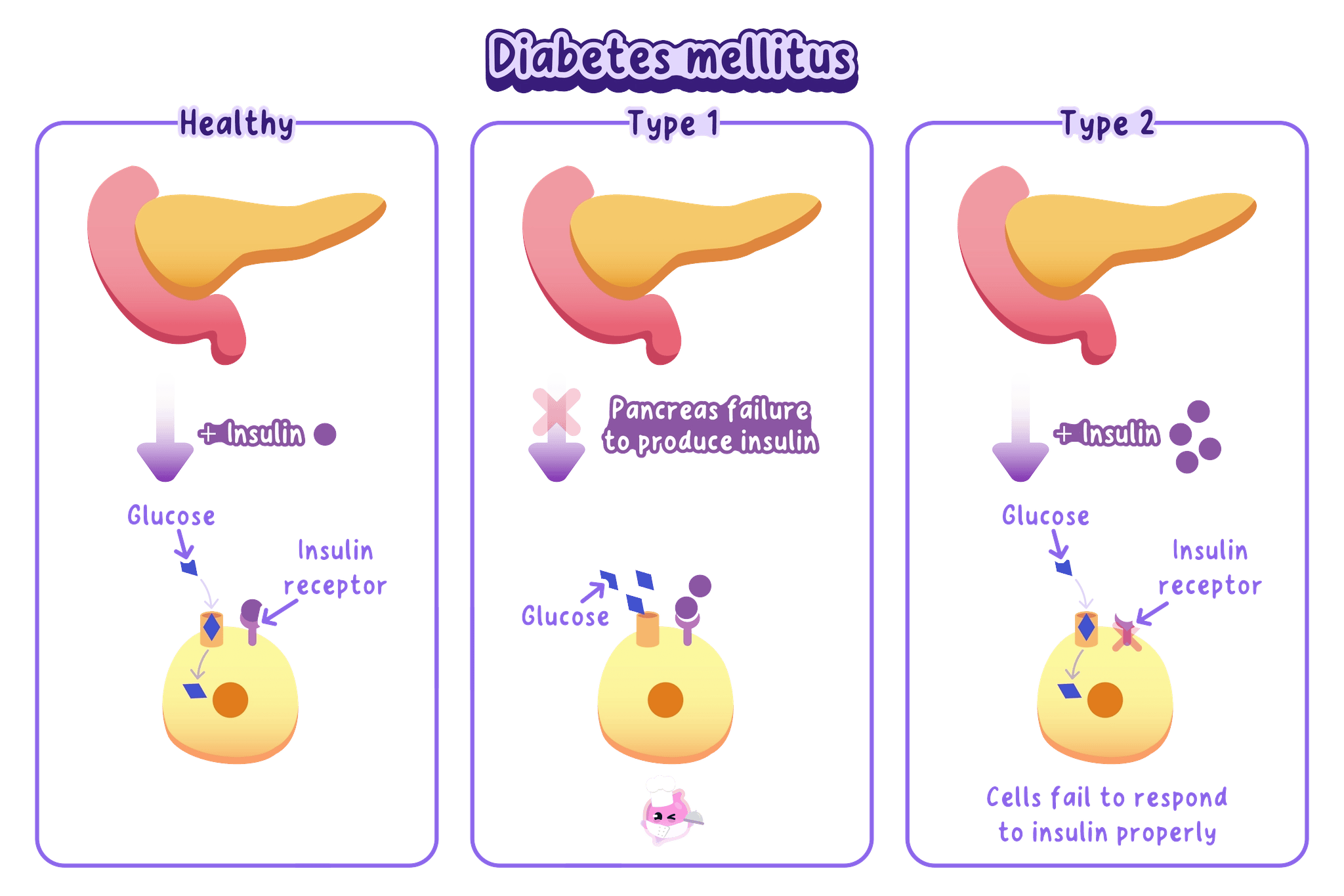
Do not describe type 2 diabetes as a failure to make any insulin, because its defining problem is a reduced response of target cells to insulin.
BMI relates mass to height
Body mass index (BMI)
A measure that relates body mass to height, calculated using BMI=mass (kg)[height (m)]2\mathrm{BMI}=\dfrac{\mathrm{mass\ (kg)}}{[\mathrm{height\ (m)}]^2}BMI=[height (m)]2mass (kg).
- Body mass index is calculated using BMI=mass (kg)[height (m)]2\mathrm{BMI}=\dfrac{\mathrm{mass\ (kg)}}{[\mathrm{height\ (m)}]^2}BMI=[height (m)]2mass (kg).
- Mass must be in kilograms and height must be in metres before the values are substituted.
- BMI estimates weight category but it does not distinguish between muscle mass and body fat.
Waist-to-hip ratio estimates fat distribution
Waist-to-hip ratio
The waist circumference divided by the hip circumference, measured in the same units.
- Waist-to-hip ratio is calculated using waist:hip ratio=waist circumferencehip circumference\mathrm{waist{:}hip\ ratio}=\dfrac{\mathrm{waist\ circumference}}{\mathrm{hip\ circumference}}waist:hip ratio=hip circumferencewaist circumference.
- Both circumferences need the same unit so centimetres divided by centimetres gives a ratio with no unit.
- A larger ratio indicates that more body mass is carried around the abdomen and is associated with a greater risk of type 2 diabetes.
Calculating two body measurements
- BMI data: a person has a mass of 72 kg72\,\mathrm{kg}72kg and a height of 1.80 m1.80\,\mathrm{m}1.80m.
- BMI calculation: BMI=72(1.80)2=22.2 kg m−2\mathrm{BMI}=\dfrac{72}{(1.80)^2}=22.2\,\mathrm{kg\,m^{-2}}BMI=(1.80)272=22.2kgm−2 to three significant figures.
- Ratio data: the same person has a waist circumference of 84 cm84\,\mathrm{cm}84cm and a hip circumference of 100 cm100\,\mathrm{cm}100cm.
- Ratio calculation: waist:hip ratio=84100=0.84\mathrm{waist{:}hip\ ratio}=\dfrac{84}{100}=0.84waist:hip ratio=10084=0.84.
- Interpretation: the measurements describe body size and fat distribution, so they estimate risk rather than diagnose diabetes.
Correlation does not prove cause
Correlation
A relationship in which two variables change together in a pattern, which does not by itself show that one variable causes the other.
- A positive correlation exists when the incidence of type 2 diabetes tends to increase as body mass or waist-to-hip ratio increases.
- Correlation is evidence of an association but it does not show by itself that increased body mass directly caused diabetes in every person.
- Other variables such as diet, physical activity, age and inherited variation may affect both body mass and diabetes risk.
- Evaluation should consider the trend, the amount of variation, the sample size and whether important variables were controlled.
Evaluating diabetes risk data
- Describe the pattern: state the direction of the correlation and support it with values from the data.
- Avoid a causal claim: use increases risk or is associated with rather than always causes.
- Identify a limitation: BMI may be high because of muscle, and another variable may explain part of the pattern.
- Reach a measured judgement: the data can support a link without proving that body mass is the only cause.
A glucose test can model urine screening
- Glucose in urine may indicate that blood glucose has been high enough for glucose to remain in the filtrate instead of all being reabsorbed.
- A chemical test can compare simulated urine samples for reducing sugar without handling human body fluid.
Testing simulated urine for glucose
- Aim: compare simulated urine samples for reducing sugar using Benedict's solution.
- Apparatus: labelled simulated urine samples, distilled water, glucose solution, test tubes and rack, pipettes, measuring cylinder, Benedict's solution, a 707070 to 80 ∘C80\,^{\circ}\mathrm{C}80∘C water bath, timer, test-tube holder and eye protection.
- Method:
- Label one tube for each sample, one distilled-water negative control and one glucose positive control
- add 2 cm32\,\mathrm{cm^3}2cm3 of the correct liquid to each tube with clean equipment
- add 2 cm32\,\mathrm{cm^3}2cm3 of Benedict's solution
- place every tube in the water bath for about 5 min5\,\mathrm{min}5min
- remove the tubes with a holder and record the final colour; 6) repeat each sample and compare the results
- Variables: change the simulated urine sample, measure the final colour, and keep the sample volume, Benedict's volume, heating time and water-bath temperature constant.
- Results: a blue result is negative, while green, yellow, orange or brick-red shows an increasing concentration of reducing sugar.
- Controls: distilled water should remain blue and the glucose solution should change colour, showing that the method can distinguish a negative result from a positive result.
- Watch out: use a clean pipette for every liquid because glucose carried between tubes can produce a convincing false positive.
- Safety: wear eye protection because Benedict's solution is an irritant, use a water bath rather than a flame, and handle hot glassware with a test-tube holder.
The result supports a diagnosis, not proves it
- A positive simulated result shows that reducing sugar is present in that sample.
- A real screening result would need confirmation with controlled blood glucose measurements and clinical assessment.
- The strongest comparison links the result to insulin action while recognising that one observation does not identify the diabetes type.
- What causes type 1 diabetes?
- What causes type 2 diabetes?
- How is each type controlled?
- How are BMI and waist-to-hip ratio calculated?
- Why does a correlation between body mass and type 2 diabetes not prove causation?
7.4.4 Structure of the urinary system
The urinary system removes urine
Urinary system
The organ system formed by the kidneys, ureters, bladder and urethra that makes, stores and removes urine.
- The urinary system consists of two kidneys, two ureters, the bladder and the urethra.
- Its route can be written as kidneys→ureters→bladder→urethra→outside the body\text{kidneys} \rightarrow \text{ureters} \rightarrow \text{bladder} \rightarrow \text{urethra} \rightarrow \text{outside the body}kidneys→ureters→bladder→urethra→outside the body.
- Each structure has a distinct position so a labelled diagram should show the kidneys above the bladder and one ureter joining each kidney to the bladder.
Kidneys receive and filter blood
Kidney
An organ that filters the blood, removes urea and regulates the water and ion content of the blood by forming urine.
- Each kidney is a bean-shaped organ supplied by a renal artery and drained by a renal vein.
- The renal artery carries blood containing urea, water, ions, glucose and other dissolved substances into the kidney.
- The renal vein carries blood away after the kidney has removed urea and adjusted water and ion contents.
- Urine leaves separately through the ureter rather than through a blood vessel.
Ureters carry urine to the bladder
Ureter
A tube that carries urine from a kidney to the bladder.
- A ureter is a tube that carries urine from one kidney to the bladder.
- There are two ureters because each kidney has its own connection to the bladder.
- The ureters do not carry blood and they do not carry urine directly out of the body.
The bladder stores urine
Bladder
A muscular organ that stores urine before it leaves the body.
- The bladder is a muscular sac that stores urine until urination.
- Storage separates continuous urine production by the kidneys from periodic removal from the body.
- The bladder is not a filter because filtration and urine formation occur in the kidneys.
The urethra leads outside
Urethra
The tube that carries urine from the bladder to the outside of the body.
- The urethra is the single tube that carries urine from the bladder to the outside of the body.
- Ureter and urethra have similar names but different connections and functions.
A ureter connects a kidney to the bladder, while the urethra connects the bladder to the outside.
Blood and urine follow different routes
Renal artery
The blood vessel that carries blood containing urea and other dissolved substances to a kidney.
- Blood enters through the renal artery and leaves through the renal vein.
- Urine leaves through a ureter and then passes through the bladder and urethra.
- The two routes meet inside the kidney because substances are removed from blood to form urine without blood itself entering the ureter.
Tracing a urea molecule
- Arrival: a urea molecule reaches a kidney in the blood carried by the renal artery.
- Removal: the kidney transfers the urea from the blood into the forming urine.
- Urine route: the molecule passes through a ureter, is stored in the bladder and leaves through the urethra.
- Filtered blood: blood with less urea leaves the kidney through the renal vein.
Diagram labels need exact connections
Urine
The liquid formed by the kidneys that contains urea, excess water and excess mineral ions.
- Urine is the liquid formed by the kidneys that contains urea, excess water and excess mineral ions.
- A correct diagram distinguishes the two ureters from the one urethra and keeps the blood vessels separate from the urine pathway.
- A label line should end on the named structure rather than the space beside it.
Describing the urinary system
- Name every main structure: kidneys, ureters, bladder and urethra.
- Use carries and stores accurately: ureters carry urine, the bladder stores it and the urethra carries it outside.
- Separate the blood supply: the renal artery enters a kidney and the renal vein leaves it.
- Check the common swap: ureter is kidney to bladder, while urethra is bladder to outside.
- Which structures form the urinary system?
- What is the route taken by urine?
- How do the ureter and urethra differ?
- Which vessels carry blood into and away from a kidney?
- Which organ stores urine?
7.4.5 Structure and function of the nephron
Nephrons form urine
Nephron
A microscopic kidney tubule in which blood is filtered and useful substances are reabsorbed to form urine.
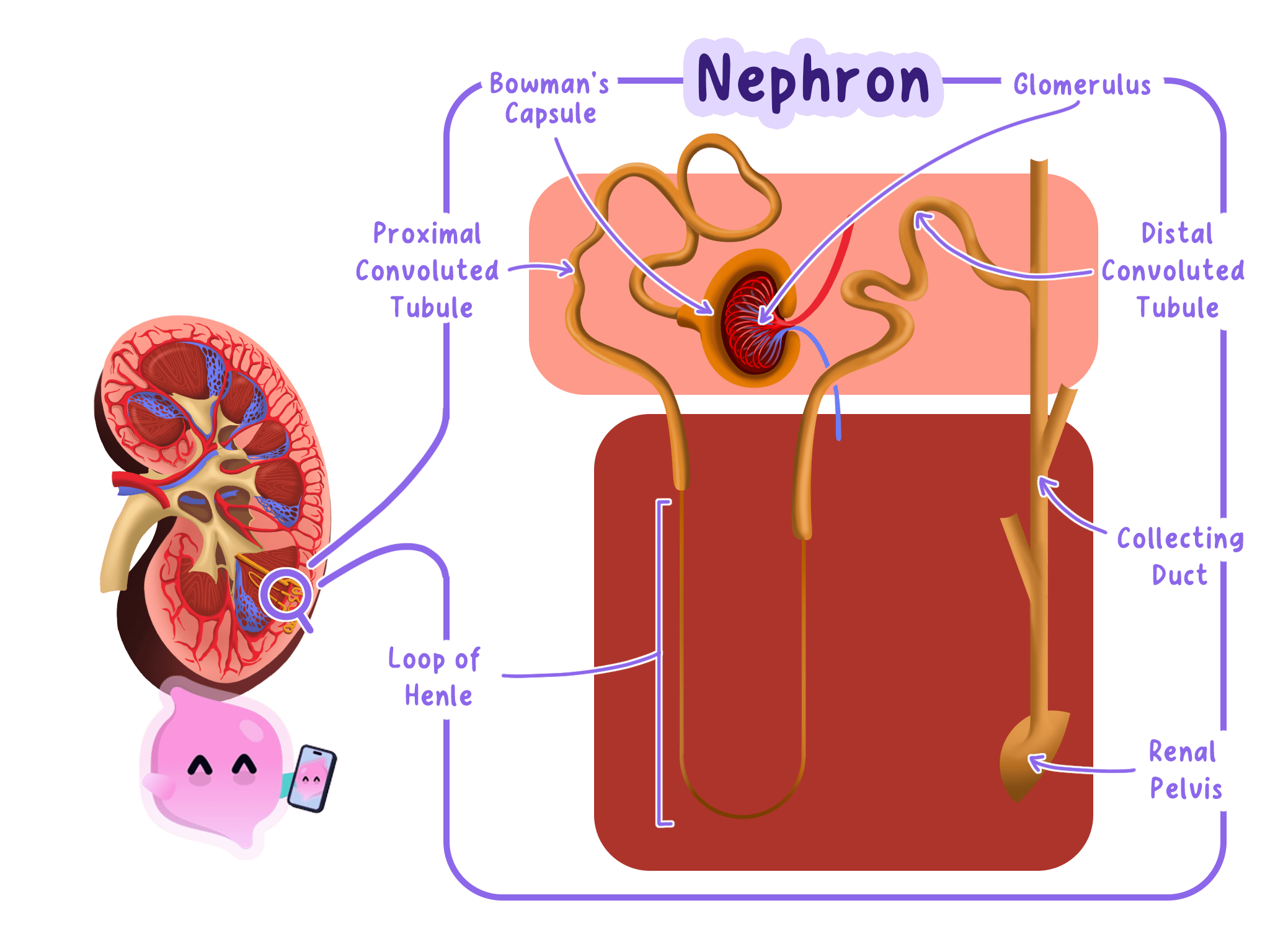
- A nephron is a microscopic kidney tubule that filters the blood and then reabsorbs useful substances.
- The nephron begins with a glomerulus enclosed by Bowman's capsule.
- The filtrate then travels along a tubule before entering a collecting duct.
- Capillaries surround the tubule so substances can move from the filtrate back into the blood.
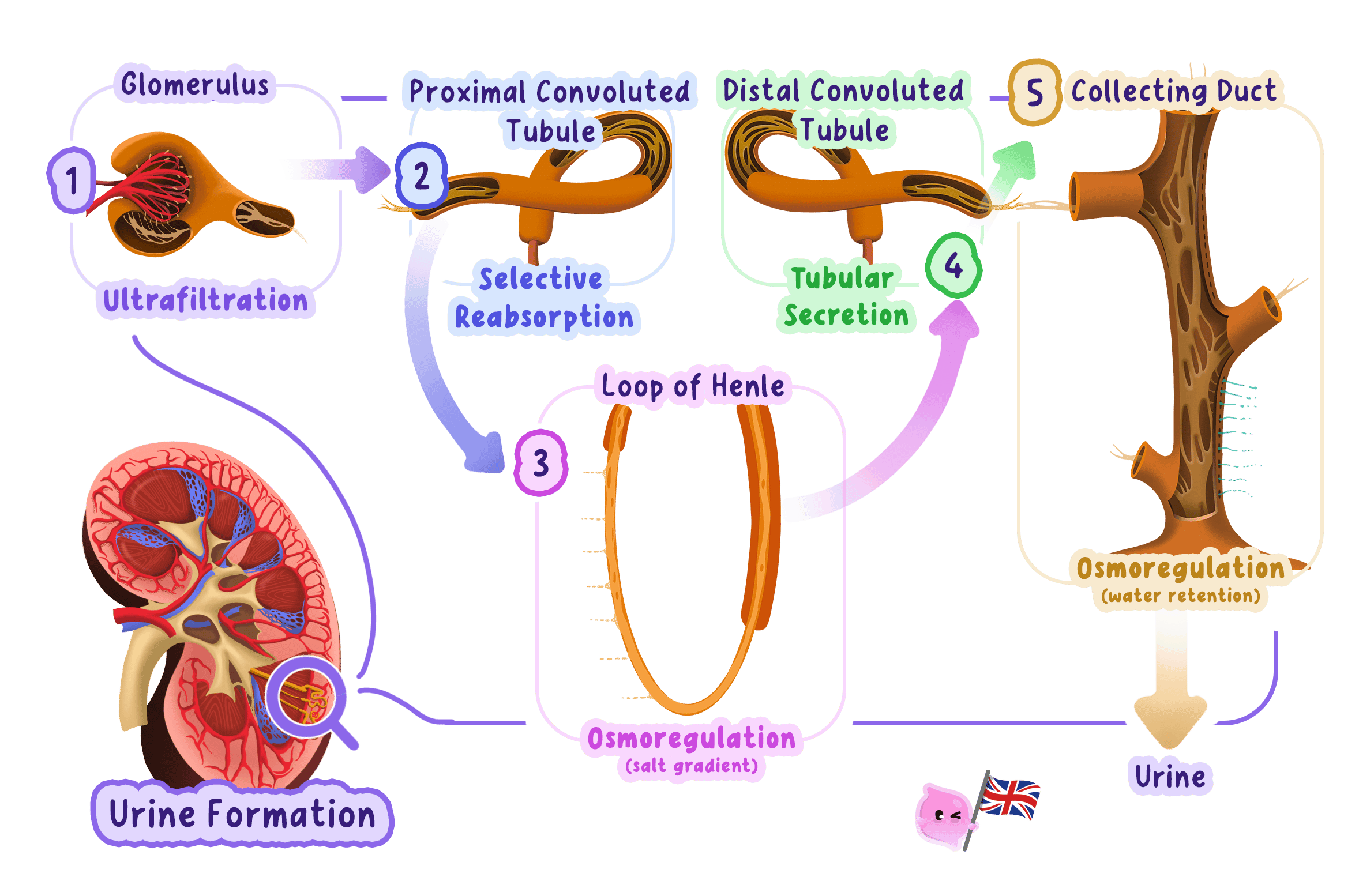
- Urine formation has two linked stages: ultrafiltration followed by selective reabsorption.
High pressure filters small molecules
Ultrafiltration
High-pressure filtration in the glomerulus that moves water and other small dissolved molecules into Bowman's capsule while blood cells and large proteins remain in the blood.
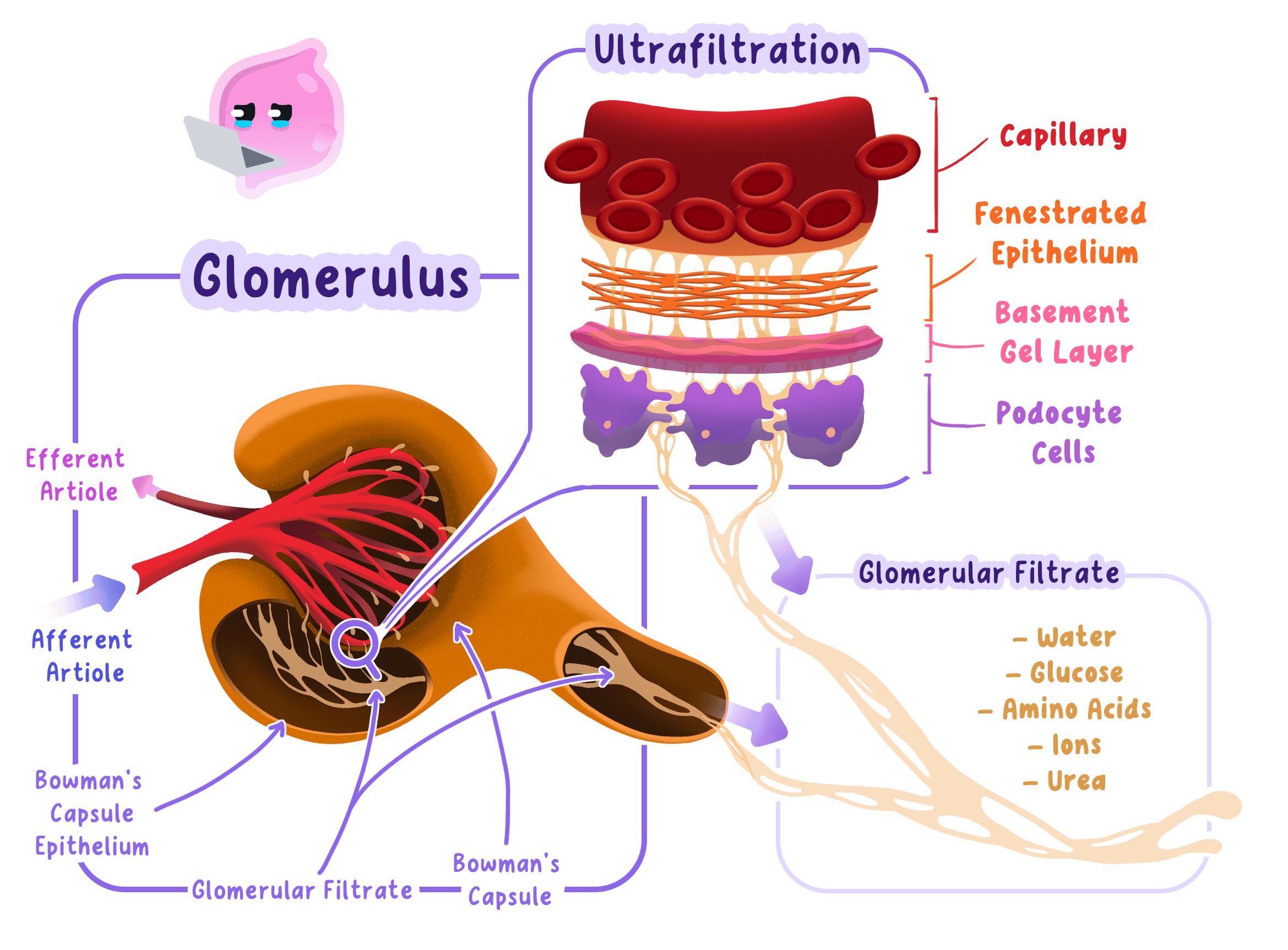
- Ultrafiltration is high-pressure filtration of the blood at the glomerulus.
- Blood pressure forces water and small dissolved molecules through the filtration barrier into Bowman's capsule.
- The filtrate contains water, glucose, urea and mineral ions because these molecules are small enough to pass.
- Blood cells remain in the capillaries because they are too large to cross the filtration barrier.
- Large plasma proteins remain in the blood because their molecules are too large to pass through the barrier.
The glomerulus produces filtrate
Glomerulus
A knot of capillaries inside Bowman's capsule where high blood pressure filters small molecules out of the blood.
- The glomerulus is a knot of capillaries inside Bowman's capsule.
- Its capillary network provides a large surface area for filtration.
- Bowman's capsule collects the liquid and small solutes that leave the glomerular blood.
- The first liquid is filtrate rather than urine because it still contains useful glucose, water and ions.
Protein or blood cells should not normally appear in the filtrate, because the filtration barrier retains them in the blood.
Selective reabsorption returns glucose
Selective reabsorption
The movement of useful substances from the nephron filtrate back into the blood.
- Selective reabsorption returns useful substances from the nephron filtrate to the blood.
- All glucose is normally reabsorbed from the first part of the tubule into the surrounding capillaries.
- Active transport moves glucose against its concentration gradient when necessary and requires energy released by respiration.
- Tubule cells contain many mitochondria to supply energy for active transport.
- Microvilli increase surface area so glucose can be reabsorbed rapidly.
Water returns to the blood
Collecting duct
A kidney tubule whose permeability to water is controlled by ADH as urine passes towards the renal pelvis.
- Water is reabsorbed from the filtrate into the blood by osmosis.
- The amount reabsorbed varies according to the water content of the blood and the permeability of the collecting duct.
- Useful quantities of ions are reabsorbed while excess ions remain in the tubule.
- Urea remains in the filtrate together with excess water and ions, forming urine.
Predicting substances in each fluid
- Blood entering the glomerulus contains blood cells, plasma proteins, glucose, urea, ions and water.
- Bowman's capsule filtrate contains glucose, urea, ions and water but normally no blood cells or large plasma proteins.
- Urine contains urea with excess water and ions but normally no glucose because glucose has been selectively reabsorbed.
- Reasoning follows molecule size during filtration and usefulness during reabsorption.
Structure explains each movement
Bowman's capsule
A cup-shaped structure around the glomerulus that collects the small molecules filtered from the blood.
- Bowman's capsule surrounds the glomerulus and receives the filtrate directly.
- A long tubule provides time and surface area for selective reabsorption.
- A close capillary supply carries reabsorbed glucose and water away and helps maintain concentration gradients.
- The collecting duct provides a final route in which water reabsorption can be adjusted before urine leaves the kidney.
Explaining nephron function
- For filtration: state that high pressure forces small molecules into Bowman's capsule while cells and proteins stay in the blood.
- For glucose: state that all glucose is selectively reabsorbed from the tubule into surrounding capillaries.
- For water: state that water returns to the blood by osmosis and that the amount varies.
- For urine: identify urea with excess water and ions as the substances left in the tubule.
- Where does ultrafiltration occur?
- Which substances enter Bowman's capsule?
- Why do blood cells and large proteins remain in the blood?
- How is glucose selectively reabsorbed?
- What remains in the tubule to form urine?
7.4.6 ADH and water regulation
ADH changes collecting duct permeability
Antidiuretic hormone (ADH)
A hormone released by the pituitary gland that increases the permeability of the collecting duct to water, so more water is reabsorbed into the blood.
- Antidiuretic hormone is released from the pituitary gland into the blood.
- Its target is the kidney where it increases the permeability of the collecting duct to water.
- A more permeable collecting duct allows more water to move by osmosis from the filtrate into the blood.
- More water reabsorption produces a smaller volume of more concentrated urine.
- Less water reabsorption produces a larger volume of more dilute urine.
The brain detects low water content
Osmoregulation
The control of the water and ion content of the body within narrow limits.
- Osmoregulation keeps the water and ion content of the body within narrow limits.
- The hypothalamus detects when the blood contains too little water and is therefore too concentrated.
- The pituitary gland responds by releasing more ADH into the blood.
- More ADH makes the collecting duct more permeable to water.
- More water returns to the blood by osmosis, so less water remains in the urine.
- The result is a small volume of concentrated urine and restoration of the blood water content.
High water content lowers ADH
Collecting duct
A kidney tubule whose permeability to water is controlled by ADH as urine passes towards the renal pelvis.
- When the blood contains too much water the hypothalamus detects that it is too dilute.
- The pituitary releases less ADH so the collecting duct becomes less permeable to water.
- Less water returns to the blood and more water remains in the filtrate.
- The result is a large volume of dilute urine and a fall in the water content of the blood towards normal.
Predicting urine after exercise
- Situation: sweating during a long run removes water from the body, so the blood becomes more concentrated.
- Detection: the hypothalamus detects the low water content and the pituitary releases more ADH.
- Kidney response: the collecting duct becomes more permeable and more water is reabsorbed by osmosis.
- Prediction: the runner produces a smaller volume of more concentrated urine.
Negative feedback reverses each change
Negative feedback
A control mechanism in which a change away from the normal level causes responses that reverse the change and restore the normal level.
- Too little water causes more ADH and more water reabsorption, which reverses the original decrease.
- Too much water causes less ADH and less water reabsorption, which reverses the original increase.
- As normal water content returns the corrective change in ADH release becomes smaller.
More ADH means more permeability, more water reabsorption and less water in the urine.
Urine volume and concentration move oppositely
- A small urine volume is usually more concentrated because more water has returned to the blood.
- A large urine volume is usually more dilute because less water has returned to the blood.
- Concentrated urine contains less water per quantity of dissolved waste, not more urea produced by the liver.
Writing the complete ADH chain
- State the water condition: identify whether the blood contains too little or too much water.
- Name the control organs: the hypothalamus detects the change and the pituitary alters ADH release.
- Link ADH to permeability: more ADH makes the collecting duct more permeable, while less ADH makes it less permeable.
- Link permeability to osmosis: a more permeable duct allows more water to return to the blood.
- Finish with both urine properties: state the correct volume and concentration.
Every direction must stay consistent
- For low blood water write more ADH, greater permeability, more reabsorption, smaller urine volume and more concentrated urine.
- For high blood water write less ADH, lower permeability, less reabsorption, larger urine volume and more dilute urine.
- Which gland releases ADH?
- How does ADH change the collecting duct?
- What happens when the blood contains too little water?
- What urine forms when ADH concentration is low?
- Why is ADH control an example of negative feedback?
7.4.7 Treatments for kidney failure
Kidney failure disrupts blood composition
Kidney failure
A condition in which the kidneys cannot filter the blood adequately or regulate its water and ion content.
- Kidney failure occurs when the kidneys cannot filter the blood adequately or regulate its water and ion content.
- Urea accumulates because it is not removed from the blood quickly enough.
- Water and ion balance changes because selective reabsorption and urine formation no longer work effectively.
- Untreated failure is dangerous because toxic waste and abnormal fluid balance disrupt cell and organ function.
- The two main treatments are dialysis and a kidney transplant from an organ donor.
Dialysis removes wastes by diffusion
Dialysis
A treatment in which blood flows beside dialysis fluid across a partially permeable membrane so urea and excess ions diffuse out of the blood.
- During dialysis blood is taken from the patient and passed through a machine before being returned.
- A partially permeable membrane separates the blood from dialysis fluid.
- The dialysis fluid contains no urea so urea diffuses from a higher concentration in the blood to a lower concentration in the fluid.
- The dialysis fluid contains a normal glucose concentration so there is no net loss of glucose from healthy blood.
- Its ion concentrations are controlled so excess ions can diffuse out while useful concentrations are maintained.
- Fresh dialysis fluid flows continuously to maintain steep concentration gradients for diffusion.
- The cleaned blood returns to the patient with less urea and a safer balance of dissolved substances.
Dialysis has repeated costs
- Advantages include avoiding donor shortage and providing treatment without placing a donor organ in the patient.
- Regular sessions are required because dialysis works only while the blood is connected to the machine.
- Time and travel can restrict work, school, diet, fluid intake and daily life.
- Risks include infection, blood clotting and sudden changes in blood pressure.
- Dialysis can bridge the wait by keeping a patient alive until a suitable donor kidney becomes available.
A transplant replaces kidney function
Kidney transplant
An operation in which a healthy donor kidney is placed in a person whose kidneys have failed.
- A kidney transplant places a healthy kidney from a living or deceased donor into the patient.
- A successful kidney filters the blood continuously, so regular dialysis is no longer needed.
- Benefits can include fewer dietary restrictions, more freedom and lower long-term treatment costs.
- Limits include donor shortage, a major operation and the possibility that the transplanted kidney will fail.
Tissue matching reduces rejection
Tissue rejection
The destruction of transplanted cells or tissue by the patient's immune system because their antigens are recognised as foreign.
- Tissue rejection occurs when the immune system recognises antigens on the donor kidney as foreign and attacks the organ.
- Tissue matching selects a donor whose antigens are as similar as possible to those of the recipient.
- Immunosuppressant drugs reduce the immune response and lower the chance of rejection.
- Reduced immunity increases susceptibility to infectious disease and makes long-term monitoring necessary.
Choosing between two treatments
- Patient information: a person can attend regular hospital sessions but has not yet found a suitable donor.
- Immediate choice: dialysis can remove urea and control ions while the patient waits.
- Long-term possibility: a matched transplant could remove the need for repeated sessions if the operation succeeds.
- Balanced judgement: dialysis avoids transplant surgery now, while a transplant offers more continuous kidney function but adds surgical, rejection and infection risks.
Treatment choice depends on evidence
- Dialysis may be preferred when no suitable donor is available or transplant surgery would carry a high risk.
- A transplant may be preferred when a suitable kidney is available and the patient can tolerate surgery and immunosuppressant treatment.
- A justified decision weighs effectiveness, availability, risks, lifestyle effects and long-term consequences for that patient.
Comparing kidney treatments
- Cover both treatments: describe dialysis and transplantation rather than writing everything about one.
- Link each feature to a consequence: regular dialysis restricts time, while a functioning transplant removes that schedule.
- Develop rejection: explain tissue matching and immunosuppressants, then connect immunosuppression to infection risk.
- End with a supported conclusion: choose the better option for the stated patient and base the decision on the evidence.
- Why does urea build up during kidney failure?
- How does dialysis remove urea from the blood?
- Why does dialysis fluid contain a normal glucose concentration?
- How are tissue matching and immunosuppressants used after a transplant?
- What evidence should guide a comparison of dialysis and transplantation?
7.4.8 Production of urea in the liver
The liver makes urea
Urea
A nitrogen-containing waste substance produced in the liver from the breakdown of excess amino acids and removed from the blood by the kidneys.
- Urea is a nitrogen-containing waste substance produced in the liver from the breakdown of excess amino acids.
- The liver is the production organ while the kidneys remove urea from the blood.
- Urea enters the blood after it is produced and travels dissolved in the plasma.
- The kidneys filter urea into the nephron so it can leave the body in urine.
- The route can be written as excess amino acids→liverurea→bloodkidneys→urine\text{excess amino acids} \xrightarrow{\text{liver}} \text{urea} \xrightarrow{\text{blood}} \text{kidneys} \rightarrow \text{urine}excess amino acidsliverureabloodkidneys→urine.
Excess amino acids cannot be stored
Amino acid
The small molecule that is the building block of a protein, of which there are twenty different kinds.
- An amino acid is a small molecule used to build proteins.
- Dietary proteins are digested into amino acids that are absorbed into the blood.
- Cells use amino acids to make the proteins required for growth, repair, enzymes and other cell structures.
- Excess amino acids cannot be stored as amino acids, so the liver breaks them down.
- Nitrogen-containing waste from this breakdown is converted into urea for transport to the kidneys.
Following excess dietary protein
- Digestion: protein in a meal is broken down into amino acids and the amino acids enter the blood.
- Use: cells take the amino acids needed for protein synthesis.
- Excess: amino acids that are not needed cannot be stored, so the liver breaks them down and produces urea.
- Excretion: blood carries the urea to the kidneys and it leaves the body in urine.
The liver and kidneys have different roles
- The liver produces urea from excess amino acids.
- The blood transports urea dissolved in plasma from the liver to the kidneys.
- The kidneys remove urea from the blood and include it in urine.
- The urinary system then carries, stores and releases the urine.
Do not write that the kidneys make urea, because urea is made in the liver and removed by the kidneys.
Explaining urea production
- Use the correct starting substance: excess amino acids, not glucose, lipids or mineral ions.
- Use the correct production organ: the liver.
- Use the correct removal organ: the kidneys filter urea from the blood.
- Keep the sequence clear: production happens before transport in plasma and excretion in urine.
- What substance is broken down to produce urea?
- In which organ is urea produced?
- Why are excess amino acids broken down?
- How does urea travel to the kidneys?
- What is the difference between the liver and kidney roles in urea excretion?
