5.3.1 The specific immune system
Antigens Trigger a Specific Immune Response
Antigen
A molecule on the surface of a pathogen that the immune system recognises as foreign.
Lymphocyte
A type of white blood cell that produces antibodies and antitoxins against a specific pathogen.
- An antigen is a molecule on a pathogen that the immune system recognises as foreign.
- A lymphocyte with a complementary receptor recognises the antigen and is activated.
- The activated lymphocyte divides to form many identical cells.
Specificity comes from the complementary match between one antigen and one lymphocyte receptor.
Antibodies Bind Specific Antigens
Antibody
A protein made by lymphocytes that binds to a specific antigen, marking the pathogen for destruction.
- Activated lymphocytes produce antibodies with binding sites complementary to the antigen.
- Antibodies bind pathogens, neutralise them or mark them for destruction by phagocytes.
- The first response is slow because the correct lymphocyte must be selected and cloned.
- Each lymphocyte carries receptors with one binding shape, so only cells complementary to the antigen are selected.
- Selected lymphocytes divide by mitosis, producing a clone that can generate a large concentration of one specific antibody.
- Antibody binding can neutralise a pathogen, cause pathogens to clump or mark them for phagocytosis.
an antibody against a measles antigen will not bind an influenza antigen because the binding-site shapes are different.
Memory Cells Produce a Faster Second Response
Memory cell
A white blood cell that stays in the body after an infection so the body can respond faster and more strongly if the same pathogen returns.
- Some activated lymphocytes become long-lived memory cells after the infection is controlled.
- If the same antigen enters again, memory cells divide rapidly and produce many antibody-secreting cells.
- Antibody concentration rises faster and higher, often destroying the pathogen before symptoms develop.
- The primary response takes time, so symptoms may develop before antibody concentration is high enough to control the pathogen.
- Memory lymphocytes persist after recovery and undergo rapid clonal expansion on re-exposure, producing a faster, larger secondary response.
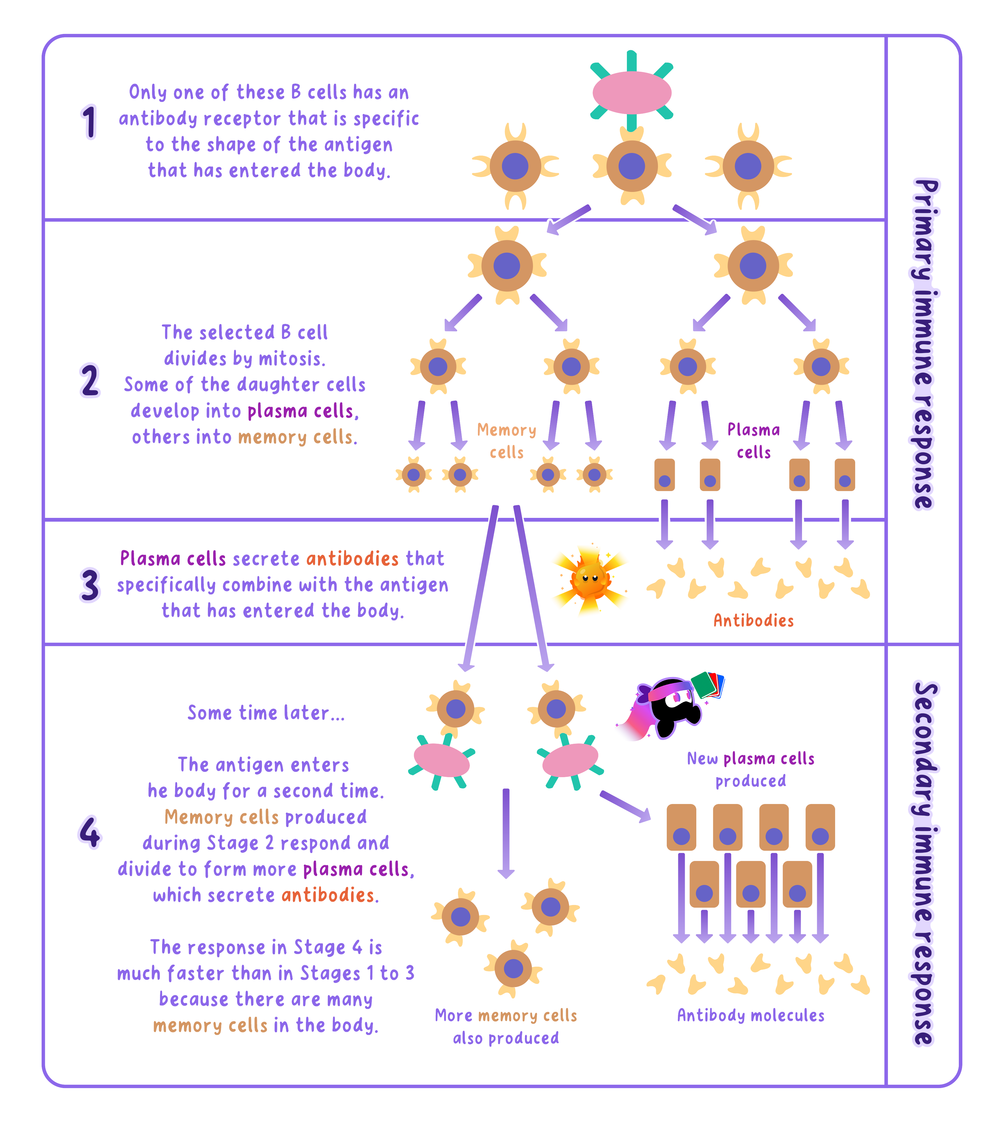
- For a secondary response, include memory cells, faster antibody production and pathogen destruction before serious symptoms.
- Do not say antibodies remember the pathogen because memory lymphocytes provide the memory.
The Response Is Antigen-specific
- Memory cells for one antigen do not protect against an unrelated antigen.
- A pathogen with changed surface antigens may escape an existing immune response.
- What is an antigen?
- How are antibodies specific?
- Why is the primary response slow?
- Why is the secondary response faster?
5.3.2 Response to immunisation
Vaccination Creates Memory Without Disease
Vaccine
A substance containing dead or inactive forms of a pathogen, or its antigens, that is given to make a person immune to a disease.
- A vaccine contains an inactive or harmless form of a pathogen, or antigens from it.
- The vaccine antigens cannot cause the full disease but are recognised as foreign.
- An inactive pathogen retains antigens that lymphocytes can recognise but cannot reproduce and cause the full disease.
Immunisation produces a primary immune response before exposure to the active pathogen.
Lymphocytes Produce Antibodies and Memory Cells
Vaccination
Introducing a harmless form of a pathogen into the body so the immune system produces antibodies and memory cells, giving protection against future infection.
- Lymphocytes with complementary receptors are activated and divide.
- Some cells produce specific antibodies, while others remain as memory cells.
Later Exposure Triggers a Rapid Response
Memory cell
A white blood cell that stays in the body after an infection so the body can respond faster and more strongly if the same pathogen returns.
- Memory cells recognise the same antigen when the live pathogen enters later.
- They divide rapidly and produce a larger concentration of antibodies in less time.
- The pathogen is destroyed before it causes serious symptoms.
- On later exposure, memory cells recognise the same antigen and produce antibodies before pathogen numbers become high enough to cause symptoms.
After vaccination, measles antigens activate memory cells, so later exposure produces antibodies quickly enough to prevent measles developing.
Booster Doses Restore Protection
- Memory-cell numbers or antibody concentrations can decline over time.
- A booster exposes the immune system to the antigen again and strengthens the memory response.
- A booster repeats antigen exposure, increasing memory-cell numbers and restoring protection when immunity has declined.
A vaccine does not act by killing pathogens directly because it trains the immune system to respond.
Write the Full Immunisation Chain
- Clonal selection and mitosis produce antibody-secreting cells and memory lymphocytes during the primary response.
- Use the order: inactive pathogen or antigen, lymphocyte activation, antibody production, memory-cell formation, faster secondary response.
- Include specificity by stating that antibodies bind the same antigen.
Immunity Is Produced Actively
Active immunity
Long-lasting protection that comes from the body making its own antibodies after meeting a pathogen, through infection or vaccination.
- Vaccination gives active immunity because the person's own lymphocytes produce antibodies and memory cells.
- This protection can last much longer than receiving ready-made antibodies.
- What does a vaccine contain?
- Why does it not cause the full disease?
- How do memory cells protect after vaccination?
- Why might a booster be needed?
5.3.3 Advantages and disadvantages of immunisation
Immunisation Protects Individuals and Populations
Vaccination
Introducing a harmless form of a pathogen into the body so the immune system produces antibodies and memory cells, giving protection against future infection.
- Vaccination produces memory cells, so later infection triggers a faster and stronger antibody response.
- Fewer vaccinated people develop disease, severe complications or long-term damage.
- Reducing infections also lowers healthcare demand and time lost from school or work.
- Vaccination reduces an individual's probability of infection and severe disease, but protection is not complete and differs between people.
An evaluation balances benefits, limitations and the circumstances that affect them.
Herd Immunity Reduces Transmission
Herd immunity
When enough of a population is immune to a disease that it cannot spread easily, which protects even those who are not immune.
- Herd immunity occurs when enough people are immune that an infected person is unlikely to meet a susceptible host.
- Transmission chains are interrupted, protecting people who cannot be vaccinated or who do not respond strongly.
- The proportion needed depends on how easily the disease spreads and how effective the vaccine is.
- Herd immunity works only when immunity is common enough to reduce the pathogen's reproductive spread through the population.
- People who cannot receive a vaccine benefit because they are less likely to contact an infected person, not because they personally become immune.
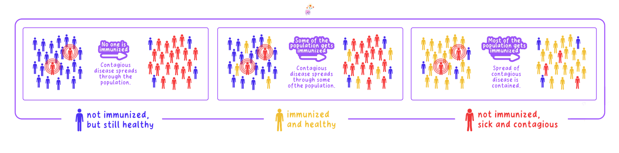
if most contacts of an infected person are immune, the pathogen reaches few susceptible people and an outbreak is less likely to grow.
Immunisation Has Limitations
- Vaccines can cause short-lived pain, swelling or fever, while serious adverse reactions are rare.
- No vaccine protects every recipient, and immunity may decline so booster doses are needed.
- Some pathogens change their antigens, reducing protection from an older vaccine.
- Developing, storing and delivering vaccines costs money, and low uptake prevents herd immunity.
- A fair evaluation compares the frequency and severity of vaccine side effects with the frequency and severity of the disease.
- Low uptake, reduced vaccine effectiveness or antigen changes can allow outbreaks despite an immunisation programme.
Do not treat a rare side effect and the disease risk as equally likely without comparing their probabilities and severity.
Reach a Justified Judgement
- For diseases that cause serious harm and have effective vaccines, the individual and population benefits usually outweigh the small risk of side effects.
- The judgement may change if effectiveness is low, protection is short-lived or the disease is uncommon and mild.
- For 'discuss', develop both advantages and disadvantages before reaching a judgement.
- Link herd immunity to interrupted transmission, not merely to a high percentage being vaccinated.
Use Evidence Carefully
- Compare infection rates in similar vaccinated and unvaccinated groups while considering age, exposure and health differences.
- A correlation between vaccination and lower disease incidence supports effectiveness but other variables must be considered.
- How does vaccination protect an individual?
- How does herd immunity protect susceptible people?
- Give two limitations of immunisation.
- What makes an evaluation judgement justified?
5.3.4 Antibiotics and bacterial infections
Antibiotics Target Bacterial Processes
Antibiotic
A medicine that kills bacteria or stops them growing inside the body, curing a bacterial disease. Antibiotics have no effect on viruses.
- An antibiotic is a medicine that kills bacteria or stops them reproducing.
- Different antibiotics inhibit processes such as bacterial cell-wall synthesis, protein synthesis or DNA replication.
- These targets differ from the host's cell processes, so a suitable dose harms bacteria more than body cells.
- Other antibiotics bind bacterial ribosomes or inhibit bacterial DNA replication, stopping growth and reproduction.
- Human cells lack peptidoglycan walls and have different ribosomes, which provides selective toxicity at a therapeutic dose.
- Penicillin inhibits enzymes that join bacterial cell-wall material, so a growing bacterium cannot resist internal pressure and may burst.
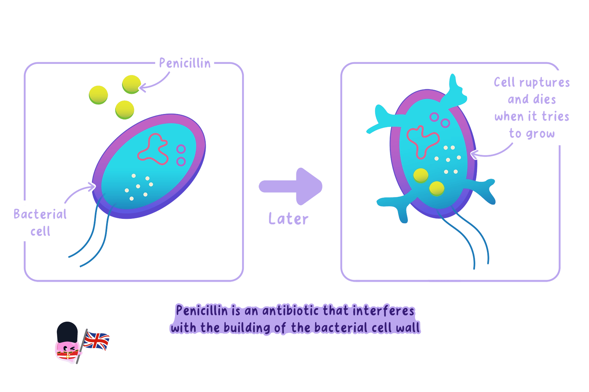
Selective toxicity depends on a bacterial target that is absent from, or sufficiently different in, human cells.
Antibiotics Do Not Treat Viruses
Virus
A non-cellular infectious particle made of a strand of genetic material enclosed in a protein coat, which can reproduce only inside a living host cell.
- Viruses have no cell wall, ribosomes or independent metabolism for an antibiotic to target.
- They reproduce using host-cell machinery, so antibiotics do not stop viral replication.
- Painkillers may reduce viral symptoms but do not kill the virus.
Do not prescribe antibiotics for colds or influenza because both are viral infections.
Correct Use Slows Resistance
Antibiotic resistance
When bacteria are no longer killed by an antibiotic that used to work, because resistant bacteria have been selected for and have spread.
- Random mutations can make some bacteria resistant to an antibiotic.
- Treatment kills susceptible bacteria, while resistant bacteria survive, reproduce and pass on resistance alleles.
- Using antibiotics only when necessary reduces selection pressure, and completing the prescribed course reduces surviving bacteria.
- Resistance alleles arise by mutation before treatment; the antibiotic selects resistant bacteria rather than causing directed mutation.
- Resistant survivors reproduce and pass resistance alleles to descendants, increasing the resistant proportion of the population.
penicillin inhibits bacterial cell-wall formation, so a growing bacterium takes in water and bursts while human cells are unaffected because they have no cell wall.
Choose Evidence-based Treatment
- A culture can be tested against antibiotic discs, and a larger zone of inhibition indicates greater bacterial growth inhibition under the test conditions.
- The chosen antibiotic must also reach the infection site safely inside the patient.
- Explain why an antibiotic works by naming the bacterial process it inhibits and stating why the host is not affected in the same way.
- For resistance, write the sequence variation, selection, survival and reproduction.
Separate Treatment From Symptom Relief
- Antibiotics treat the bacterial cause, while painkillers lower pain or fever without removing the pathogen.
- The immune system still removes dead bacteria and damaged cells.
- Why can antibiotics treat bacterial infections?
- Why do antibiotics not treat viruses?
- How does antibiotic resistance increase?
- What does a zone of inhibition show?
5.3.5 Aseptic techniques in culturing
Growing microorganisms
Culture medium
A nutrient jelly (agar) or liquid broth that supplies microorganisms with the nutrients they need to grow.
- A culture medium supplies water and nutrients so microorganisms can grow. It may be a liquid broth or nutrient agar in a Petri dish.
- On solid agar, one microorganism can divide repeatedly to form a visible colony. A spread of bacteria across the agar is called a bacterial lawn.
Aseptic technique
Aseptic technique
A set of precautions used when culturing microorganisms to prevent contamination of the culture and to stop unwanted microbes spreading.
- Aseptic technique prevents unwanted microorganisms contaminating the culture and reduces the chance that cultured microorganisms escape into the surroundings.
- An autoclave uses pressurised steam at a high temperature to sterilise culture medium and Petri dishes before use. Sterilisation kills microorganisms and resistant spores.
- A sterile inoculating loop is used to transfer microorganisms. If a metal loop is used, it is heated in a Bunsen flame until red hot and allowed to cool before touching the culture.
- The Petri-dish lid is opened only slightly and briefly. After inoculation, it is secured at two or three points so it stays covered but is not sealed completely.
State the purpose as well as the step: sterilising equipment kills contaminating microorganisms, while keeping the lid covered reduces entry and escape of airborne microorganisms.
Antimicrobial cultures
Inhibition zone
The clear area around an antibiotic or antiseptic where bacteria have been killed or stopped from growing.
- A paper disc containing an antibiotic, antiseptic or plant extract is placed on a bacterial lawn. The substance diffuses through the agar.
- Where bacterial growth is killed or inhibited, a clear zone of inhibition forms. A larger zone usually indicates a more effective antimicrobial substance when all control variables are kept constant.
Investigating antimicrobial substances
- Aim: Compare the effectiveness of antiseptics, antibiotics or plant extracts by measuring the clear zones they produce in a bacterial culture.
- Apparatus: Sterile nutrient-agar plate, safe bacterial culture, sterile inoculating loop, sterile forceps, filter-paper or prepared antibiotic discs, test substances, sterile-water control, marker pen, ruler, sticky tape, incubator, Bunsen burner, ethanol and eye protection.
- Method:
- Disinfect the bench and work beside a lit Bunsen burner.
- Flame a metal inoculating loop until red hot, then let it cool without touching anything.
- Lift the Petri-dish lid only slightly and spread the bacteria evenly over the agar to form a lawn.
- Sterilise the forceps, allow them to cool, and place equal-sized discs containing equal volumes of the test substances onto the agar. Add a disc containing sterile water as a control.
- Label the base of the plate.
- Secure the lid with two or three pieces of tape, but do not seal it all the way around.
- Incubate the plate upside down at 25 ∘C25\,^{\circ}\mathrm{C}25∘C for 242424 to 484848 hours.
- Without opening the plate, measure each clear zone twice at right angles and calculate the mean diameter.
- Variables: Change the antimicrobial substance. Measure the area of the zone of inhibition. Keep the bacterium, agar depth, disc diameter, volume and concentration of substance, spacing of discs, incubation time and temperature constant.
- Results: A clear ring forms where bacteria have been killed or prevented from reproducing. The sterile-water control should have no clear zone, showing that any inhibition is caused by the test substance rather than the paper disc.
- Maths: For a circular zone, calculate the mean diameter, use r=d2r=\frac{d}{2}r=2d, then calculate A=πr2A=\pi r^2A=πr2. Measure the whole clear zone, including the disc, and give the area in mm2\mathrm{mm}^2mm2.
- Watch out: Unequal disc sizes, different volumes of substance, overlapping zones or an uneven bacterial lawn make the comparison invalid. Repeat each treatment and calculate a mean.
- Safety: Incubate at 25 ∘C25\,^{\circ}\mathrm{C}25∘C rather than body temperature, never reopen an incubated plate, keep ethanol away from the flame, and autoclave cultures after use.
Interpreting the investigation
- The plate is inverted during incubation so condensation collects on the lid rather than dripping onto the agar and spreading colonies.
- School cultures are incubated at 25 ∘C25\,^{\circ}\mathrm{C}25∘C because temperatures closer to 37 ∘C37\,^{\circ}\mathrm{C}37∘C would favour microorganisms adapted to the human body, including potential pathogens.
- Why must culture medium and Petri dishes be sterilised before use?
- Why is a sterile-water disc included?
- Why is the plate incubated upside down at 25 ∘C25\,^{\circ}\mathrm{C}25∘C?
- A clear zone has a mean diameter of 18 mm18\,\mathrm{mm}18mm. Calculate its area.
5.3.6 Cross-sectional areas of cultures
Measuring circular areas
- Bacterial colonies and zones of inhibition are often approximately circular, so their cross-sectional area is calculated from the radius.
- Measure the diameter through the centre of the circle. If the edge is uneven, measure two perpendicular diameters and calculate their mean.
- Calculate the radius using r=d2r=\frac{d}{2}r=2d, then use A=πr2A=\pi r^2A=πr2.
****A clear zone has a mean diameter of 16 mm16\,\mathrm{mm}16mm. Its radius is r=162=8 mmr=\frac{16}{2}=8\,\mathrm{mm}r=216=8mm. Therefore A=π×82=201.1 mm2A=\pi\times 8^2=201.1\,\mathrm{mm}^2A=π×82=201.1mm2, which is 201 mm2201\,\mathrm{mm}^2201mm2 to three significant figures.
Comparing antimicrobial effects
- When the same microorganism and experimental conditions are used, a larger zone of inhibition provides evidence that the antimicrobial substance was more effective.
- Area is proportional to the square of the radius. A zone with twice the radius has four times the area, not twice the area.
- Use the same units throughout. If the radius is measured in millimetres, the area must be recorded in mm2\mathrm{mm}^2mm2.
Show every stage: write r=d2r=\frac{d}{2}r=2d, substitute into A=πr2A=\pi r^2A=πr2, keep extra calculator digits during the calculation, and round only the final answer with the correct squared unit.
Evaluating measurements
- An irregular zone is a source of measurement uncertainty. Taking two diameters at right angles and using their mean gives a more representative radius.
- A larger clear area does not by itself prove that a medicine will work better inside a person. Diffusion through agar, concentration and experimental conditions also affect zone size.
- State the equation for the area of a circular culture or clear zone.
- Why should two perpendicular diameters be measured for an uneven zone?
- A colony has a diameter of 12 mm12\,\mathrm{mm}12mm. Calculate its area.
- Explain why comparing diameters alone can be misleading.
5.3.7 Stages of developing new medicines
Discovering medicines
- Medicine development begins with the discovery of a substance that may prevent or treat disease. Candidates may come from microorganisms, plants or chemical synthesis.
- Scientists investigate how the candidate acts, then develop a suitable formulation and dose before testing whether it is safe and effective.
Preclinical testing
- Preclinical testing takes place before a medicine is given to human volunteers.
- The substance is tested on cells and tissues to examine its biological effect and identify obvious toxicity.
- It is then tested in living organisms to investigate how the whole body absorbs, distributes, breaks down and removes it, and to check for harmful effects.
- These tests provide evidence about toxicity, efficacy and a possible dose. A candidate that is ineffective or unacceptably harmful does not progress.
Clinical trials
Clinical trial
A stage of drug testing in which a new medicine is tested on human volunteers to check it is safe, that it works, and to find the right dose.
Placebo
A dummy treatment with no active drug, used in a trial so the real medicine's effect can be compared fairly.
- Clinical trials test the medicine in people. Early trials use a small number of healthy volunteers and a very low dose, which is increased carefully to investigate safety and side effects.
- Later trials use larger groups of patients to test whether the medicine treats the condition and to refine the effective dose.
- A placebo contains no active medicine. Comparing a treatment group with a placebo group helps show whether improvement is caused by the medicine rather than expectation or natural recovery.
- In a double-blind trial, neither the participants nor the researchers assessing them know who receives the medicine until the results are analysed. This reduces conscious and unconscious bias.
- Random allocation and large sample sizes reduce the effect of individual differences and make the groups more comparable.
For a question about drug testing, link each stage to its purpose: preclinical tests investigate toxicity and efficacy before human exposure; early clinical trials focus on safety and dose; later trials test effectiveness and uncommon side effects in more patients.
Review and monitoring
- Results are checked by independent scientists through peer review. Regulators examine the evidence before deciding whether the medicine can be licensed.
- After licensing, the medicine continues to be monitored because rare or long-term side effects may only appear when it is used by many people.
- Put discovery, preclinical testing and clinical testing in the correct order.
- What are the purposes of preclinical testing?
- Why are placebos and double-blind trials used?
- Why must a licensed medicine continue to be monitored?
5.3.8 Production of monoclonal antibodies
Monoclonal antibodies
Monoclonal antibody
An antibody made in the laboratory from identical cells, so all the antibodies are the same and bind to one specific antigen.
- A monoclonal antibody is one of many identical antibodies made from a single clone of cells, so every antibody binds to the same specific antigen.
- Their specificity allows a particular molecule or type of cell to be detected, located or targeted.
Producing hybridomas
Hybridoma
A cell made by fusing a lymphocyte with a tumour cell; it divides rapidly and produces large amounts of a single type of antibody.
- A lymphocyte that makes the desired antibody is selected. It produces the correct antibody but does not divide repeatedly.
- The lymphocyte is fused with a tumour cell, which divides rapidly, to form a hybridoma.
- Hybridoma cells combine both useful properties: they divide to form a clone and each cell produces the desired antibody.
- The clone is cultured, and the identical antibodies are collected and purified.
A complete production explanation must state both halves of the fusion: the lymphocyte provides the ability to make the desired antibody, while the tumour cell provides the ability to divide repeatedly.
Pregnancy tests
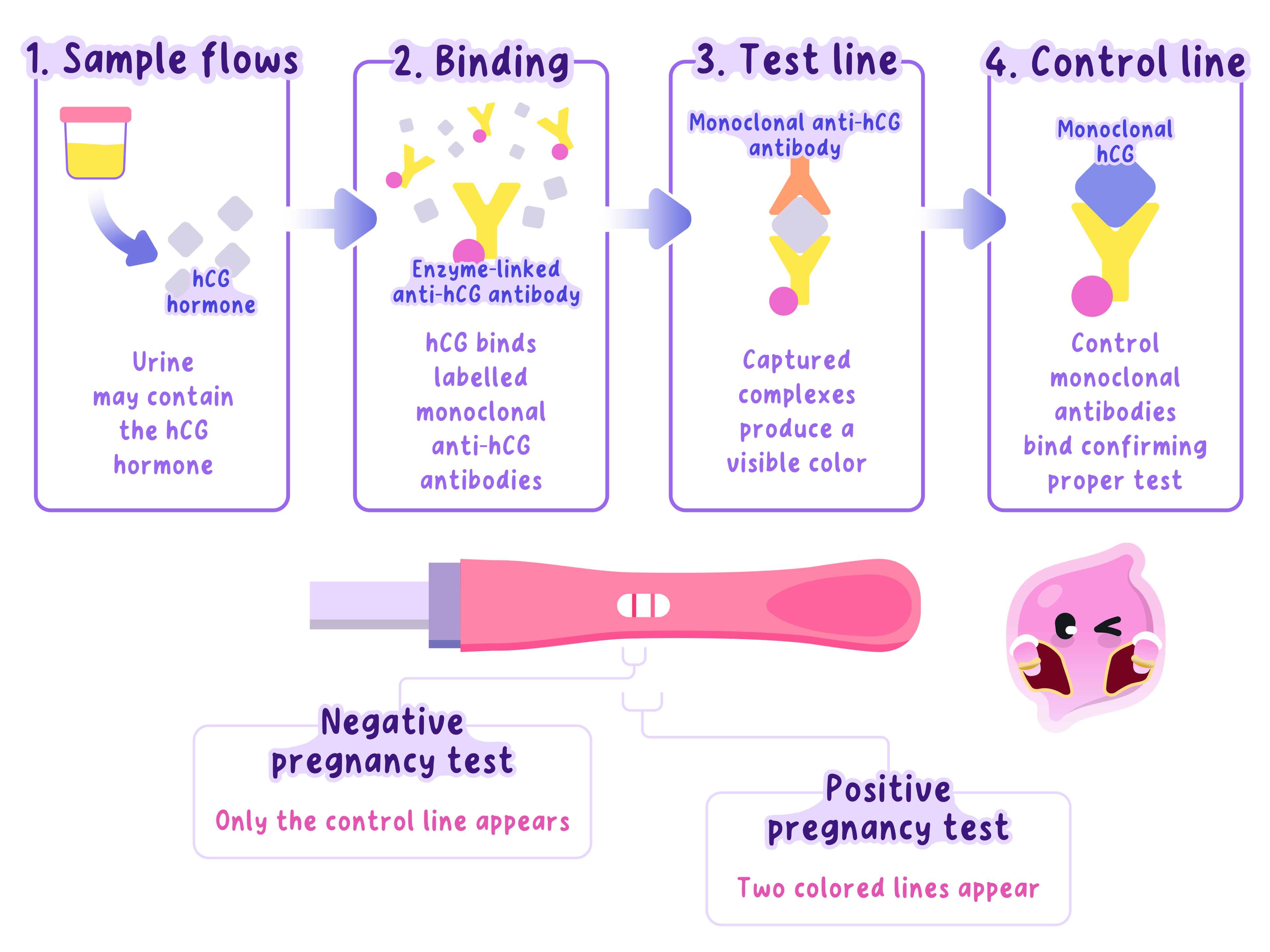
- Pregnancy tests detect the hormone hCG in urine using monoclonal antibodies that bind specifically to hCG.
- Urine carries coloured antibodies along the strip. If hCG is present, it binds to them and the complex is trapped by fixed antibodies at the test line, producing a coloured line.
- A separate control line traps excess coloured antibodies whether or not hCG is present. It shows that the liquid moved through the strip and that the test worked.
Diagnosis and treatment
- Monoclonal antibodies can be attached to fluorescent markers or radioactive substances. After binding to a target antigen, the marker reveals the position of cancer cells or a blood clot.
- For treatment, an antibody can bind to an antigen found mainly on cancer cells. It may block a growth signal or deliver a toxic drug or radioactive substance directly to the target cells.
- This targeting can damage fewer healthy cells than non-specific drug treatment or radiotherapy, so it may reduce side effects.
- Limitations include immune reactions, unexpected side effects, high production costs and the possibility that not every target cell carries the antigen.
****A scan shows radioactivity concentrated in one region after a labelled monoclonal antibody is injected. The evidence suggests that cells in this region carry the antigen complementary to the antibody, so the labelled antibodies accumulated there.
- Explain how a hybridoma combines the useful properties of two different cells.
- Why does a pregnancy test need both a test line and a control line?
- How can monoclonal antibodies locate a blood clot or cancer cells?
- Give one advantage and one limitation of targeted monoclonal-antibody treatment.
